Rotator Cuff Tear vs. Frozen Shoulder: A Doctor’s Guide to Spotting the Difference

Rotator Cuff Tear vs. Frozen Shoulder | Dr. Manu Mengi

Rotator Cuff Tear vs. Frozen Shoulder confusion is something I encounter frequently in my practice, with many patients mistakenly self-diagnosing their shoulder pain. These two conditions are often confused with each other, yet they present distinct symptoms and have significantly different recovery timelines.

In my experience, understanding the key differences can help you identify your condition more accurately. Rotator cuff tears typically have a sudden onset, particularly after activities like lifting something heavy or falling. At the same time, frozen shoulder develops gradually with pain that slowly worsens over weeks or months. The most telling difference? If you have a rotator cuff tear, you can likely lift your shoulder without assistance. However, with a frozen shoulder, you’ll face difficulty raising your arm by yourself.

Throughout this guide, I’ll walk you through the specific symptoms, causes, and treatments for both conditions. We’ll explore why frozen shoulder involves progressive stiffness accompanied by dull, aching pain (especially at night), while rotator cuff tears cause sharper, more localised pain when lifting or rotating the arm. By the end, you’ll understand why that stiffness in your shoulder joint might indicate one condition over the other.

This self-diagnosis often leads to anxiety. If you're in Mohali and struggling with shoulder pain, the most important step is to get a professional evaluation. Book an appointment with a specialist to get an accurate diagnosis and peace of mind.

Understanding the Shoulder: Anatomy and Function

The shoulder represents one of the most complex and mobile joints in the human body. To fully understand the differences between rotator cuff injuries and frozen shoulder, we must first examine the intricate architecture that allows for this remarkable range of motion. Unlike other joints that prioritise stability, the shoulder sacrifices some stability for mobility, making it uniquely susceptible to certain conditions.

The role of the rotator cuff in shoulder movement

The rotator cuff forms the cornerstone of shoulder function – a group of four muscles and their tendons that surround the shoulder joint like a protective collar. This muscular quartet connects your shoulder blade (scapula) to your upper arm bone (humerus) and works together to enable precise movement while maintaining joint stability.

The four muscles that make up this critical structure include:

  • Supraspinatus: Initiates the first 15 degrees of arm abduction (lifting your arm away from your body)
  • Infraspinatus: Powers external rotation of the arm away from your body
  • Teres minor: Assists with external rotation and stabilisation
  • Subscapularis: Enables internal rotation (turning your arm toward your body)

A helpful mnemonic for remembering these muscles is “SITS” – Supraspinatus, Infraspinatus, Teres minor, and Subscapularis.

The primary biomechanical role of these muscles extends beyond their movement function. They compress the head of the humerus against the shallow glenoid cavity of the scapula, creating what doctors call “dynamic stability”. Without this muscular support, the disproportionate size between the humeral head and the glenoid fossa (a 4:1 surface area ratio) would make the joint highly unstable.

Furthermore, these muscles don’t work in isolation. For example, during abduction, the supraspinatus handles the initial 0-15 degrees of movement, then the deltoid muscle takes over from 15-90 degrees. Finally, the scapula must rotate to achieve full range of motion, which is accomplished by the trapezius and serratus anterior muscles.

What is the shoulder capsule, and why does it matter

Surrounding the glenohumeral joint is a fibrous sheath called the shoulder capsule. This structure extends from the anatomical neck of the humerus to the rim of the glenoid fossa, completely enveloping the joint. Unlike other joint capsules in your body, the shoulder capsule has a unique characteristic – it’s remarkably loose and lax, allowing the bones to separate by more than 2.5 cm.

This laxity isn’t a design flaw but rather a deliberate feature that permits the extensive freedom of movement characteristic of the shoulder joint. Nevertheless, this same feature also makes the joint more vulnerable to instability and injury.

The capsule doesn’t work alone in providing joint stability. It’s reinforced by:

  • The glenoid labrum – a fibrocartilaginous ring that deepens the glenoid cavity
  • Ligaments that connect bones to bones
  • Tendons that connect muscles to bones
  • The rotator cuff muscles themselves, which fuse with the capsule

Additionally, fluid-filled sacs called bursae surround the capsule, facilitating smooth movement by reducing friction between the structures. The synovial membrane inside the capsule produces lubricating fluid that further aids motion.

Interestingly, when the shoulder capsule becomes damaged or inflamed, it can lead to adhesive capsulitis – commonly known as frozen shoulder. In this condition, the capsule thickens and becomes stiff, significantly restricting movement and causing pain.

How shoulder mobility is affected by injuries

Given the complexity of the shoulder’s structure, it’s not surprising that injuries can substantially impact its function. Shoulder pain affects approximately 18 million Americans each year, with rotator cuff tears accounting for a significant number of these cases.

Rotator cuff tears can occur through trauma, overuse, or age-related degeneration. These tears affect one in every four adults aged 60 years and older, with the supraspinatus tendon being most commonly involved. Risk factors include smoking, high cholesterol, and family history.

When the rotator cuff is damaged, patients typically experience:

  • Pain during overhead activities
  • Limited range of motion
  • Weakness in the shoulder
  • Difficulty with daily activities

Studies using three-dimensional motion analysis have shown that individuals with symptomatic rotator cuff tears exhibit abnormal shoulder motion when performing common daily tasks compared to those without tears. This dysfunction occurs because rotator cuff injuries compromise the joint’s dynamic stability, creating muscle force imbalances.

Alternatively, when the shoulder capsule becomes inflamed and thickened in adhesive capsulitis (also known as frozen shoulder), patients experience progressive stiffness and significant pain. This condition not only limits one movement but can also restrict motion in multiple directions simultaneously.

Research shows that patients with rotator cuff-related shoulder pain demonstrate significantly less shoulder flexion, internal and external rotation range of motion compared to individuals without shoulder problems. This finding helps explain why these conditions have such a profound impact on everyday activities.

Both conditions can make simple tasks challenging – whether reaching for objects on high shelves, putting on a coat, or even combing your hair. Understanding the underlying anatomy helps explain why these different conditions can sometimes present with similar symptoms despite having distinct causes.

As we've seen, both conditions severely impact your daily life. The key to effective treatment is a correct diagnosis. If you're experiencing limited motion or pain, consider visiting a 
Specialised Shoulder Clinic in Mohali for a comprehensive assessment.

Rotator Cuff Tear: Causes, Symptoms, and Diagnosis

Rotator cuff tears represent one of the most common shoulder injuries I diagnose in my practice. These tears are the leading cause of shoulder pain and shoulder-related disability, affecting millions of people each year. In fact, more than 2 million Americans experience some rotator cuff problem annually. While these injuries can affect people of all ages, they’re particularly common in adults over 40, with prevalence increasing significantly with age.

As a physician, I find that many patients are surprised to learn that some individuals have rotator cuff tears without experiencing any symptoms whatsoever. This silent presentation can make diagnosis challenging in certain cases.

Common causes: trauma, overuse, and ageing

Rotator cuff tears typically fall into two distinct categories: acute traumatic tears and degenerative tears. Understanding this distinction is crucial for proper treatment.

Traumatic (Acute) Tears occur suddenly as a result of a specific injury. In my experience, patients often report:

  • Falling on an outstretched arm
  • Lifting something heavy with a jerking motion
  • Experiencing sudden force while pushing or pulling
  • Sustaining an injury during a shoulder dislocation

These traumatic tears account for approximately 8% of all rotator cuff injuries. They tend to stem from higher energy mechanisms and typically cause full-thickness tears, whereas degenerative tears often begin as partial-thickness injuries. Moreover, traumatic tears generally are larger and more likely to involve the subscapularis muscle.

Degenerative (Wear-Related) Tears develop gradually over time due to natural ageing processes. These constitute the majority of rotator cuff injuries I see in my practice. Several factors contribute to these tears:

  1. Repetitive stress – Certain activities that involve repeated shoulder motions put significant strain on the rotator cuff. Sports like baseball, tennis, rowing, and weightlifting carry an increased risk of overuse. Likewise, many occupations requiring repetitive overhead movements can lead to progressive damage.
  2. Decreased blood supply – As we age, blood flow to our rotator cuff tendons diminishes. Without adequate blood supply, the body’s natural ability to repair tendon damage becomes impaired, ultimately leading to tissue breakdown.
  3. Bone spurs, also known as age-related bone overgrowth (osteophytes), can develop on the acromion, creating friction against the rotator cuff tendons when you move your arm.

Essentially, the supraspinatus tendon bears the brunt of most rotator cuff injuries, as it endures the majority of shoulder-stabilising strain. Approximately 53% of patients with supraspinatus tears also have involvement of the infraspinatus.

Concerning symptoms, patients typically experience:

  • Pain localised to the anterior shoulder or greater tuberosity area
  • Discomfort when lying on the affected shoulder
  • Pain with overhead activities
  • Night pain that interrupts sleep
  • Weakness in the affected arm
  • Crackling or popping sensations during certain movements
  • A positive painful arc sign during clinical examination

Diagnosing rotator cuff tears involves several components. First, I take a detailed history, including the mechanism of injury and activities that aggravate or relieve symptoms. Next, I perform a thorough physical examination, checking for tenderness, range of motion, and specific clinical tests designed to stress the rotator cuff tendons. Additionally, diagnostic imaging plays a crucial role in confirming the diagnosis and determining the extent.

Specifically, when patients present with an inability to elevate their arm after a traumatic event, despite normal radiographs, I maintain a high index of suspicion for rotator cuff pathology, as these injuries can often be missed during initial assessment.

While this guide helps you understand the differences, only a physical examination can confirm your condition. For expert rotator cuff tear treatment in Mohali or frozen shoulder treatment in Mohali, seeking help from a qualified orthopaedic doctor is crucial. Learn more about your Shoulder Pain Treatment Options in Mohali.

Key Takeaways

Understanding the differences between rotator cuff tears and frozen shoulder can help you identify your condition and seek appropriate treatment more effectively.

• Onset patterns differ significantly: Rotator cuff tears typically occur suddenly after trauma or lifting, while frozen shoulder develops gradually over weeks to months with progressively worsening pain.

• Movement ability is the key differentiator: With rotator cuff tears, you can usually lift your arm without assistance, but frozen shoulder severely limits your ability to raise your arm independently.

• Pain characteristics vary between conditions: Rotator cuff tears cause sharp, localised pain during lifting or rotation, while frozen shoulder presents as dull, aching pain that’s particularly troublesome at night.

• Age and activity patterns matter for diagnosis: Rotator cuff tears affect over 25% of adults aged 60+, often from repetitive overhead activities or sudden trauma. At the same time, a frozen shoulder involves inflammation and stiffness of the capsule.

• The rotator cuff’s four muscles (SITS) – Supraspinatus, Infraspinatus, Teres minor, and Subscapularis – work together to provide dynamic shoulder stability, making tears particularly disabling for daily activities.

Both conditions significantly impact quality of life; however, accurate identification of symptoms, such as pain location, movement limitations, and onset patterns, enables more targeted treatment approaches and better outcomes.

FAQs

Q1. How can I differentiate between a frozen shoulder and a rotator cuff tear?

A1. The key difference lies in your ability to move your arm. If you can lift your arm independently, it’s more likely to be a rotator cuff tear. Frozen shoulder severely limits your ability to raise your arm without assistance. Additionally, rotator cuff tears often have a sudden onset, while frozen shoulder develops gradually over time.

Q2. Is it possible for a rotator cuff tear to be misdiagnosed as frozen shoulder?

A2. Yes, these conditions can be confused. Some partial rotator cuff tears may not cause pain and can mimic symptoms of a frozen shoulder. However, a thorough medical examination, including imaging tests, can help differentiate between the two conditions.

Q3. What are the most effective treatments for frozen shoulder?

A3. Most cases of frozen shoulder improve on their own within 12 to 18 months. For severe or persistent symptoms, treatments may include physical therapy, corticosteroid injections, and in some cases, surgery. Steroid injections can be particularly effective when administered early in the progression of the condition.

Q4. What other conditions can be mistaken for a rotator cuff tear?

A4. Several shoulder conditions can present similar symptoms to rotator cuff tears, including biceps tendinitis, shoulder impingement syndrome, labral tears, arthritis, bursitis, and tendinitis. Each of these conditions affects different parts of the shoulder structure but may cause similar pain or movement limitations.

Q5. How does age affect the likelihood of developing rotator cuff tears?

A5. The risk of developing a rotator cuff tear increases significantly with age. They are particularly common in adults over 40, with more than 25% of adults aged 60 and older experiencing some form of rotator cuff tear. This is often due to natural wear and tear, decreased blood supply to the tendons, and the development of bone spurs that can irritate the rotator cuff over time.

Still unsure about your shoulder pain? Don't let it limit your life any longer. If you're looking for an Affordable Shoulder Treatment in Mohali or simply want to consult with a trusted expert, reach out to us today. Schedule a consultation to get a clear diagnosis and start your journey to recovery.
📞 Call Us: +91 87250 11661 | 📍 Find Our Clinic: Google My Business

📖 Learn More About Shoulder Pain:

Dr. Manu Mengi – Shoulder Pain Specialist in Mohali

With extensive experience in diagnosing and treating complex shoulder conditions, Dr. Manu Mengi is a leading Shoulder Pain Specialist in the Mohali and Tricity region. He is dedicated to providing personalised care, utilising accurate diagnosis and evidence-based treatment plans to help patients regain pain-free movement and improve their quality of life.

How Physiotherapy Actually Fixes Shoulder Pain: A Physiotherapist’s Guide

Shoulder pain affects approximately 18% of people worldwide, with women experiencing it more frequently than men. As a physiotherapist specialising in shoulder rehabilitation, I’ve seen firsthand how physiotherapy for shoulder pain offers one of the most effective, drug-free approaches to treatment. In fact, with 18 to 26 percent of the population struggling with persistent shoulder discomfort, understanding proper rehabilitation techniques has never been more important.

When it comes to non-surgical management for shoulder pain, the right exercises and therapy protocols can make a significant difference. Research consistently shows that both home-based and supervised exercise programs are more effective than no intervention. Throughout this guide, I’ll share the exact techniques we use in our practice, from mobilisation techniques for frozen shoulder to progressive strengthening exercises that actually restore function.

Whether you’re dealing with a rotator cuff injury, frozen shoulder, or chronic pain from poor posture, this comprehensive guide will walk you through how physiotherapy effectively addresses the root causes of shoulder problems—not just the symptoms. Let’s explore how we can get your shoulders moving freely again, without surgery or medication.

Understanding the Root Causes of Shoulder Pain

The shoulder is among the most complex and mobile joints in our body, making it particularly vulnerable to various issues. Behind most shoulder pain cases lies a combination of anatomical, lifestyle, and medical factors that physiotherapists must identify before developing effective treatment plans.

Common injuries and overuse patterns

Overuse injuries account for a significant portion of shoulder problems, especially among athletes and people with repetitive work movements. In baseball players, shoulder injuries represent 12% to 19% of all injuries, while swimmers experience an even higher rate—between 23% and 38% within a single year. These statistics highlight how repetitive overhead movements can gradually damage shoulder structures.

Overhead athletes typically develop specific modifications in their shoulders, including increased external rotation and decreased internal rotation. Furthermore, these changes can occur not just after years of practice but sometimes after just a single season.

Common overuse conditions include:

  • Rotator cuff tears, which affect the four small muscles that stabilise your shoulder joint
  • Impingement syndrome, where tendons get pinched between bones during arm movement
  • Bursitis, involving inflammation of the fluid-filled sacs that cushion joint movement
  • Tendinitis, particularly affecting the rotator cuff and biceps tendons

Scapular dyskinesis (abnormal shoulder blade movement) is notably prevalent among overhead athletes (61%) compared to non-overhead athletes (33%). This condition often accompanies other shoulder problems and contributes to pain during movement.

Medical conditions like arthritis and frozen shoulder

Arthritis significantly impacts shoulder function, with osteoarthritis being the most common type. This “wear-and-tear” arthritis gradually breaks down cartilage, eventually allowing bone-on-bone contact that causes pain and stiffness.

Other arthritis types affecting the shoulder include:

  • Rotator cuff tear arthropathy, developing after large rotator cuff tears
  • Rheumatoid arthritis, an autoimmune condition that causes joint inflammation
  • Post-traumatic arthritis, occurring after shoulder injuries or dislocations

Frozen shoulder (adhesive capsulitis) presents another challenging condition. Although its exact cause remains unclear, certain groups face a higher risk. Women between 40-60 years old are more susceptible, as are individuals with diabetes (10%-20% of diabetic patients develop this condition). Additionally, heart disease, thyroid disorders, and Parkinson’s disease correlate with increased frozen shoulder risk.

Postural habits and lifestyle triggers

Poor posture has emerged as a significant yet often overlooked contributor to shoulder dysfunction. Studies consistently show a strong association between posture and shoulder pain. Forward head posture, rounded shoulders, and increased thoracic kyphosis (mid-back rounding) all contribute to shoulder impingement by altering normal shoulder mechanics.

Postural deviations change how shoulder muscles function by:

  • Affecting the positioning of the shoulder blade
  • Reducing the efficiency of the shoulder muscles
  • Limiting available space for tendon movement

Metabolic factors also play a surprising role in shoulder health. Research indicates connections between shoulder pain and waist circumference, smoking, and metabolic syndrome. In men specifically, type 2 diabetes and carotid intima-media thickness (an indicator of atherosclerosis) show associations with shoulder pain.

Understanding these root causes is essential for proper physiotherapy treatment planning. Instead of merely addressing symptoms, identifying the underlying factors allows us to create targeted interventions that provide lasting relief and functional improvement.

Is your shoulder pain rooted in overuse, posture, or an underlying condition? Stop guessing and get a professional diagnosis. Book your assessment with Dr. Aayushi, a Leading Physiotherapist in Mohali, at the Best Physio Clinic in Mohali. Call 0172 - 3137922 to schedule your visit.

How Physiotherapy Approaches Shoulder Pain

When a patient arrives at my clinic with shoulder discomfort, a methodical approach is essential for effective treatment. Proper physiotherapy for shoulder pain involves a three-stage process that begins with a comprehensive assessment, moves to personalised planning, and considers optimal timing for intervention.

Initial assessment and diagnosis

A thorough diagnostic process forms the foundation of successful shoulder rehabilitation. My assessment typically starts with a detailed patient history, gathering information about how and when the pain began, any previous shoulder issues, and how symptoms affect daily activities. This anamnestic evaluation helps rule out red flags and guides my subsequent physical examination.

The physical examination involves several key components:

  • Visual observation for asymmetry, swelling, or postural abnormalities
  • Careful palpation to identify tender points, tissue texture changes, and temperature differences
  • Range of motion assessment in all functional planes (flexion, extension, abduction, adduction, internal/external rotation)
  • Strength testing of shoulder muscle groups
  • Neurological screening if warranted

For complex cases, I may recommend diagnostic imaging. X-rays can reveal osteoarthritis, acromioclavicular joint issues, or calcific tendinitis. MRIs prove valuable for evaluating soft tissue structures like muscles and ligaments, whereas CT scans provide detailed bone imaging. Ultrasound offers real-time visualisation of shoulder structures during movement.

Nevertheless, it’s worth noting that relying solely on special tests for diagnosis has limitations. Recent research demonstrates that many shoulder special tests lack sufficient sensitivity or specificity when used in isolation. Consequently, I interpret test outcomes within the context of the complete clinical picture.

Creating a personalised treatment plan

Once I’ve established a clear understanding of the underlying issues, I develop a customised treatment approach. The goal is straightforward—to restore movement and functionality throughout the affected area.

Treatment plans typically incorporate multiple elements based on individual needs. For most shoulder conditions, I recommend relative rest initially, followed by a gradual return to normal activities within pain limitations. This approach prevents the detrimental effects of prolonged immobility.

For evidence-based interventions, research suggests equivalent short-term benefits between supervised exercise programs and steroid injections. However, patients receiving physiotherapy are less likely to require additional medical consultations afterwards.

My personalised plans often include:

  • Manual therapy techniques to improve joint mobility
  • Targeted strengthening exercises for stability
  • Modalities like ultrasound or TENS for pain management
  • Education on proper posture and movement patterns
  • Progressive rehabilitation protocols matched to the specific condition

When to start shoulder physiotherapy after injury

Regarding timing, the consensus is clear—sooner is better. Early intervention prevents conditions from worsening and reduces the likelihood of requiring surgery. This is particularly crucial for overuse injuries, which often become apparent only after significant damage has occurred.

Patients who delay treatment typically face extended recovery times and potentially higher treatment costs. Moreover, early physiotherapy helps identify and correct problematic movements or habits that might otherwise lead to recurring injuries.

For acute injuries causing intense pain, immediate medical care is essential. However, with less severe discomfort, waiting a few days may be reasonable to see if rest resolves the issue. If symptoms persist beyond two weeks despite self-care attempts, professional evaluation becomes necessary.

After surgery, a structured conditioning program helps patients return to daily activities safely. These programs typically continue for 4-6 weeks but can be maintained long-term for ongoing shoulder health. Generally, performing exercises 2-3 days weekly maintains strength and range of motion effectively.

Ready for a personalised treatment plan designed by Mohali's Top Physiotherapist? Don't wait for the pain to worsen. Start your journey to recovery today at our Physiotherapy Clinic in Mohali. Contact us at 0172 - 3137922 or visit our website to learn more.

Core Physiotherapy Treatments That Work

After proper assessment, physiotherapists employ several evidence-based treatments that effectively address shoulder pain. The right combination of these therapies can significantly improve outcomes, often preventing the need for surgical intervention.

Manual therapy and mobilisation techniques

Manual therapy forms the cornerstone of shoulder rehabilitation. I frequently use joint mobilisation to improve range of motion and reduce pain by gently manipulating the glenohumeral, scapulothoracic, sternoclavicular, and acromioclavicular joints. This technique has proven particularly effective for conditions like adhesive capsulitis and subacromial pain syndrome.

Soft tissue mobilisation targets muscle tightness and adhesions, focusing on the rotator cuff muscles, deltoid, pectoralis major/minor, and upper trapezius. Meanwhile, muscle energy techniques require patient participation—I provide resistance as patients contract specific muscles to enhance rotator cuff strength and scapular stability.

For immediate pain relief, strain-counterstrain positioning effectively addresses trigger points and muscle guarding. These techniques collectively improve joint mobility, muscle flexibility, tissue quality, and neuromuscular control.

Electrotherapy and interferential therapy for frozen shoulder

Electrotherapy modalities deliver energy (electrical, sound, light, thermal) into body tissues to reduce pain and enhance function. For frozen shoulder, interferential therapy (IFT) shows promising results. IFT uses medium-frequency electrical currents that penetrate deeply with less skin resistance, making treatment more comfortable.

Research demonstrates that both electroacupuncture and interferential electrotherapy significantly improve Constant Murley Assessment scores and reduce visual analogue scale pain scores in frozen shoulder patients, with benefits maintained at least six months post-treatment. Similarly, low-level laser therapy (LLLT) combined with exercise proves more effective than placebo plus exercise, reducing pain scores by 19 points and improving function by 12 points.

Heat and cold therapy for pain relief

The timing of temperature therapy is crucial for optimal results. For acute injuries (within 48-72 hours), ice therapy is preferred. Cold constricts blood vessels, reducing inflammation and numbing pain. Apply ice wrapped in a towel to the affected shoulder for 15-20 minutes every 2-3 hours.

Conversely, heat therapy works best for chronic conditions or after the acute phase (post-72 hours). Heat increases blood circulation, relaxes muscles, and improves flexibility. Apply a heating pad for 15-20 minutes before exercises to enhance their effectiveness.

Alternating between hot and cold treatments can also provide effective relief for persistent shoulder pain.

Non-surgical management for shoulder pain

Beyond the aforementioned treatments, comprehensive non-surgical management includes several other approaches. Physical therapy strengthens shoulder muscles, increases flexibility, and corrects joint positions that may aggravate pain.

For temporary relief and protection, shoulder braces or slings can aid recovery by restricting movement and providing compression. Furthermore, injection-based procedures like corticosteroid injections help reduce inflammation in conditions such as frozen shoulder, rotator cuff tears, and subacromial bursitis.

The combination of these evidence-based treatments, customised to each patient’s specific condition, offers effective relief without surgery for most shoulder conditions.

Experience the difference expert manual therapy and electrotherapy can make. If you're seeking the best non-surgical management for shoulder pain, trust the experts at the Best Physiotherapist Clinic in Mohali. Call 0172 - 3137922 to book your session with Dr. Aayushi and start your treatment.

Essential Exercises for Shoulder Recovery

Physical exercise forms the foundation of successful shoulder rehabilitation. Throughout my practice, I’ve found certain exercises consistently deliver remarkable results across various shoulder conditions.

Pendulum exercises for frozen shoulder

Pendulum exercises, often called Codman exercises, use gravity to create passive motion without stressing shoulder muscles. These gentle movements help relax the shoulder and neck muscles while enabling passive range of motion. For frozen shoulder patients, I recommend starting with small circular motions (about 1 foot in diameter) and gradually increasing to larger movements. Perform these exercises 5 times daily, allowing your arm to hang naturally while leaning forward.

Wand exercises for shoulder mobility

Wand exercises utilise a simple cane or stick to improve mobility through assisted movements. For shoulder flexion, hold the wand with both hands and use your unaffected arm to guide the affected arm upward. External and internal rotation can be practised by holding the wand horizontally with bent elbows, moving side-to-side. These exercises effectively increase the range of motion across multiple planes, making them perfect for post-surgical recovery.

Isometric shoulder exercises for early rehab

Early in rehabilitation, isometric exercises provide strength benefits without requiring joint movement. Standing facing a wall, place your fist against it with a folded towel for comfort, then press gently for 5 seconds. Repeat this process for flexion, abduction, extension, and rotation directions, performing 10-15 repetitions per direction. These exercises prove particularly beneficial after surgery when movement remains restricted.

Scapular stabilisation and retraction exercises

Scapular control remains crucial for proper shoulder function. Scapular retraction (shoulder blade squeezes) strengthens stabilising muscles by bringing the shoulders back and down. Beyond that, the ITYW sequence targets different aspects of scapular control—lie facedown and perform arm movements resembling each letter shape. Proper stabilisation reduces impingement risk by maintaining ideal joint positioning.

Shoulder abduction with the theraband

Theraband abduction strengthens the shoulder muscles that lift your arm sideways. Standing with good posture, grasp both ends of the band with palms facing upward and elbows bent at 90°. Next, squeeze the band while lifting both elbows outward, holding briefly before returning to the starting position.

Resistance band exercises for strength

Resistance bands offer versatile, joint-friendly strengthening options. For comprehensive shoulder rehabilitation, incorporate front raises, lateral raises, and band pull-aparts. Typically, perform 2-3 sets of 10-15 repetitions for each exercise. Bands become more resistant as they stretch, allowing natural progression as strength improves.

Performing these exercises correctly is key to your recovery. Get expert guidance to ensure you're on the right track. Schedule a consultation at our Physio Clinic in Mohali with Dr. Aayushi for a supervised and effective exercise regimen. Call 0172 - 3137922 now.

Rehabilitation Protocols and Long-Term Care

Structured rehabilitation programs offer a roadmap for optimal shoulder recovery. These evidence-based protocols guide the progression from initial injury through complete functional restoration.

Frozen shoulder physiotherapy protocol

Successful frozen shoulder management requires a stage-specific approach. Throughout the freezing phase (painful stage), gentle mobilisation exercises within pain tolerance coupled with short-duration stretches (1-5 seconds) work best. Subsequently, as patients transition to the frozen stage, strengthening exercises like scapular retraction and isometric shoulder rotations become crucial for maintaining muscle strength. Finally, during the thawing phase, both stretching and strengthening intensify with longer holding durations. Applying heat before exercises enhances effectiveness by increasing tissue flexibility.

Physiotherapy after rotator cuff repair

Following rotator cuff surgery, recovery progresses through distinct phases. Initially, a 6-week immobilisation period with sling protection promotes tendon-to-bone healing. According to research, mature tendon-to-bone healing takes approximately 15 weeks, with Sharpey fibres developing around 12 weeks post-surgery. Hence, excessive tension must be avoided during this critical period. At 10-14 weeks, gentle active-assisted movements begin, followed by active motion at 14-18 weeks. Resistance training starts only after 18 weeks.

Shoulder strengthening after immobilisation

Post-immobilisation strengthening requires gradual progression. Starting with isometric exercises provides strength benefits without risking muscle integrity. Thereafter, incorporate progressive loading through resistance bands and lightweight exercises. Beginning with 3 sets of 8 repetitions and advancing to 3 sets of 12 demonstrates effective progression. Exercising 2-3 days weekly maintains optimal strength gains.

Posture correction in rotator cuff dysfunction

Scapular positioning fundamentally affects shoulder function. Indeed, alterations in scapular position appear in 68-100% of shoulder injury cases. Proper scapular movement preserves central humeral head positioning. Address associated impairments through scapular neuromuscular education in side-lying positions before initiating rotator cuff exercises.

Home physiotherapy for frozen shoulder

For home-based management, always warm shoulders with a 10-15 minute shower or moist heat before exercises. Effective home exercises include pendulum stretches, towel stretches, finger walks, cross-body reaches, and armpit stretches performed 10-20 times daily. Alongside stretches, incorporate outward and inward rotation exercises with resistance bands for complete rehabilitation.

Need a structured, stage-specific rehabilitation protocol for your shoulder condition? Our clinic offers expert-guided programs for frozen shoulder, post-surgical rehab, and more. Trust your recovery to the Best Physio Clinic in Mohali. Book your appointment with Dr. Aayushi today: 
0172 - 3137922.

Conclusion

Shoulder rehabilitation requires patience and consistency, yet the results make the journey worthwhile. Throughout this guide, we’ve explored how physiotherapy effectively addresses shoulder pain through targeted interventions rather than simply masking symptoms. Most shoulder conditions respond well to proper assessment and personalised treatment plans, especially when started early after injury.

Physiotherapy offers a comprehensive approach that combines manual techniques, therapeutic modalities, and progressive exercises to restore function. Certainly, the exact protocol depends on your specific condition – whether you’re dealing with frozen shoulder, rotator cuff injury, or postural dysfunction.

The exercises outlined here form the foundation of most shoulder rehabilitation programs. Pendulum movements provide gentle mobilisation, while resistance training builds the strength necessary for long-term stability. Additionally, addressing scapular control proves essential for preventing future problems.

Remember that recovery happens in stages. Initially, pain management and gentle movement take priority. Subsequently, we focus on rebuilding strength and endurance. Finally, functional activities help transition back to normal daily movements without pain or restriction.

Home exercise programs play a crucial role in your recovery. Though I guide patients through proper techniques in the clinic, their commitment to consistent practice at home ultimately determines success. Therefore, incorporating these exercises into your daily routine accelerates healing and prevents recurrence.

Physiotherapy represents the gold standard for non-surgical shoulder pain management. With proper guidance and dedication to your rehabilitation program, you can expect significant improvement in pain levels and functional ability. Your shoulders support countless daily activities – investing in their proper care through physiotherapy ensures they continue serving you well for years to come.

Key Takeaways

Understanding how physiotherapy addresses shoulder pain can help you make informed decisions about treatment and recovery. Here are the essential insights every patient should know:

• Early physiotherapy intervention prevents conditions from worsening and reduces the likelihood of requiring surgery

• Proper assessment identifies root causes like overuse patterns, medical conditions, and postural habits rather than just treating symptoms

• Manual therapy, electrotherapy, and temperature treatments provide evidence-based pain relief without medication

• Progressive exercises from pendulum movements to resistance training restore both mobility and strength effectively

• Stage-specific rehabilitation protocols ensure safe recovery, especially after surgery or during frozen shoulder phases

• Consistent home exercise programs accelerate healing and prevent recurrence when combined with professional guidance

The key to successful shoulder rehabilitation lies in addressing underlying causes through personalised treatment plans. With proper physiotherapy guidance and patient commitment to prescribed exercises, most shoulder conditions can be effectively managed without surgery, leading to lasting pain relief and restored function.

You don't have to live with shoulder pain. Take the first step towards a pain-free life. Consult with Dr. Aayushi, widely regarded as the Best Physiotherapist in Mohali, for a comprehensive assessment and a personalised treatment plan. Visit us at our Physiotherapy Clinic in Mohali or 
call 0172 - 3137922 to schedule your appointment.

FAQs

Q1. How long does it typically take to recover from shoulder pain with physiotherapy?

A1. Recovery time varies depending on the specific condition and severity, but most patients see significant improvement within 6-12 weeks of consistent physiotherapy. Some complex cases may require several months of treatment for full recovery.

Q2. Can physiotherapy help avoid shoulder surgery?

A2. Yes, in many cases, physiotherapy can effectively treat shoulder conditions without the need for surgery. Early intervention with proper exercises and manual therapy techniques often resolves issues and restores function, potentially avoiding more invasive procedures.

Q3. What are the most effective exercises for shoulder pain relief?

A3. Some of the most effective exercises include pendulum movements for gentle mobilisation, scapular stabilisation exercises, and progressive resistance training with bands. The specific exercises recommended will depend on your condition and stage of recovery.

Q4. Is it normal to experience pain during shoulder physiotherapy exercises?

A4. Mild discomfort during exercises is common, but sharp or severe pain should be avoided. It’s important to communicate with your physiotherapist about any pain you experience so they can adjust your treatment plan accordingly.

Q5. How often should I do my home exercises for shoulder rehabilitation?

A5. For most shoulder conditions, performing prescribed exercises two to three times daily is recommended. Consistency is key, so aim to incorporate these exercises into your daily routine. Your physiotherapist will provide specific guidance based on your individual needs and progress.

Still have questions? Get all your answers directly from an expert. Reach out to Mohali's Top Physiotherapist, Dr. Aayushi, for a consultation. Call 0172 - 3137922 or read patient reviews on our Google My Business page.

About the Physiotherapist – Dr. Aayushi

Dr. Aayushi is a highly skilled and dedicated physiotherapist practicing in Mohali, with a special interest in musculoskeletal and sports rehabilitation. With extensive experience in treating shoulder injuries, from complex rotator cuff tears to adhesive capsulitis (frozen shoulder), she is committed to providing evidence-based, non-surgical treatment to her patients. Dr. Aayushi believes in a holistic and personalised approach to physiotherapy, focusing on identifying the root cause of pain and dysfunction to deliver lasting results. Her clinic is equipped with modern modalities and she is known for her compassionate care, helping individuals regain their mobility, strength, and quality of life.

Your Complete Patient Guide to Orthopedic Management of Achilles Tendon Tear

Despite being the largest tendon in your body and capable of withstanding forces up to 10 times your body weight, the Achilles tendon is surprisingly vulnerable to complications. Understanding the Orthopedic Management of Achilles Tendon Tear is the first step toward a successful recovery. Achilles tendon ruptures are very common sports injuries, occurring most frequently in people ages 30 to 40 and affecting men more often than women.

If you’re among the “weekend warriors” who exercise intensely without regular training, you face a higher risk of tearing your Achilles than younger, well-trained athletes. Additionally, these injuries can be missed in up to 25% of cases, making proper diagnosis crucial. Whether you’re dealing with a partial or complete tear, understanding your treatment options is essential for recovery. This comprehensive guide will walk you through everything you need to know about Achilles tendon tears—from identifying symptoms and risk factors to exploring both conservative and surgical treatment approaches that can help you return to your normal activities.

For expert diagnosis and a personalised treatment plan, 
Consult the Best Orthopedic Doctor in Mohali or call +91 87250 11661

Understanding Achilles Tendon Tear

The Achilles tendon is a critical structure that plays a significant role in your daily movements. Understanding its function and how it can tear will help you better recognise and manage this injury.

What is the Achilles tendon?

The Achilles tendon, also called the calcaneal tendon, is the thickest and strongest tendon in your body. This fibrous band of tissue connects your calf muscles (gastrocnemius and soleus) to your heel bone (calcaneus). Located at the back of your lower leg, this powerful tendon spans approximately 6 to 10 inches in adults.

What makes the Achilles tendon remarkable is its incredible strength—it can support forces up to four times your body weight. This impressive capacity allows you to perform essential movements like walking, running, and jumping. When your calf muscles contract, they pull on the Achilles tendon, causing your foot to point downward (plantarflexion), which helps lift your heel off the ground during physical activities.

Partial vs complete Achilles tendon tear

Achilles tendon tears exist on a spectrum from minor damage to complete rupture. Understanding the difference between partial and complete tears is crucial for proper treatment.

In a partial tear, only a portion of the tendon fibres is damaged. Think of this like a frayed rope where some strands remain intact while others are torn. With a partial tear, you’ll experience:

  • Localised soreness around the tendon
  • Some swelling that gradually decreases
  • Full function of the ankle despite pain

A complete tear occurs when the tendon ruptures entirely, separating into two distinct parts. This severe injury results in:

  • A noticeable “pop” or snapping sound at the moment of injury
  • Complete loss of strength and function in the ankle
  • Extreme difficulty walking or exercising
  • Significant swelling around the ankle
  • In some cases, visible displacement of calf muscles

The Thompson test is commonly used by doctors to diagnose a complete rupture—when squeezing the calf fails to produce normal foot movement, it indicates a tear.

Common causes and overuse injuries

Most Achilles tendon tears happen during recreational sports or athletic activities. The tendon typically ruptures when exposed to sudden, powerful forces, especially during activities requiring quick stops, starts, and pivots—such as soccer, football, basketball, tennis, or squash.

Several specific scenarios commonly lead to tears:

  • Landing awkwardly from a jump
  • Cutting movements during sports
  • Sudden acceleration or forceful pushing off with the foot
  • Direct trauma to the tendon
  • Tripping or falling, particularly when the foot is forced upward[18]

Certain factors increase your risk of experiencing an Achilles tendon tear. As you age, the tendon naturally becomes stiffer and weaker. “Weekend warriors”—adults who exercise intensely without regular training—face a higher risk than consistently trained athletes.

Furthermore, medical conditions like inflammatory diseases, diabetes, obesity, and certain medications (including fluoroquinolone antibiotics and corticosteroids) can weaken the tendon structure. Mechanical issues such as tight calf muscles, improper footwear, and training errors also contribute to vulnerability.

Overall, Achilles tendon ruptures affect approximately 12 per 100,000 individuals, most commonly occurring between ages 40 and 50, with men experiencing them 2 to 12 times more frequently than women.

If you're active and experiencing heel pain, visit a Sports Injury Clinic Mohali for a risk evaluation. Book your appointment or call +91 87250 11661

Recognising Symptoms and Risk Factors

Recognising an Achilles tendon tear promptly can make a significant difference in your treatment outcomes. By understanding the tell-tale signs and knowing your risk profile, you might prevent a delayed diagnosis that occurs in up to 25% of cases.

Sudden pop in the back of the ankle

The most distinctive sign of an Achilles tendon rupture is experiencing (and sometimes hearing) a sudden “pop” or “snap” at the back of your ankle. This sensation is so pronounced that many mistake it for being struck from behind. The sound represents the actual moment when your tendon fibres separate.

After this characteristic pop, you’ll likely feel immediate, intense pain. The sensation resembles what would happen if someone kicked you forcefully in the lower leg. Unlike gradual onset injuries, this moment is unmistakable and marks a clear point when damage occurred.

Call your doctor immediately if you experience this sudden snap during physical activity. This symptom alone strongly suggests a complete tear rather than a minor strain, consequently requiring proper medical evaluation.

Heel pain & swelling

Following a tear, sharp, sudden pain typically develops near your heel. Initially, this pain might be unbearable, though it sometimes subsides slightly after the acute injury. The area around your Achilles tendon will swell noticeably, often accompanied by bruising along the back of the ankle.

The discomfort pattern differs from other foot conditions. With an Achilles tendon tear, pain tends to be localised specifically to the back of the ankle where the tendon attaches to your heel bone. Throughout the day, this pain might intensify with activity.

Morning stiffness presents another common symptom, where the affected area feels particularly tight and sore when you first get up. As you move around, this stiffness sometimes improves temporarily.

Calf pain after injury

Beyond the immediate heel area, pain often radiates upward into your calf muscle. This happens because the calf muscles connect directly to the Achilles tendon, creating a continuous pain pathway.

The discomfort in your calf typically worsens during activities that engage these muscles, such as climbing stairs or walking uphill. Furthermore, weakness in the leg becomes apparent when attempting to push off with the affected foot.

For partial tears, you might maintain some function despite the pain. However, with complete ruptures, standing on tiptoes or performing basic foot movements becomes nearly impossible.

Risk factors for Achilles tendon tear

Several factors increase your vulnerability to Achilles tendon tears:

Age and Gender: The peak age for ruptures occurs between 30-40 years, though some sources identify 40-50 as the highest risk period. Men experience these injuries four to five times more frequently than women.

Activity Patterns: “Weekend warriors” face significantly higher risk than regularly trained athletes. Sports involving quick stops, starts, and pivots present the greatest danger—particularly soccer, basketball, tennis, football, and racquet games.

Medical Factors: Certain conditions predispose you to tendon injuries:

  • Previous Achilles tendinopathy
  • Diabetes
  • High cholesterol or blood pressure
  • Psoriasis
  • End-stage kidney disease
  • Inflammatory arthritis

Medication Usage: Some medications weaken tendon structure, notably:

  • Fluoroquinolone antibiotics[28]
  • Corticosteroid injections
  • Oral bisphosphonates

Biomechanical Issues: Physical characteristics matter too. Having tight calf muscles, flat arches, overpronation (ankles rolling inward), or legs of different lengths all increase your risk[30].

Understanding these symptoms and risk factors helps you identify potential problems early and seek appropriate orthopaedic management for Achilles tendon tears before complications develop.

Experienced a pop or snap in your ankle? Seek immediate consultation with an Orthopedic Doctor in Mohali for Achilles Tear, call +91 87250 11661

How Achilles Tendon Tears Are Diagnosed

Getting an accurate diagnosis is essential for proper orthopaedic management of an Achilles tendon tear. Medical professionals use several examination techniques combined with imaging to confirm your injury.

Physical exam and Thompson test

Your doctor will begin by asking about your symptoms and medical history, followed by a thorough physical examination of your lower leg. During this examination, they’ll check for tenderness, swelling, and potentially feel for a gap in your tendon if it has ruptured completely.

The Thompson test (sometimes called the calf squeeze test) is a key diagnostic procedure with 96-100% sensitivity and 93-100% specificity for detecting Achilles ruptures. Here’s how it works:

  • You’ll lie face down with your feet hanging over the edge of the exam table
  • Your doctor will gently squeeze your calf muscle
  • In a healthy tendon, this causes your foot to point downward naturally
  • If your foot doesn’t move during the squeeze, it indicates a likely rupture

To confirm the diagnosis, doctors often use additional clinical signs like checking for a palpable gap in the tendon (typically 3-6cm above the heel) and assessing plantar flexion strength.

MRI vs Ultrasound in Achilles tendon tear

Both MRI and ultrasound provide valuable diagnostic information, albeit with different strengths:

Ultrasound shows the tendon in real-time and demonstrates how it responds to movement. It’s highly accurate with 95% sensitivity and 99% specificity for detecting full-thickness tears. Ultrasound is generally:

  • More cost-effective
  • Readily available
  • Excellent for detecting tendinopathy and complete ruptures

MRI creates detailed images of soft tissues and is particularly valuable for:

  • Detecting partial tears (superior to ultrasound)
  • Assessing the distance between torn tendon ends
  • Postoperative evaluation
  • Ruling out other injuries with similar symptoms

Most specialists recommend ultrasound over MRI for initial diagnosis and monitoring, though your doctor may order both depending on your specific situation.

When to see a doctor

Seek immediate medical attention if you experience:

  • A popping or snapping sound at the time of injury
  • Suddenly, severe pain in the back of your ankle
  • Difficulty walking or standing on tiptoes
  • Visible swelling around the heel area

Even if you can walk with a ruptured Achilles (which many people can), it’s crucial to see a healthcare provider promptly. Using your ankle and putting full weight on it before diagnosis can worsen the injury. Importantly, up to 20% of Achilles tendon ruptures are initially misdiagnosed, often confused with ankle sprains, making proper medical evaluation essential for effective treatment.

For advanced diagnostic imaging and expert interpretation, visit the Best Ortho Doctor in Mohali. Schedule your visit.

Treatment Options: Conservative and Surgical

Treatment decisions for Achilles tendon tears depend on several factors, including your age, activity level, and the severity of your injury. Both non-surgical and surgical approaches offer viable pathways to recovery, each with distinct advantages.

Achilles tendon tear – conservative management

Conservative treatment involves non-surgical approaches that typically include rest, immobilisation, and controlled rehabilitation. This option is often suitable for older patients, those with limited activity goals, or individuals with health conditions that increase surgical risks.

For partial tears with less than 5mm gap between ruptured tendon edges, conservative management can be particularly effective. The traditional approach involves wearing a below-knee cast in an equinus (pointed down) position for four weeks without weight-bearing, followed by a neutral position cast with weight-bearing for another four weeks.

Surgical treatment of Achilles tendon tear

Surgical intervention appears to be the preferred method for athletes and younger, active individuals. The primary benefit of surgery is a lower re-rupture rate compared to non-surgical treatment.

The procedure typically involves making an incision in the back of your leg and stitching the torn tendon together. In cases of severe degeneration, surgeons may remove damaged portions and repair the remaining healthy tendon.

Minimally invasive Achilles tendon tear surgery

This advanced technique involves a small 3-4cm incision instead of the traditional 10cm cut. Through this smaller opening, specialised instruments guide sutures into the tendon to complete the repair.

The minimally invasive approach offers several advantages:

  • Reduced wound healing issues
  • Lower infection rates
  • Less scar tissue formation
  • Faster return to normal activities

Immobilisation vs early mobilisation in Achilles tendon tear

Historically, rigid cast immobilisation for six weeks was standard practice. Nevertheless, recent research strongly supports early functional rehabilitation and mobilisation.

Studies demonstrate that early mobilisation doesn’t increase re-rupture rates. Moreover, it offers superior benefits:

  • Decreases excessive adhesion formation
  • Improves the biomechanical properties of healing tissue
  • Enhances tendon gliding function
  • Reduces joint stiffness and muscle atrophy

Medication for tendon inflammation

Pain management typically begins with over-the-counter options like ibuprofen or naproxen sodium. For persistent discomfort, prescription medications might include COX-2 inhibitors, which potentially cause fewer gastrointestinal side effects than traditional NSAIDs.

PRP Achilles tendon tear therapy

Platelet-rich plasma (PRP) therapy involves injecting a concentrated solution of your own platelets into the injured area. These platelets contain growth factors that may promote tissue repair and regeneration.

Currently, evidence regarding PRP effectiveness remains mixed. Some studies show improvements in ankle dorsiflexion angle and calf circumference, whereas others found no significant differences in patient-reported outcomes at two years post-injury.

Explore all treatment options, including Minimally Invasive Achilles Surgery in Mohali, with the Best Orthopedician in Mohali. Discuss your choices at https://jointpaindoctor.in/ or 
Call *+91 87250 11661* to learn more about the Achilles Tear Surgery Cost Mohali.

Recovery, Rehab, and Return to Activity

Full healing from an Achilles tendon tear requires a comprehensive rehabilitation approach tailored to your specific needs. The recovery journey typically spans four to six months, regardless of whether you underwent surgical or non-surgical treatment.

Physical therapy and strengthening

Physical therapy serves as the cornerstone of Achilles tendon rehabilitation. The duration varies based on injury severity—from a few weeks to several months. Your therapist will focus on three primary goals: pain relief through various modalities, restoring proper movement patterns, and rebuilding muscle strength and balance.

Eccentric exercises stand out as the most evidence-based intervention for Achilles rehabilitation. This approach, typically performed twice daily for at least 11 weeks, has been shown to reduce pain by an average of 60% across multiple clinical trials. The Alfredson protocol remains the gold standard, gradually progressing from bilateral to single-leg heel raises.

For optimal recovery, maintain a consistent exercise regimen alongside gradually increasing weight-bearing activities. Initially, you’ll use a walking boot with progressively decreasing heel wedges until reaching a neutral position, usually around 6-8 weeks post-injury.

Custom orthotics post Achilles repair

Bespoke orthotics play a valuable role in recovery by providing proper foot alignment, enhancing shock absorption, and correcting biomechanical issues that might stress your healing Achilles tendon. These devices primarily keep your heel raised, reducing the workload on the tendon while protecting against re-rupture.

Studies have demonstrated that custom foot orthoses can significantly improve symptoms in athletes with Achilles tendinopathy, with participants reporting an average 92% improvement when using high-density EVA orthotics.

Equinus contracture after Achilles tendon tear

Equinus contracture—excessive tightness limiting ankle dorsiflexion—often develops following Achilles injuries. Conservative management through physical therapy, stretching, and night splints should be attempted first. For refractory cases, surgical options include gastrocnemius lengthening, soleus fascial release, or Achilles tendon lengthening procedures.

Return to sports after Achilles tendon tear

Returning to sports requires patience—full athletic activities should be avoided for at least 6 months post-injury. The return process follows a carefully structured progression: controlled strengthening, followed by plyometric training, and finally sport-specific movements.

Before resuming competitive activities, you should achieve specific milestones: single-leg heel raise at 90% height compared to your uninjured side, normal gait mechanics, and pain-free performance of sport-specific movements. Even with optimal rehabilitation, expect some persistent strength deficits (10-30%) in the affected leg beyond the one-year mark.

Access comprehensive rehabilitation programs at our Orthopedic Hospital in Mohali for the Achilles Rupture facility. Start your recovery journey with the Best Achilles Tendon Surgeon in Mohali by calling +91 87250 11661

Conclusion

Achilles tendon tears represent serious injuries that require prompt diagnosis and appropriate treatment for optimal recovery. Throughout this guide, we’ve explored how these tears happen, their symptoms, and the available treatment approaches. Whether you choose conservative management or surgical intervention, your recovery journey demands patience and commitment to rehabilitation protocols.

Most patients can expect a full recovery period of four to six months, though some strength deficits might persist beyond the one-year mark. During this time, physical therapy will become your essential ally, particularly through eccentric strengthening exercises that have proven highly effective for tendon healing.

Remember that each case differs based on factors like age, activity level, and tear severity. Therefore, working closely with healthcare professionals remains crucial for developing a personalised treatment plan. Custom orthotics might benefit your recovery by improving foot alignment and reducing stress on your healing tendon.

Though returning to sports and normal activities takes time, a structured approach to rehabilitation significantly improves your outcomes. Above all, don’t rush this process. Your body needs adequate time to rebuild the strongest tendon in your body.

Armed with this knowledge about Achilles tendon tears, you can now make informed decisions about your care if faced with this injury. Early recognition of symptoms, prompt medical attention, and dedication to your rehabilitation program will ultimately determine your successful return to the activities you enjoy.

For a successful recovery under expert guidance, book your final consultation at https://jointpaindoctor.in/ or call +91 87250 11661

Key Takeaways

Understanding Achilles tendon tears and their proper management can significantly impact your recovery outcomes and help you make informed treatment decisions.

• Recognise the warning signs early: A sudden “pop” sound, severe heel pain, and inability to stand on tiptoes indicate a potential Achilles rupture requiring immediate medical attention.

• Both surgical and conservative treatments work: Your age, activity level, and tear severity determine the best approach—athletes often benefit from surgery while older patients may succeed with non-surgical management.

• Early mobilisation beats prolonged immobilisation: Modern rehabilitation emphasises controlled movement over extended casting, leading to better outcomes and faster functional recovery.

• Recovery takes 4-6 months minimum: Patience is crucial as rushing back to activities increases re-rupture risk—expect some strength deficits even after one year.

• Physical therapy is non-negotiable: Eccentric strengthening exercises, particularly the Alfredson protocol, form the foundation of successful rehabilitation regardless of treatment method chosen.

The key to successful Achilles tendon recovery lies in prompt diagnosis, appropriate treatment selection, and unwavering commitment to structured rehabilitation. Don’t underestimate this injury—proper management now prevents long-term complications and ensures your return to normal activities.

Ready to start your treatment? Contact the Best Orthopedic Doctor Mohali today 
or call +91 87250 11661

FAQs

Q1. What are the main symptoms of an Achilles tendon tear?

A1. The primary symptoms include a sudden “pop” or snapping sensation in the back of the ankle, intense heel pain, swelling around the affected area, and difficulty walking or standing on tiptoes.

Q2. How long does it typically take to recover from an Achilles tendon tear?

A2. Recovery usually takes 4-6 months, regardless of whether surgical or non-surgical treatment is chosen. However, some strength deficits may persist for over a year.

Q3. Is surgery always necessary for an Achilles tendon tear?

A3. Not always. The decision between surgical and conservative treatment depends on factors like age, activity level, and tear severity. Athletes often benefit from surgery, while older patients may succeed with non-surgical management.

Q4. What role does physical therapy play in Achilles tendon tear recovery?

A4. Physical therapy is crucial for recovery, focusing on pain relief, restoring proper movement, and rebuilding strength. Eccentric exercises, particularly the Alfredson protocol, are considered highly effective for rehabilitation.

Q5. When can I return to sports after an Achilles tendon tear?

A5. Full athletic activities should be avoided for at least 6 months post-injury. Before returning to competitive sports, you should achieve specific milestones like single-leg heel raises at 90% height compared to the uninjured side and pain-free performance of sport-specific movements.

About the Doctor – Dr. Manu Mengi

Dr. Manu Mengi is a highly skilled and renowned Orthopedic Surgeon in Mohali, specialising in the management and treatment of sports injuries, particularly complex Achilles tendon tears. With extensive experience and a commitment to adopting the latest surgical techniques, including minimally invasive procedures, Dr. Mengi provides personalised care to each patient. He leads a state-of-the-art Ortho Clinic in Mohali that is equipped with advanced diagnostic technology to ensure accurate assessments and the most effective treatment plans. Dedicated to helping patients return to their active lifestyles, Dr. Mengi is considered one of the best orthopedic doctors in the region for Achilles tendon repair and rehabilitation.


Book an Appointment today - 
📱 +91 87250 11661 | 🌐 Read Reviews | 📍 Get Directions

The Expert Guide to Physiotherapy Management of Achilles Tendon Tear

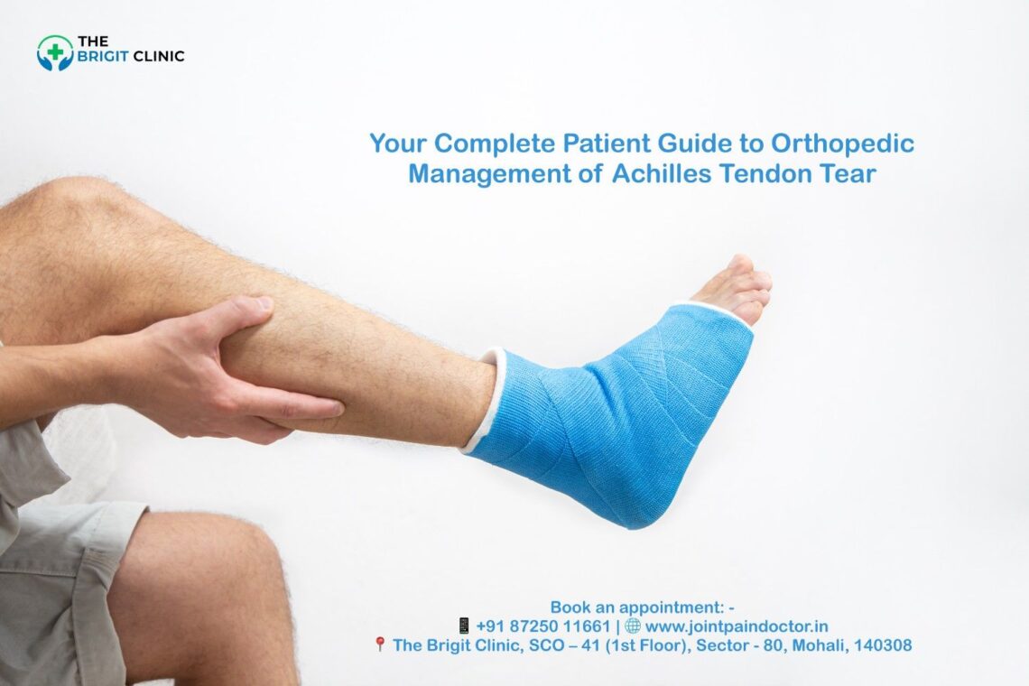
Physiotherapy management of Achilles tendon tear is crucial when dealing with the most commonly ruptured tendon in the human body. For patients seeking expert Physiotherapy for Achilles Tear in Mohali, understanding this structured rehabilitation process is the first step toward a successful recovery. This debilitating injury affects 1 in 15,000 people, increasing to 1 in 8,000 in competitive athletes, and represents 6-18% of all sporting injuries. If you’re among the “middle-aged weekend warriors” who account for approximately 70% of these cases, understanding proper rehabilitation is essential for your recovery.

The journey to full healing after an Achilles rupture is undoubtedly challenging. Your tendon requires at least two months to heal, with several additional months needed to regain strength and flexibility. Moreover, the return to sport typically takes between 4-12 months, depending on your activity level and rehabilitation progress. Without proper physiotherapy for Achilles tendon tears, you face a 12.1% probability of rerupture with nonoperative management, highlighting why structured Achilles tendon tear recovery exercises and clear rehabilitation goals after tendon rupture are critical.

Throughout this comprehensive guide, you’ll discover evidence-based protocols for managing an Achilles tendon tear effectively. From early intervention strategies to advanced conditioning for return to sport, we’ll walk you through each phase of rehabilitation with expert guidance. Whether you’re recovering from surgery or managing a conservative treatment approach, this 2025 protocol will equip you with the knowledge to navigate your recovery successfully.

Ready to start your recovery with Mohali's Top Physiotherapy Clinic? Schedule a consultation with our Achilles tendon specialist today.

Setting the Foundation: Early Goals of Physiotherapy

The initial phase following an Achilles tendon tear sets the critical foundation for successful rehabilitation. During these early weeks, your physiotherapy management focuses on three crucial goals that protect your healing tendon while preventing complications that could derail your recovery journey.

Protecting the repair site

Protection of the repair site is paramount during the first 2-3 weeks after an Achilles tendon tear. This phase allows initial tendon healing to begin while minimising the risk of complications. Initially, your foot will be immobilised in a position that promotes optimal healing of the tendon.

For surgical repairs, you’ll typically wear a splint that cannot be removed, as it serves to protect the newly repaired tendon. For non-surgical management, your foot is placed in a rigid cast or functional brace with your ankle positioned in full equinus (approximately 30° of plantarflexion) to maintain contact between the torn tendon ends.

Regarding weight-bearing, protocols vary based on your surgeon’s preference and the type of intervention:

  • For traditional approaches, you’ll remain non-weight bearing (NWB) with crutches, a walker, or a wheelchair for at least 6 weeks while wearing your splint or CAM boot
  • Some accelerated protocols may allow immediate partial weight-bearing with a CAM boot containing heel lifts

The use of heel lifts serves an important purpose beyond comfort—they can reduce plantar flexor muscle activity by up to 57% during normal gait, consequently decreasing strain on your healing tendon. During this critical protection phase, avoid any activities that stretch or stress the Achilles tendon.

Controlling swelling and pain

Effective management of swelling and pain accelerates healing and improves your comfort during the early rehabilitation phase. The RICE method (Rest, Ice, Compression, Elevation) forms the cornerstone of this approach:

Rest: Stop activities that stress your tendon and switch to low-impact alternatives that don’t strain your Achilles. Your physiotherapist will advise you on appropriate activity modifications.

Ice: Apply ice packs to your tendon for 15-20 minutes every 2 hours during the first 3-4 days after injury or surgery. Always use a waterproof barrier between the ice and your dressing or splint to keep it dry.

Compression: Use an athletic wrap or surgical tape to compress the injured area, which helps reduce swelling. Be careful not to wrap too tightly, as this could impair circulation.

Elevation: Perhaps the most crucial element for swelling control—keep your injured leg elevated above heart level whenever possible. This is particularly important during the first few days post-injury or surgery.

Additionally, your healthcare provider may prescribe pain medications. Use narcotic medications sparingly and try to gradually decrease the amount and frequency over the first two weeks. For milder pain, acetaminophen may be sufficient, although ibuprofen should be avoided as it can delay healing.

Maintaining strength in surrounding joints

While protecting your Achilles tendon, it’s essential to maintain strength in the surrounding joints and muscles to prevent deconditioning and facilitate a faster return to function later. Furthermore, this approach helps minimise the detrimental effects of immobilisation.

During the immediate post-operative phase, you can safely begin:

  • Proximal and core strengthening exercises as part of your home exercise program
  • Hip and knee muscle exercises to maintain lower extremity strength
  • Muscle pump exercises on your uninjured ankle to promote circulation
  • Submaximal plantarflexion isometrics in your boot or cast (if approved by your surgeon) to stimulate calf activity while in a protected position

At this point, your physiotherapist will also work to maintain a full range of motion in your hip and knee joints. Throughout this early phase, your therapist will assess your progress using specific criteria, including pain levels (should be less than 5/10) and swelling measurements.

To sum up, the initial goals of physiotherapy after an Achilles tendon tear focus on creating the optimal environment for healing while preventing complications that could prolong your recovery. With proper protection, swelling control, and maintenance of surrounding joint strength, you establish a solid foundation for the subsequent phases of rehabilitation.

Don't let pain and immobility slow you down. Our team at the Best Physio Clinic in Mohali can create a personalised early-stage recovery plan for you. Call 0172-3137922 to book an appointment.

Pain Management Techniques in Achilles Rehab

Effective pain management serves as a cornerstone of successful Achilles tendon rehabilitation, enabling you to progress through therapy milestones while maintaining comfort and function. Understanding the physiological mechanisms behind various pain control techniques helps optimise your recovery journey.

Cryotherapy and its timing

Cold therapy (cryotherapy) remains a fundamental approach for managing pain and inflammation following Achilles tendon tears. This technique works through several mechanisms that directly impact your healing process.

First, cryotherapy significantly reduces local blood flow to your injured Achilles tendon. Research shows that intermittent cold application decreases mid-portion capillary blood flow by an impressive 71%. This reduction helps control the inflammatory cascade that contributes to pain and swelling.

The timing and application method of cryotherapy substantially influence its effectiveness. Notably, intermittent applications of 3×10 minutes produce better clinical outcomes than a single 20-minute session for similar injuries. This protocol maximises the beneficial effects while allowing for tissue reperfusion between applications.

Within just 2 minutes after removing ice, tendon oxygen saturation returns to normal levels. This reperfusion pattern creates a beneficial environment for healing, as repetitive short periods of ischemia (reduced blood flow) followed by reperfusion have been shown to enhance oxygen delivery to tissues.

Beyond pain relief, cryotherapy provides additional physiological benefits:

  • It reduces postcapillary venous filling pressures, which improves venous outflow from the tendon
  • It decreases production of prostaglandin E2 (PGE2), a key inflammatory mediator in tendon pain
  • It lowers COX-2 protein expression, which contributes to the anti-inflammatory effect

For optimal results, apply ice to your Achilles region for 15-20 minutes every 2 hours during the initial 3-4 days post-injury or surgery, gradually decreasing frequency as acute symptoms subside.

Use of TENS or ultrasonics in tendon healing

The factual key points do not provide specific information about TENS or ultrasonic therapy for Achilles tendon healing. Your physiotherapist might incorporate these modalities based on individual assessment and the latest evidence available in clinical practice.

Manual therapy for pain modulation

Manual therapy techniques offer significant pain relief and functional improvements for Achilles tendon injuries through biomechanical, neurophysiological, and psychosocial mechanisms. Though historically underutilised in Achilles rehabilitation, recent evidence supports its effectiveness.

Joint mobilisation and manipulation techniques produce immediate improvements in several measurable outcomes:

  • Decreased pain levels during single-leg heel raises
  • Increased pressure pain thresholds (PPT), indicating reduced sensitivity
  • Improved joint mobility and ankle motion
  • Enhanced performance in functional tests like single-leg heel raises

Remarkably, these benefits occur not only when treatment targets the ankle complex but also when applied to remote body sites. This suggests that manual therapy influences central pain processing mechanisms rather than simply addressing local tissue dysfunction.

The neurophysiological effects of joint mobilisation include:

  • Decreased nociceptive reflex excitability (reducing pain signals)
  • Enhanced conditioned pain modulation (improving your body’s natural pain control)
  • Reduction of bilateral hyperalgesia following unilateral treatment

Studies tracking patients with chronic Achilles tendinopathy demonstrate that adding manual therapy to standard eccentric exercise programs leads to significant improvements in self-reported function measured by the Victorian Institute for Sport Assessment questionnaire (VISA-A). These improvements persist at 9-month follow-up assessments, suggesting long-term benefits.

To maximise outcomes, your physiotherapist will likely incorporate joint mobilisation techniques directed at both the ankle complex (talocrural and subtalar joints) and potentially remote sites that influence pain-processing pathways. This comprehensive approach addresses both local mechanical issues and systemic pain mechanisms for optimal recovery.

Struggling with pain from your Achilles injury? Our Achilles Tendon Specialist in Mohali uses advanced pain modulation techniques to accelerate healing. Contact our clinic now.

Restoring Mobility: Range of Motion and Joint Work

Restoring proper mobility represents a critical turning point in your Achilles tendon rehabilitation journey. As you progress beyond the initial protection phase, a carefully structured range of motion work and joint mobilisation techniques become essential for optimal healing without compromising the repair.

Range of motion exercises after Achilles tendon surgery

The introduction of ankle range of motion (ROM) exercises follows a specific timeline based on healing phases. Generally, ankle ROM exercises begin around 4-6 weeks post-surgery, coinciding with the transition to full weight-bearing in a CAM boot.

Initially, ROM work focuses on these key movements:

  • Ankle pumps (avoiding dorsiflexion beyond neutral/0 degrees)
  • Ankle circles (staying within safe ranges)
  • Ankle inversion and eversion
  • Seated heel-slides for ankle dorsiflexion (limited to neutral)

Early initiation of active range of motion (AROM) plays a crucial role in facilitating proper collagen fibril formation. Research indicates that early mobilisation specifically helps reduce Achilles tendon elongation and improves clinical outcomes.

From weeks 7-8, you can safely progress to active assisted range of motion (AAROM) and passive range of motion (PROM) techniques as your repair strengthens. Throughout this phase, dorsiflexion limitations gradually ease—starting with restriction to neutral (0 degrees) until approximately week 8, after which you can gently progress dorsiflexion ROM according to tolerance.

Importantly, a sensation of tightness throughout early rehabilitation phases is both expected and often preferred, potentially indicating appropriate tendon elongation rates. In fact, patients rarely complain about their Achilles being “too tight” at long-term follow-up appointments.

Joint mobilisation techniques for ankle & subtalar joints

Joint mobilisation refers to specialised manual therapy techniques used to modulate pain and treat joint dysfunctions that limit the range of motion. For Achilles rehabilitation, assessment and treatment of joint mobility dysfunctions should begin within protected ranges to improve joint mobility without passively stretching the Achilles complex.

Specific joint mobilisation techniques include:

  • Talocrural joint mobilisations (anterior-posterior glides) to immediately improve dorsiflexion ROM
  • Subtalar joint mobilisations to address compensatory pronation patterns
  • Midfoot and metatarsophalangeal (MTP) mobilisations as indicated

The clinical rationale behind these techniques extends beyond mechanical benefits. AP talocrural joint mobilisations have been demonstrated to immediately improve dorsiflexion ROM, which may decrease compensatory subtalar joint pronation as the lower limb advances over the ankle during gait. This reduction in abnormal mechanics decreases abnormal loading through the Achilles tendon.

Furthermore, joint-based mobilisation creates immediate improvements in strength through both peripheral and central mechanisms. Patients with Achilles tendinopathy who receive joint-directed manual therapy as part of a comprehensive treatment plan demonstrate clinically significant improvements in functional measures and pain reduction.

Stretching protocols and precautions

Regarding stretching, a fundamental principle must be emphasised: avoid forceful active and passive range of motion of the Achilles for 10-12 weeks. This precaution prevents excessive strain on the healing tendon fibres.

The stretching protocol typically follows this progression:

  1. Weeks 4-6: No direct Achilles stretching; focus on toe mobility with great toe dorsiflexion and plantarflexion stretching (not exceeding neutral)
  2. Weeks 7-8: Continue seated heel-slides for dorsiflexion ROM to tolerance, as dorsiflexion restrictions begin to ease
  3. After week 8: Progress to standing ankle dorsiflexion stretch on a step

Throughout all phases, carefully monitor your tendon and incision sites for mobility and signs of scar tissue formation. Regular soft tissue treatments, including scar mobilisation (starting 4 weeks post-op) and friction massage, help decrease fibrosis. However, avoid any instrument-assisted soft tissue mobilisation (IASTM) directly on the tendon until at least 16 weeks post-operation.

Equally essential is the stretching of proximal muscle groups. As rehabilitation advances, incorporate gentle stretching of quadriceps, hamstrings, hip flexors, and piriformis as indicated to maintain optimal lower extremity mechanics.

Regaining full ankle mobility is critical. Our Physiotherapists in Mohali are experts in safe and effective joint mobilisation. Visit us to restore your movement.

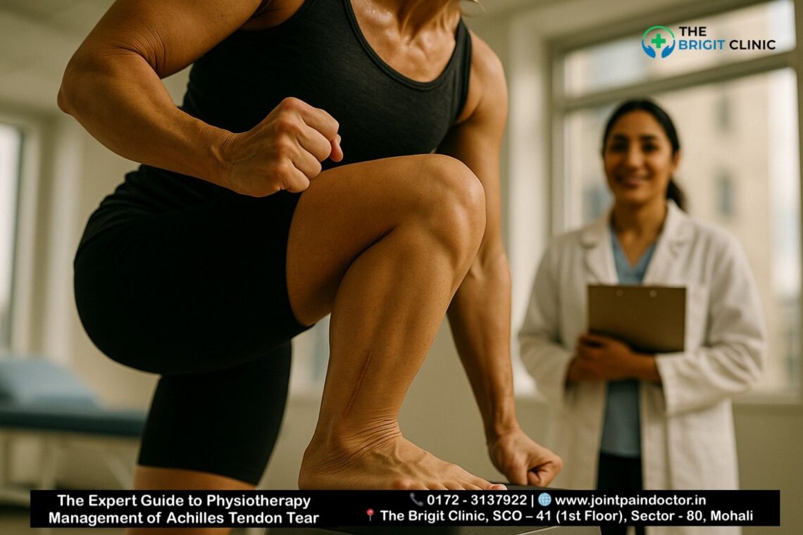
Building Strength: Progressive Loading Strategies

Progressive strength building represents the foundation of successful Achilles rehabilitation once basic mobility has been restored. This critical phase introduces graduated loading to stimulate tendon healing and restore function to weakened calf muscles. Research consistently demonstrates that carefully structured strengthening protocols improve clinical outcomes and accelerate return to activity.

Isometric calf exercises post rupture

Isometric exercises—contractions where muscle length remains unchanged—serve as the ideal starting point for strengthening after Achilles tendon rupture. These exercises produce minimal tendon stress yet provide significant therapeutic benefits.

Isometric training should begin in the initial stages of rehabilitation to activate calf muscles with a focus on pain control. For optimal results:

  • Perform submaximal, non-painful isometric contractions at end-range plantarflexion, which puts the gastrocsoleus complex in a shortened position and minimises elongation stress on the repair
  • Start with gentle calf isometrics throughout the day, where you push the ball of your foot into the ground at a tolerable effort level
  • Aim for 2-3 sets of 15-45 second holds, performed 1-3 times daily depending on your tolerance

Recent research indicates that isometric plantarflexion holds can provide approximately 50% immediate reduction in Achilles tendon pain during functional loading tests. Interestingly, performing isometrics with the knee extended may produce a 20% larger reduction in symptoms compared to knee-flexed positions, though this difference wasn’t statistically significant.

Unlike patellar tendinopathy, where immediate pain relief follows isometric exercise, Achilles tendinopathy doesn’t consistently show the same immediate analgesic response. Nevertheless, isometrics remain valuable for their ability to promote muscle activation without overstressing the healing tendon.

Theraband exercises for Achilles rehabilitation

Following successful isometric training, resistance band exercises provide the next logical progression in strengthening your Achilles complex. These exercises introduce controlled resistance while maintaining protection of the healing tendon.

To implement Theraband exercises effectively:

  • Begin seated with a moderate to heavy resistance band placed around the ball of your foot (not the toes)
  • Hold each end with your hands, ensuring appropriate tension by removing any slack from the band
  • Straighten your leg while holding the straps, then point your toes as if pushing a gas pedal
  • Maintain straight leg position and slow, controlled motion without ankle wobbling
  • Perform 10-15 repetitions for 2-3 sets on each leg

As your rehabilitation advances, plantar flexion isometrics can progress to limited range isotonic progressive resisted exercises with blood flow restriction (BFR) as range of motion and contraction tolerance improve. The use of BFR has shown promising results in post-operative Achilles tendon rupture rehabilitation, with one randomised controlled trial demonstrating greater isokinetic strength in the operative calf muscle at 3 months when using BFR compared to standard rehabilitation.

Eccentric and concentric loading phases

The introduction of eccentric and concentric loading represents a pivotal advancement in your strength progression. Eccentric exercise—where the muscle lengthens under tension—has been associated with significant clinical improvements in pain and function for patients with Achilles tendinopathy.

Eccentric loading provides several unique benefits:

  • It improves tendon structure, which historically was considered a mechanism for improvement in some persons with Achilles tendinopathy
  • It reduces tendon thickness, with studies showing localised decrease in tendon thickness correlated with patient satisfaction
  • It potentially increases tendon stiffness, enhancing its response to strain

A standard progressive loading protocol typically follows this pattern:

  • Begin with double-leg heel raises standing on flat ground (3 sets of 10-15 repetitions)
  • Progress to single-leg seated heel raises (3 sets of 15)
  • Advance to two-legged heel raises at the edge of a stair (3 sets of 15)
  • Move to one-legged heel raises at the edge of a stair (3 sets of 15)
  • Finally, incorporate quick-rebounding heel raises (3 sets of 20)

For eccentric training specifically, the classic Alfredson protocol recommends 3 sets of 15 repetitions twice daily with an extended knee, followed by 3 sets of 15 repetitions twice daily with a flexed knee. Resistance should be added once you can perform these exercises without discomfort.

As strength improves, gravity-assisted devices (such as AlterG) and aquatic therapy can be valuable when developing single-leg strength and the ability to perform heel raises without compensation. By progressively increasing load as the tendon and muscle develop strength and show fewer symptoms, you create the optimal environment for complete rehabilitation and eventual return to full function.

Need a structured strengthening program for your Achilles Rupture Rehab in Mohali? We use evidence-based protocols for maximum results. Get expert guidance today.

Improving Balance and Proprioception

Balance and proprioception training form a vital bridge between basic strength building and functional movement in your Achilles rehabilitation journey. Given that proprioception provides essential information needed to modify ankle position during complex motor tasks, restoring this neural sense becomes indispensable for preventing future injuries.

Ankle proprioception training

Proprioception—the neural process by which your body takes in sensory input from the environment and integrates that information to produce appropriate motor responses—dramatically affects recovery outcomes. After Achilles injury, this neural feedback system often becomes compromised, making targeted retraining essential.

For optimal rehabilitation, physiotherapists typically incorporate ankle proprioceptive neuromuscular facilitation (PNF) techniques using the contract-relax approach. Research indicates these techniques should be performed with ten repetitions for one set, twice daily. Early enhancements in joint proprioception through these interventions aid in earlier mobilisation, creating a positive cycle of improvement.

The most compelling evidence supports beginning proprioception training once you’ve established basic weight-bearing tolerance. Physiotherapists design specific exercises based on orthopaedic loading recommendations coupled with your clinical condition. One study demonstrated that athletes with functional ankle instability showed significant improvements in eversion, plantarflexion, dorsiflexion, and inversion joint position sense following eight weeks of ankle proprioceptive exercises.

Balance & stability training for Achilles injury

Effective balance training encompasses exercises that challenge your neuromuscular system’s ability to maintain stability. Studies examining proprioceptive training’s prophylactic effectiveness found a significant 35% reduction in ankle sprain risk for individuals who completed such programs.

Common balance exercises include:

  • Double limb standing balance on uneven surfaces like wobble boards
  • Single-leg balance exercises, first on flat surfaces, then progressing to unstable surfaces
  • Balance training with perturbation challenges—where balance is deliberately disrupted
  • BAPS (Biomechanical Ankle Platform System) board exercises in the standing position
  • Walking on mini trampolines

First thing to remember is that balance training should start with simple, supported exercises before advancing to more challenging variations. As per research findings, compliance with rehabilitation protocols substantially affects outcomes—studies showed noncompliance levels between 10-40%, which aligned with real-world clinical practice. Hence, exercises should be engaging yet manageable to ensure adherence.

Progression from bilateral to single-leg stance

The systematic progression from bilateral to unilateral stance represents a crucial advancement in your rehabilitation. Initially, you’ll begin with double-leg balance activities on stable surfaces, henceforth progressing to more challenging environments and positions.

A structured progression typically follows this pattern:

  1. Double leg balance on firm ground with eyes open
  2. Double leg balance on air pads or balance boards (aiming for 10-second holds)
  3. Single leg stance with eyes open (10-second holds)
  4. Single leg stance while throwing and catching a ball against a wall
  5. Single leg stance with eyes closed (10-second holds)
  6. Single leg balance on air pads or balance boards
  7. Advanced training on BOSU balls or trampolines

Throughout this progression, focus on proper ankle mechanics rather than compensating through hip movements. For patients transitioning out of the protective boot phase, single-leg balance exercises help normalise walking patterns and eliminate limping. Practising for 3 sets of 60 seconds can significantly improve stability.

Studies reveal that implementing balance and proprioception training can prevent one ankle sprain for every 17 patients who complete the protocol, regardless of prior injury history. Above all, those with previous Achilles injuries show even greater benefits, with a 36% reduction in reinjury risk.

Prevent future injuries with our specialised balance training programs at our Physiotherapy Clinic in MohaliBook your assessment now.

Gait Training and Functional Movement Re-education

Regaining natural walking patterns stands as a crucial milestone in your Achilles tendon rehabilitation. Studies reveal that gait abnormalities often persist for more than a year after surgery, including increased dorsiflexion range of motion, co-activation of lower leg muscles, and decreased step length. These lingering issues directly impact your overall quality of life and physical activity levels.

Gait training after Achilles tear

Following an Achilles rupture, your tendon typically elongates during healing, causing increased dorsiflexion during walking. Research shows this anatomical change forces your calf muscles to work harder—EMG studies demonstrate significantly increased muscle activity in the triceps surae on the affected side. This heightened muscle activation represents your body’s attempt to compensate for increased tendon slack during walking.

Your rehabilitation should follow a structured progression for weight bearing:

  • Weeks 0-2: Non-weight bearing in protective splint
  • Weeks 3-6: Begin partial progressive weight bearing with an assistive device and boot with three heel wedges
  • Weeks 6-7: Full weight bearing in boot with gradually decreasing heel wedges
  • Weeks 8-12: Gait training to wean off assistive devices while normalising gait pattern

Interestingly, aquatic therapy and unweighted treadmills prove especially beneficial for restoring proper mechanics simultaneously with gradual weight-bearing progression.

Correcting compensatory patterns

Patients with Achilles injuries typically develop specific compensatory patterns to reduce tendon loading. Research identifies common adaptations, including reduced ankle dorsiflexion and knee flexion during heel drop exercises. Another study revealed that even after 4.5 years, patients still exhibit 13.4% larger peak dorsiflexion in stance on the injured leg.

As a physiotherapist, identifying these compensations through careful observation remains essential. Look for prolonged stance phase and avoidance of push-off in terminal stance. In tandem with visual assessment, techniques like in-line tandem walking effectively highlight and correct remaining asymmetries.

Use of assistive devices and weaning off

Initially, crutches or a walker with strict non-weight bearing protect your surgical repair. The transition away from these devices follows a methodical approach coordinated with your weight-bearing status. By week 4, you’ll typically begin partial progressive weight bearing on crutches in an Achilles boot with three wedges.

An often-overlooked tool—the “Even Up” shoe leveller for your uninvolved foot—prevents secondary musculoskeletal problems by equalising leg lengths. Correspondingly, gait training with assistive devices should emphasise that your weight-bearing progression aligns with ideal mechanics, as poor patterns lead to joint dysfunction and adverse muscle tone.

By week 8, you should achieve full weight bearing in the boot without crutches, demonstrating a normalised gait pattern. Subsequently, progress to walking in athletic shoes with a heel lift around weeks 8-10 under clinical supervision before community ambulation.

Walk with confidence again. Our Best Physiotherapist for Achilles Tear in Mohali will correct your gait and eliminate limping. Start your functional re-education.

Advanced Conditioning and Return to Sport

The final phase of Achilles tendon rehabilitation focuses on advanced conditioning and sport reintegration. Despite successful surgical repair, studies reveal that 20-25% of patients cannot return to sport after an Achilles tendon tear, making this transition particularly challenging yet crucial for athletes and active individuals.

Return to activity guidelines after Achilles repair

The journey back to sports typically begins 6+ months post-surgery, with return to play ranging from 61-100% in elite athletes. Before advancing to sport-specific activities, you must meet several objective criteria:

  • Standing heel rise test >90% compared to the uninjured side
  • Lower extremity functional tests ≥90% compared to the contralateral side
  • Completion of both phases of a return-to-running program without pain/swelling
  • No major deficits with core and single-leg squat testing

Research indicates plantar flexion strength deficits often persist between 10-30% even after one year post-operative. Due to this, a target of >90% Limb Symmetry Index (LSI) for strength assessment is recommended before sport reintegration.

Sport-specific drills and plyometrics

Plyometric training becomes essential for developing the stretch-shortening cycle of your tendons—a key requirement for running and jumping activities. Remarkably, research shows jumping exercises can create forces exceeding seven times your bodyweight through the Achilles tendon.

Your plyometric progression should follow this sequence:

  • Double-leg hops with slightly bent knees
  • Double-leg hops with stiff knees (isolating Achilles work)
  • Single-leg hops with gradual intensity increases
  • Sport-specific movement patterns

For optimal tendon adaptation, limit plyometric sessions to 1-2 weekly with adequate recovery between strength training days. Sport-specific conditioning typically initiates around 18-20 weeks post-surgery, with formal testing including the Vail Sport Test, agility T-test, and three-cone drill to assess readiness.

Psychological readiness and functional testing

In essence, psychological factors significantly impact recovery during rehabilitation and return to sport. Fear of reinjury emerges as the primary barrier, reported by 41.30% of patients who didn’t return to their previous activity levels.

The Ankle Ligament Reconstruction-Return to Sport Injury (ALR-RSI) score provides a valid assessment of psychological readiness. This tool demonstrates strong correlation with functional outcomes and excellent discriminant validity—patients who returned to sport scored significantly higher (83.2) than those who didn’t (60.7).

Before full clearance, comprehensive functional testing should include:

  • Sport-specific movement assessments
  • Reactive strength index testing for explosive sports
  • Single-leg hop tests with >95% LSI compared to the uninjured leg
Ready to return to your sport? Our Achilles Tendon Tear Treatment Mohali program includes advanced sport-specific conditioning. Achieve your comeback with us.

Home Exercise Program and Long-Term Maintenance

Mastering long-term self-management marks the final frontier in your Achilles tendon tear rehabilitation. Even after formal physiotherapy concludes, your commitment to consistent home exercises and vigilant monitoring determines the ultimate success of your recovery journey.

Home exercises program for the Achilles tendon

A well-structured home exercise program remains fundamental to your continued progress. Most patients can return to normal activity in 4-6 months with proper home exercise adherence. Your program should evolve as you heal:

Early Phase (Weeks 2-4):

  • Seated calf stretch with knee straight: Hold 15-30 seconds, repeat 2-4 times
  • Passive toe stretch: Gently bend your toe forward and backwards, holding each position for 15 seconds
  • Submaximal plantarflexion isometrics in a protected position

Intermediate Phase:

  • Calf stretch on a step: Lower heels below step edge, hold 15-30 seconds
  • Heel raises: Progress from seated to standing exercises

Advanced Phase:

  • Single-leg heel raises: 3 sets of 8-12 repetitions
  • Lower calf strengthener: 30 repetitions with knees slightly bent

For instance, the calf stretch involves standing facing a wall, placing one leg behind with the heel down, then gently leaning forward until you feel a stretch. Exercise frequency should be consistent—aim for daily stretching and strengthening sessions to maximise recovery outcomes.

Monitoring for signs of overuse or re-rupture

In parallel with your exercise program, vigilant monitoring for warning signs prevents setbacks. After healing, you face a greater risk of re-injuring your Achilles tendon. Key warning signs include:

  • Sudden sharp pain in the tendon area
  • New swelling or redness
  • Inability to rise onto tiptoes
  • Altered gait mechanics
  • Pain that persists more than 24 hours after activity

The contralateral limb likewise requires attention—studies show a higher incidence of contralateral Achilles rupture following initial ATR compared to general population rates. Ultimately, maintaining awareness of both tendons safeguards your long-term function.

Patient education and lifestyle modifications

Beyond exercises, specific lifestyle adjustments support lasting recovery. Essential modifications include:

  • Footwear selection: Avoid high-heeled shoes, which increase tendon stress
  • Activity preparation: Always stretch before exercise and incorporate a proper warm-up
  • Sport considerations: Consult your provider before returning to sports involving rapid starts/stops like tennis, racquetball or basketball
  • Progressive loading: Increase exercise intensity gradually—never more than 10% weekly

As you continue rehabilitation, swimming, cycling, jogging, or walking effectively enhances muscle strength and range of motion. Gradually introducing low-impact activities before returning to high-impact exercises protects your tendon for the initial 6 months post-injury. Following these protocols not only prevents re-rupture but also creates optimal conditions for lifelong tendon health.

Get a personalised home exercise program from the Best Physio Clinic in Mohali. We ensure you recover fully and stay healthy. Contact us for long-term support.

Conclusion

Rehabilitation after an Achilles tendon tear undoubtedly represents a lengthy process requiring patience, dedication, and expert guidance. Throughout this comprehensive guide, you’ve learned the essential components of effective physiotherapy management—from initial protection strategies to advanced sport-specific training. Accordingly, your recovery journey progresses through distinct phases, each building upon the previous while addressing specific rehabilitation goals.

The early phase focuses on protecting your healing tendon while managing pain and swelling. Subsequently, careful mobility work prepares your ankle for progressive loading, which stimulates proper tendon healing. Balance training and gait re-education, then restore normal movement patterns before sport-specific exercises, prepare you for return to activity.

Despite following optimal protocols, most patients still experience some strength deficits even a year after rupture. Nevertheless, these deficits rarely impact daily function when properly managed. Your commitment to home exercises after formal physiotherapy concludes significantly determines your long-term outcomes.

Many patients wonder about their ultimate recovery potential. Research shows that while complete recovery takes time, most individuals return to previous activities, albeit sometimes at modified levels. Your dedication to rehabilitation directly correlates with recovery quality—those who adhere strictly to protocols generally achieve better functional outcomes than those who don’t.

Remember that healing continues well beyond the initial repair phase. Therefore, maintaining vigilance for warning signs while gradually increasing activity levels safeguards your recovery investment. Though challenging at times, proper physiotherapy management after an Achilles tendon tear provides your best path toward restored function and return to the activities you enjoy.

Your journey to recovery starts with a single step. Trust Mohali's Top Physiotherapy team to guide you every step of the way. Schedule your comprehensive assessment at our clinic today.

Key Takeaways

This comprehensive guide reveals evidence-based strategies for successful Achilles tendon tear rehabilitation, from initial injury through complete recovery and return to sport.

• Early protection is critical: Maintain non-weight bearing for 6+ weeks with proper immobilisation to prevent re-rupture (12.1% risk without proper care)

• Progressive loading accelerates healing: Begin with isometric exercises, advance to eccentric training using the Alfredson protocol (3 sets of 15 reps twice daily)

• Balance training prevents future injury: Proprioception exercises reduce ankle sprain risk by 35% and reinjury risk by 36% in previously injured patients

• Strength deficits persist long-term: Expect 10-30% plantar flexion weakness even one year post-surgery; achieve >90% limb symmetry before sport return

• Home exercise adherence determines success: Daily stretching and strengthening exercises are essential for a 4-6 month recovery timeline and long-term tendon health

• Psychological readiness matters equally: Fear of reinjury affects 41% of patients who don’t return to sport; address mental barriers alongside physical rehabilitation

Recovery typically takes 4-12 months, depending on activity level, with formal physiotherapy progressing through distinct phases of protection, mobility restoration, strength building, and sport-specific conditioning. Success depends on strict protocol adherence and gradual activity progression.

FAQs

Q1. How long does physical therapy typically last for an Achilles tendon tear?

A1. Physical therapy for an Achilles tendon tear usually lasts 4-6 months, but can extend up to 12 months depending on the severity of the injury and the individual’s activity level. The rehabilitation process progresses through distinct phases, from initial protection to advanced sport-specific training.

Q2. What is the Alfredson protocol for Achilles tendon rehabilitation?

A2. The Alfredson protocol is a specific eccentric exercise program for Achilles tendon rehabilitation. It involves performing 3 sets of 15 repetitions twice daily with an extended knee, followed by 3 sets of 15 repetitions twice daily with a flexed knee. This protocol has shown significant clinical improvements in pain and function for patients with Achilles tendinopathy.

Q3. When can I start weight-bearing after an Achilles tendon tear?

A3. Weight-bearing typically follows a structured progression. You’ll usually remain non-weight bearing for the first 2-3 weeks, then begin partial progressive weight bearing around weeks 3-6 with an assistive device and protective boot. Full weight bearing in a boot often starts around 6-7 weeks post-injury, with gradual weaning off assistive devices in the following weeks.

Q4. How effective is balance training in preventing future Achilles injuries?

A4. Balance and proprioception training are highly effective in preventing future injuries. Research shows that implementing such training can reduce ankle sprain risk by 35% in general and decrease reinjury risk by 36% in previously injured patients. These exercises are crucial for restoring proper neuromuscular control and stability.

Q5. What are the key indicators that I’m ready to return to sports after an Achilles tear?

A5. Before returning to sports, you should meet several criteria: achieve >90% strength in the affected leg compared to the uninjured side, complete a return-to-running program without pain or swelling, demonstrate no major deficits in core and single-leg squat testing, and score well on psychological readiness assessments. Additionally, sport-specific movement assessments and functional tests should show comparable performance to the uninjured leg.

About the Physiotherapist – Dr. Aayushi

Dr. Aayushi is a renowned physiotherapist and the driving force behind one of Mohali’s leading physiotherapy clinics. With extensive experience and a deep commitment to patient care, she specialises in the management of complex musculoskeletal conditions, including Achilles tendon tears. Dr. Aayushi believes in a holistic and evidence-based approach to rehabilitation, combining advanced manual therapy techniques with tailored exercise programs to ensure optimal outcomes for every patient. Her expertise in Achilles Tendon Tear Treatment in Mohali has helped numerous athletes and active individuals successfully return to their desired levels of activity, making her a trusted name for Physiotherapy in Mohali.

Your Journey to Recovery Starts Here. Let’s Take the First Step Together.

Recovering from an Achilles tear is a marathon, not a sprint. It requires expert guidance, unwavering dedication, and a personalised plan that adapts to your unique healing process. You don’t have to navigate this challenging path alone.

At our Mohali clinic, we don’t just treat injuries; we rebuild confidence and restore function. Under the expert care of Dr. Aayushi, you will receive a comprehensive treatment program based on the latest evidence, designed to get you back to the life and sports you love, stronger and safer than before.


Stop searching for the “Best Physiotherapist for Achilles Tear in Mohali.” You’ve found her.
📞 Call us at 0172-3137922 to speak directly with our team and book your initial assessment.
📍 Visit our Google My Business profile to see reviews from patients who have successfully completed their recovery journey.
🌐 Schedule your appointment online and begin your personalised Physiotherapy for Achilles Tear in Mohali today.
Your comeback story begins now. Let’s write it together.

Role of Orthopedic Surgeon in Diabetic Foot Reconstruction & Off-Loading

Diabetic Foot complications affect approximately half of all people with diabetes who experience some form of nerve damage. These problems represent a major health concern and a common cause of hospitalisation. Without fast treatment, a toe, foot, or even part of the leg may require amputation, making timely orthopedic intervention critical for limb preservation. Dr. Manu Mengi, recognised as the Best Orthopedic Surgeon for Diabetic Foot Care in Mohali, emphasises that early specialist care can dramatically reduce the risk of amputation.

As orthopedic specialists, we regularly see how diabetic neuropathy and poor blood flow create serious risks for developing foot ulcers and infections. Additionally, conditions like Charcot foot disease can create deformities where bone is exposed directly under the skin surface, significantly increasing the risk of amputation. When diabetic patients develop an ulcer with Charcot foot, their amputation risk jumps to 50%, compared to just 10% without an ulcer.

In this comprehensive guide, we’ll explore our role as orthopedic surgeons in diabetic foot management, including reconstruction techniques, off-loading strategies, and collaborative approaches that help preserve function and prevent devastating complications.

Understanding Diabetic Foot Complications

Diabetic foot complications primarily stem from the long-term effects of persistently high blood sugar on nerves and blood vessels. These conditions develop gradually, often without noticeable symptoms until significant damage has occurred. Understanding these mechanisms is essential for early intervention and prevention of serious outcomes.

What causes diabetic foot problems?

The foundation of diabetic foot problems lies in blood sugar levels that remain elevated over time. This hyperglycemia damages both small and large blood vessels throughout the body, but the effects are particularly pronounced in the feet. Furthermore, the production of advanced glycation end products (AGEs) plays a crucial role in tissue damage. These compounds form when glucose binds to proteins and DNA, altering their structure and function.

Poor blood supply to the feet creates a perfect storm for complications. Essentially, two major pathways lead to foot problems: nerve damage that reduces sensation and blood vessel damage that impairs healing. The interaction between these factors means even minor injuries can develop into serious complications.

Nearly 15% of people with diabetes will eventually develop a foot ulcer, and damage can occur without the person ever noticing due to diminished pain sensation. Moreover, studies show that having an A1C over 7% for at least three years substantially increases your risk of developing diabetic neuropathy.

The role of diabetic neuropathy and poor circulation

Diabetic neuropathy manifests in several ways that directly impact foot health. Sensory neuropathy reduces protective sensation, making it difficult to feel cuts, blisters, or pressure points. Therefore, injuries often go unnoticed until they’ve become severe. Motor neuropathy affects muscle function, leading to foot deformities and abnormal pressure distribution when walking.

Perhaps most overlooked, autonomic neuropathy impairs sweat production, resulting in dry, cracked skin that’s more susceptible to injury. This creates easy entry points for infection. Unlike normal feet that signal pain when injured, neuropathic feet might continue bearing weight on damaged areas, worsening injuries.

Poor circulation compounds these problems by reducing blood flow to injured areas. Specifically, diabetes damages small blood vessels, limiting the delivery of oxygen, nutrients, and infection-fighting cells to wounds. This vascular insufficiency means:

  • Wounds heal more slowly
  • Infections spread more easily
  • Tissues receive inadequate nutrition
  • Antibiotics have difficulty reaching infection sites

Common conditions: ulcers, infections, Charcot foot

Diabetic foot ulcers represent one of the most serious complications, affecting about 15% of diabetics during their lifetime. Unfortunately, these open wounds often develop silently on pressure points, especially under the foot. Once an ulcer forms, the race begins to heal it before infection sets in.

Infections in diabetic feet quickly become polymicrobial, involving multiple types of bacteria. Initially, they may start with common bacteria like Staphylococcus or Streptococci, but rapidly progress to include gram-negative and anaerobic organisms. Consequently, approximately 15% of ulcers develop osteomyelitis (bone infection), and of those, 15% require amputation.

Charcot foot, though affecting less than 1% of all people with diabetes, represents one of the most destructive complications. This condition develops when small injuries go unnoticed, allowing continued walking on damaged bones and joints. Over time, the foot’s architecture collapses, creating the characteristic “rocker-bottom” deformity.

The presence of Charcot deformity dramatically increases amputation risk. Specifically, diabetics with Charcot foot have a 50% chance of amputation if they develop an ulcer, versus only 10% without an ulcer. This stark difference highlights why early recognition and appropriate off-loading are critical in preventing catastrophic outcomes.

Early detection saves limbs! Visit the Best Hospital for Diabetic Foot Problems in Mohali for a thorough evaluation.

When Orthopedic Surgeons Step In

Proper timing of orthopedic intervention often makes the crucial difference between limb salvage and amputation for patients with diabetic foot complications. My experience shows that coordinated care yields dramatically better outcomes, with studies demonstrating that multidisciplinary approaches reduce ulcer recurrence rates from 58.4% to 30.4% over two years.

Referral criteria for orthopedic evaluation

Prompt orthopedic referral becomes necessary under several specific circumstances. Primarily, patients showing these red flags require immediate orthopedic evaluation:

  • Active ulcerations, particularly those with signs of infection
  • Traumatic injuries to the diabetic foot
  • Suspected or confirmed Charcot foot
  • Presence of a foreign body
  • Signs of ischemia or gangrene

Additionally, patients with a history of previous ulceration or amputation fall into the highest risk category. Without preventive intervention, 58-83% of these patients will develop another ulcer within just one year. Nonetheless, with proper therapeutic footwear and insoles, this recurrence rate can be halved to 30-50% annually.

Besides acute presentations, orthopedic evaluation should be considered for patients showing no signs of wound healing within 4-6 weeks despite optimal management, regardless of initial vascular diagnostic test results.

Diabetic Foot in Orthopedics: scope and responsibilities

As orthopedic surgeons, our responsibilities in diabetic foot management encompass both preventive and reactive interventions. The foundation of our approach involves a thorough assessment of peripheral neuropathy, skin integrity, existing ulcers, foot deformities, vascular status, and footwear appropriateness.

Subsequently, our clinical work includes specialised surgical interventions like debridement, ray amputations, and corrective procedures for Charcot deformities. We also implement advanced wound healing technologies where appropriate. For instance, negative pressure wound therapy (NPWT) has emerged as one of the most effective current strategies for reducing amputation risk while increasing healing rates.

Orthopedic management further involves risk stratification and treatment planning based on the presence of peripheral neuropathy, foot deformity, and ulcer history. Correspondingly, we design individualised treatment plans combining patient education, orthoses, specialised footwear, and schedules for ongoing skin and nail care.

Multidisciplinary collaboration with endocrinologists and podiatrists

The complexity of diabetic foot disease necessitates collaborative care. A well-structured multidisciplinary team (MDT) typically includes specialists from medicine (endocrinology, infectious diseases, vascular surgery), podiatry, nursing, nutrition, orthotics, and physiotherapy.

In this collaborative model, podiatrists often serve as “gatekeepers” for prevention and management, with their specialised focus on biomechanics being particularly valuable. Their expertise in preventive strategies includes regular monitoring, routine care of calluses, and insole/shoe recommendations.

Meanwhile, endocrinologists manage the underlying metabolic condition, as glycemic control directly impacts wound healing. Studies demonstrate that comprehensive MDT approaches reduce hospital stays, decrease financial burden, and substantially lower amputation rates by 36-86%. Furthermore, patients managed by MDTs show faster wound healing times and lower severity of necessary amputations.

The International Working Group on the Diabetic Foot (IWGDF) recommends at least three levels of foot care management with interdisciplinary specialists. Under this framework, we coordinate with vascular surgeons for blood flow assessment, infectious disease specialists for antibiotic management, and endocrinologists for optimising glycemic control.

Overall, research consistently shows that MDT management yields superior outcomes to isolated speciality care. Accordingly, establishing clear communication channels between team members ensures seamless care coordination from hospital to community settings.

Need expert care for diabetic foot ulcers? Dr. Manu Mengi specialises in Diabetic Foot off-loading Techniques in Mohali and advanced limb-saving procedures.

Surgical Reconstruction Techniques in Diabetic Foot

Reconstructive surgery offers limb-saving options for patients with advanced diabetic foot conditions. With proper surgical intervention, we can often preserve function and mobility even in cases that might otherwise progress to major amputation.

Surgical Reconstruction in Charcot Foot

Charcot neuroarthropathy creates complex deformities that often require surgical reconstruction to preserve the foot. Although conservative management remains the first line of treatment, surgery becomes necessary when patients develop significant instability, ulceration, or deformity despite adequate offloading.

First and foremost, the timing of surgery is critical—ideally performed during the quiescent stage (Eichenholtz stage 2 or 3) of the disease process. Surgical techniques include internal fixation, external fixation, or a combination of both approaches. In essence, our goal is to achieve a stable, plantigrade foot resistant to ulceration.

Recent studies show that despite high complication rates, surgical reconstruction can achieve successful fusion in approximately 86% of cases. Nevertheless, complications directly attributable to the surgical technique occur in about 36% of patients. The post-reconstruction amputation rate remains relatively low at 5.5%, with 91% of patients returning to ambulation.

Arthrodesis of Diabetic Foot: when and why

Ankle arthrodesis (fusion) serves as a salvage procedure for diabetic patients with significant ankle instability or deformity. Indeed, tibiotalocalcaneal (TTC) fusion is commonly performed to address both ankle and hindfoot issues simultaneously.

Although considered an effective limb-salvage procedure, arthrodesis carries substantial risks. Studies report overall complication rates exceeding 75% in diabetic patients undergoing primary ankle arthrodesis. These complications include a 38.5% reoperation rate, 38.5% infection rate, and 23.1% amputation rate.

Rather than viewing these statistics as prohibitive, we use them to guide patient selection and counselling. Since patient selection significantly impacts outcomes, we carefully evaluate comorbidities and vascular status before recommending fusion procedures.

Ray Amputation in Diabetic Foot

Ray amputation involves removing a toe along with part of its corresponding metatarsal. This procedure provides an excellent option for ensuring adequate surgical debridement of infected margins while preserving foot function.

In contrast to major amputations, ray amputations show significantly lower mortality rates. Studies indicate the hazard rate is 1.6 times higher with major amputations compared to ray procedures. Furthermore, limb salvage can be achieved in almost two-thirds of patients undergoing minor amputations.

Appropriate patient selection remains crucial for success. Ideally, patients should have at least one palpable pedal pulse and adequate vascular indices. With proper selection, ray amputations show success rates of approximately 70%.

Managing Lisfranc Dislocation in Charcot Foot

Lisfranc joint involvement occurs in 45% of Charcot foot cases, making it the most common site affected in Charcot arthropathy. Although rare in the general population, Lisfranc injuries occur more frequently in diabetic patients and often follow minimal trauma.

Since these injuries can trigger or represent the first sign of an underlying Charcot process, careful evaluation is essential. MRI examination proves particularly valuable as radiographic findings are frequently subtle and easily missed. The oblique axial plane allows visualisation of the entire length of the Lisfranc ligament, providing crucial diagnostic information.

Arthrodesis vs Amputation Decision-Making in Diabetic Foot

When deciding between reconstruction and amputation, we consider several key factors:

  • Vascular status – Adequate blood flow is essential for healing after reconstruction
  • Extent of infection – Deep, uncontrollable infection may necessitate amputation
  • Functional goals – Patient’s mobility needs and lifestyle considerations
  • Overall health – Comorbidities that might affect healing potential

With this in mind, a strategic approach often involves performing the most distal amputation possible when necessary. This preserves length and function while removing infected or necrotic tissue. Although complications may occur, research shows that 80% of minor amputees remain alive after 2 years, with 64% maintaining full ambulation.

Worried about Charcot Foot Surgery in Mohali? Book a consultation with Dr. Manu Mengi to discuss minimally invasive options.

Off-Loading Strategies and Custom Orthotics

Off-loading remains the cornerstone of diabetic foot ulcer treatment, yet many clinics continue to use methods that haven’t been proven effective. Properly redistributing pressure away from vulnerable areas creates the foundation for successful healing and prevention of future complications.

What is off-loading, and why does it matter

Off-loading refers to relieving areas of elevated plantar pressure that contribute to diabetic foot ulcers. This pressure relief is crucial because studies show that uncomplicated plantar ulcers should heal within 6-8 weeks with adequate off-loading. Primarily, the goal is to redistribute weight evenly across the foot, reducing stress on bony prominences where ulcers commonly develop.

Even more importantly, effective off-loading prevents the “nutcracker effect” where continued pressure damages already vulnerable tissue. Unfortunately, patient adherence represents a major challenge—research shows patients use removable walking casts for only about 59% of their daily activity.

Custom Orthotics for Diabetic Foot

Custom orthotic devices play a vital role in managing diabetic foot by providing support, pressure relief, and protection. These specialised inserts are fabricated from breathable materials designed to cushion while supporting the ankle, arch, and heel.

Orthotic devices not only provide stability and limit joint movement but also help control foot deformities. For those with Charcot arthropathy, devices like the CROW (Charcot Restraint Orthotic Walker) orthosis completely cover the feet and legs, evenly distributing pressure to prevent ulcer development.

Total contact casting and removable boots

Total contact cast (TCC) remains the gold standard for off-loading diabetic foot ulcers. This specialised casting technique distributes weight along the entire sole by making close contact with its exact contours. A recent meta-analysis showed TCC provides higher healing rates (RR=1.22) and shorter healing time compared to removable devices.

As an alternative, removable knee-high walking boots offer similar benefits with greater convenience. The International Working Group on Diabetic Foot recommends a removable knee-high off-loading device with an adequate foot-device interface as first-choice treatment.

Preventing Joint Deformity in Diabetic Foot

Immobilisation alongside off-loading proves key to limiting structural damage. Certainly, early intervention with appropriate devices helps prevent the collapse of foot architecture commonly seen in Charcot arthropathy.

The patellar tendon weight-bearing (PTB) cast has shown promising results for stabilising joints affected by Charcot arthropathy with associated diabetic foot ulcers. This fibreglass cast reduces plantar pressure by facilitating load distribution across the entire plantar surface. Following initial treatment with PTB casting, patients typically transition to alternative removable off-loading devices over 3.5±1.9 months before gradually transferring to accommodative footwear over 6.1±1.8 months.

Looking for Custom Orthotics for Diabetic Foot in Mohali? Our clinic offers advanced solutions—schedule an appointment now!

Post-Surgical Care and Long-Term Management

Successful management of diabetic foot conditions extends far beyond the operating room. Consistent follow-up care, vigilant monitoring, and patient education form the foundation for long-term success after surgical intervention.

Diabetic Foot Debridement and Wound Care

Debridement plays a pivotal role in diabetic foot care as it removes nonviable tissue, facilitates wound healing, and helps prevent serious complications. This critical procedure can be performed through several methods, including sharp/surgical, enzymatic, autolytic, mechanical, and biological techniques.

Notably, removing devitalized tissues promotes angiogenesis and the development of granulation tissue, which accelerates healing timeframes. For optimal outcomes, we select the debridement method based on wound characteristics—surgical debridement for necrotic and infected wounds, enzymatic approaches for sloughy areas where surgery is contraindicated.

Research indicates that wounds decreasing their surface area by 20–40% within four weeks have a higher closure likelihood. Given that diabetes compromises immune function, timely debridement also limits the growth of pathologic organisms, including methicillin-resistant Staphylococcus aureus (MRSA).

Monitoring for Osteomyelitis in Diabetic Foot

Osteomyelitis (bone infection) requires vigilant monitoring as it significantly increases amputation risk. The treatment approach—surgical versus antibiotic therapy—remains individualised based on infection severity, vascular status, and patient factors.

Historically, complete surgical removal of infected bone was considered essential. Yet recent studies demonstrate that conservative treatment with prolonged antibiotic therapy can effectively promote wound healing and reduce major amputation risk. Conservative surgical approaches, where only infected bone is removed without amputation, show better outcomes in terms of ulcer healing (78% vs 57%) and healing time (181±30 days vs 462±98 days) compared to antibiotics alone.

Current guidelines suggest antibiotic therapy for 6 weeks if infected bone remains, but potentially just one week if properly resected. We generally consider osteomyelitis resolved when inflammatory markers decrease and radiological signs show positive evolution.

Education for Diabetic Foot Self-Care

Teaching patients proper self-care practices fundamentally reduces complication risks. Key recommendations include:

  • Checking feet daily for cuts, redness, swelling, sores, or blisters
  • Washing feet in warm (not hot) water and drying thoroughly
  • Never going barefoot, even inside the home
  • Wearing properly fitted shoes with socks

Temperature monitoring represents an innovative approach to early problem detection. Studies show that home temperature monitoring reduces ulcer incidence significantly—patients using infrared thermometers experience 8.5% ulcer occurrence versus 29.3% in standard therapy groups.

Preventing recurrence and future complications

Preventing recurrence requires diligent adherence to recommended practices. Primarily, therapeutic footwear and custom insoles serve as mainstays for preventing repeated ulceration. Even with optimal care, recurrence rates remain concerning—estimated at 40% within one year and 65% within three years.

Regular foot checks during healthcare visits plus annual podiatric evaluations form the cornerstone of prevention. Equally important, controlling blood glucose, blood pressure, and cholesterol helps minimise the risk of developing new complications.

Given that self-care may be the single most important factor in preventing complications, we ensure patients understand both the “how” and “why” behind recommended practices. This comprehensive approach—combining surgical intervention, careful monitoring, patient education, and preventive measures—offers patients the best chance for long-term foot health and mobility.

Prevent recurrence with expert care! Consult Dr. Manu Mengi, the Top Orthopedic Surgeon for Diabetic Neuropathy in Mohali, for lifelong foot health.

Conclusion

Diabetic foot complications represent a significant challenge in orthopedic practice, requiring specialised knowledge and a multidisciplinary approach. Throughout this article, we’ve examined how timely orthopedic intervention makes the critical difference between limb preservation and amputation. Therefore, understanding the complex interplay between neuropathy, vascular insufficiency, and mechanical stress remains essential for effective management.

The surgical techniques we’ve discussed—from ray amputations to complex arthrodesis procedures—offer viable options for patients facing serious complications. However, success depends on careful patient selection, meticulous surgical technique, and comprehensive post-operative care. Additionally, our approach must balance reconstruction against the sometimes necessary decision for amputation, always prioritising function and quality of life.

Off-loading strategies undoubtedly form the cornerstone of both prevention and treatment. Total contact casting continues to serve as the gold standard, though removable walking boots provide practical alternatives when applied correctly. Custom orthotics further enhance pressure redistribution, especially for patients with complex deformities or previous ulceration history.

Long-term management extends beyond surgical intervention, encompassing regular debridement, vigilant monitoring for osteomyelitis, and comprehensive patient education. Certainly, teaching patients proper self-care practices dramatically reduces recurrence rates and helps preserve mobility and independence.

As orthopedic surgeons, we play a crucial role within the multidisciplinary team addressing diabetic foot complications. This collaborative approach—combining our surgical expertise with specialised knowledge from endocrinologists, podiatrists, vascular surgeons, and wound care specialists—offers patients the best chance for successful outcomes. While diabetic foot complications present significant challenges, appropriate intervention combined with patient education and preventive measures can significantly reduce amputation rates and improve quality of life for patients living with diabetes.

Key Takeaways

Orthopedic surgeons play a critical role in diabetic foot management, offering specialised surgical interventions and off-loading strategies that can mean the difference between limb preservation and amputation.

• Early orthopedic referral is crucial – Patients with active ulcers, Charcot foot, or infection signs need immediate evaluation to prevent progression to amputation

• Multidisciplinary care reduces amputation rates by 36-86% – Collaboration between orthopedic surgeons, endocrinologists, and podiatrists delivers superior outcomes

• Total contact casting remains the gold standard for off-loading diabetic foot ulcers – providing higher healing rates than removable devices

• Surgical reconstruction can preserve limbs – Techniques like arthrodesis and ray amputation offer viable alternatives to major amputation in selected patients

• Patient education and self-care prevent 60% of recurrences – Daily foot checks, proper footwear, and temperature monitoring significantly reduce ulcer development

• Off-loading is the cornerstone of treatment – Proper pressure redistribution allows uncomplicated ulcers to heal within 6-8 weeks

The key to successful diabetic foot management lies in combining timely surgical intervention with comprehensive patient education and preventive strategies, ultimately preserving mobility and quality of life for patients with diabetes.

FAQs

Q1. What are the most effective off-loading techniques for diabetic foot ulcers?

A1. Total contact casting (TCC) is considered the gold standard for off-loading diabetic foot ulcers. It provides higher healing rates and shorter healing times compared to removable devices. Other effective techniques include removable knee-high walking boots and custom orthotics designed to redistribute pressure away from vulnerable areas.

Q2. How does surgical off-loading benefit patients with diabetic foot complications?

A2. Surgical off-loading can be beneficial for patients with Charcot neuroarthropathy deformities, especially when associated with infection and soft tissue loss. The goals of surgical off-loading include reducing hospital stays, minimising antibiotic therapy duration, decreasing surgical site complications, and achieving a stable, plantigrade foot.

Q3. What is the significance of the “50% rule” in diabetic foot wound management?

A3. The “50% rule” suggests that if a diabetic foot wound doesn’t decrease in size by at least 50% after four weeks of standard care (including infection control, optimised perfusion, pressure off-loading, and local wound care), it’s unlikely to heal within 12 weeks. This guideline helps clinicians determine when to consider alternative treatment strategies.

Q4. What role do orthopedic surgeons play in diabetic foot management?

A4. Orthopedic surgeons play a crucial role in diabetic foot management by providing specialised surgical interventions, implementing off-loading strategies, and participating in multidisciplinary care teams. Their expertise is particularly valuable in complex cases requiring reconstruction, managing Charcot foot deformities, and making decisions between limb salvage and amputation.

Q5. How can patients with diabetes best care for their feet to prevent complications?

A5. Patients with diabetes should check their feet daily for cuts, redness, swelling, or blisters. They should wash their feet in warm (not hot) water and dry thoroughly, never go barefoot (even at home), and wear properly fitted shoes with socks. Regular podiatric check-ups, blood glucose control, and using prescribed orthotics or therapeutic footwear are also essential for preventing complications.

About the Doctor

Dr. Manu Mengi is a highly skilled orthopaedic surgeon specialising in diabetic foot reconstruction and off-loading techniques in Mohali. With years of expertise in Charcot foot surgery, diabetic ulcer management, and limb salvage procedures, he is dedicated to providing cutting-edge treatments to prevent amputations and restore mobility.

Why Choose Dr. Manu Mengi?

✔ Best Orthopedic Surgeon for Diabetic Foot in Mohali
✔ Advanced diabetic foot reconstruction surgery at affordable costs
✔ Multidisciplinary approach for diabetic foot treatment in Mohali
✔ Personalised off-loading and orthotic solutions

Don’t let diabetic foot complications limit your life! Consult Dr. Manu Mengi, Mohali’s trusted diabetic foot specialist, today for expert care.