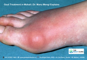
Hydrogen Infusion for Wellness – How it works & its impact on the body.

Hydrogen Drip IV Therapy represents one of the most promising wellness treatments you might not have heard about yet. This lightweight, odourless, and colourless gas rapidly diffuses into your tissues and cells, functioning as an anti-inflammatory and anti-apoptotic agent while stimulating energy metabolism. Surprisingly, the first documented medical use of hydrogen dates back to British military doctors during the 1914-1918 war, who treated 26 terminally ill patients with remarkable results—13 of these patients survived against all odds.

Furthermore, your body can benefit from hydrogen in multiple ways beyond basic wellness. Specifically, hydrogen therapy for inflammation has shown impressive clinical outcomes, with studies demonstrating its effectiveness in reducing reperfusion damage in heart attacks, strokes, and acute ischemia. Additionally, hydrogen therapy for overall wellness works through multiple mechanisms, including increasing antioxidants and decreasing oxidative stress, cell death, and inflammation. What makes hydrogen therapy for cellular repair particularly valuable is that it reduces oxidative stress not only through direct reactions with strong oxidants but also indirectly by regulating various gene expressions. Throughout this article, you’ll discover how hydrogen infusion works in your body and why it might be the missing element in your wellness routine.

What is Hydrogen Infusion Therapy?

Molecular hydrogen, the smallest molecule in existence, has been quietly making waves in the wellness and medical communities as a powerful therapeutic agent. First discovered in 1520 by Philippus Aureolus Paracelsus as a mysterious flammable gas, hydrogen remained unnamed until 1783 when Lavoisier used the French word ‘hydrogene’ to describe it. Despite its long history, hydrogen’s therapeutic potential remained largely overlooked until recent decades.

Definition and origin of hydrogen therapy

Hydrogen Infusion Therapy involves the administration of molecular hydrogen (H₂) to the body through various methods to achieve therapeutic effects. Originally appearing in medical literature as early as 1888 in the Annals of Surgery, hydrogen was used by surgeons to locate visceral injuries in the gastrointestinal tract, avoiding unnecessary surgeries.

However, the true breakthrough came in 2007 when a landmark study published in Nature Medicine demonstrated hydrogen’s neuroprotective effects in cases of cerebral ischemia. This pivotal research sparked global interest in hydrogen’s therapeutic applications, with publications exploding from fewer than 50 pre-2007 to over 2000 in the past decade. In fact, this milestone publication is widely regarded as the spark that ignited modern hydrogen medicine.

Consequently, hydrogen therapy has gained significant momentum in clinical settings. By 2017, inhalation of hydrogen gas was approved as an advanced medicine by Japan’s Ministry of Health for treating post-cardiac arrest syndrome. Currently, over 100 human studies show hydrogen’s translational potential across various conditions, including metabolic syndrome, diabetes, Parkinson’s disease, and rheumatoid arthritis.

Hydrogen therapy can be administered through several methods:

  • Inhalation of hydrogen gas
  • Consumption of hydrogen-rich water
  • Injection of hydrogen-rich saline
  • Topical applications and baths

Why is hydrogen used in wellness treatments

What makes hydrogen particularly valuable in wellness treatments is its unique set of properties. As the smallest gas molecule with a molecular weight of only 2 Da and a kinetic diameter of 289 pm, hydrogen can easily penetrate cell membranes, cross the blood-brain barrier, and access critical cellular components like mitochondria and nuclei.

Essentially, hydrogen functions as a selective antioxidant, primarily targeting harmful free radicals like hydroxyl radicals (•OH) and peroxynitrite anions (ONOO-) while preserving beneficial reactive oxygen species needed for normal cell signalling. This selective action makes hydrogen superior to conventional antioxidants that indiscriminately neutralise all reactive species.

Notably, hydrogen offers multiple therapeutic mechanisms beyond antioxidation. It demonstrates potent anti-inflammatory effects by regulating pro-inflammatory cytokines like IL-1β, IL-6, and TNF-α. Additionally, hydrogen exhibits antiapoptotic properties, helping prevent programmed cell death.

Another advantage of hydrogen therapy is its exceptional safety profile. Unlike other gaseous signalling molecules such as nitric oxide or carbon monoxide, hydrogen has no known toxicity even at high concentrations. Its low solubility in water (1.9 mL H2/100 mL H2O at 20°C) means that concentrations remain well below the 4% needed to react with oxygen, making it completely safe within the human body.

Given these characteristics—powerful permeability, selective antioxidant effects, anti-inflammatory properties, and outstanding safety profile—hydrogen has rightfully earned recognition as the fourth signalling gas molecule after nitric oxide, carbon monoxide, and hydrogen sulfide.

How Hydrogen Works in the Body

The remarkable biological effects of hydrogen stem from its unique physical and chemical properties. At just 2 Da in molecular weight, hydrogen stands as the smallest molecule in existence, enabling it to penetrate biological membranes and reach critical cellular compartments where other molecules simply cannot go.

Cellular absorption and diffusion

Once administered, hydrogen rapidly traverses throughout your body thanks to its exceptional permeability. Unlike larger antioxidant compounds, hydrogen effortlessly passes through cell membranes and diffuses into subcellular compartments, including mitochondria and nuclei. First of all, this remarkable diffusion capacity allows hydrogen to reach the primary sites of reactive oxygen species (ROS) generation, where it can exert its protective effects most efficiently.

Indeed, hydrogen’s extraordinary ability to penetrate biological barriers enables it to access areas typically off-limits to conventional therapeutic agents. It easily crosses the blood-brain barrier, placental barrier, and testis barrier without requiring special transport mechanisms. Moreover, when hydrogen-rich water is consumed, absorption begins in the stomach and continues in the small intestine, where millions of tiny finger-like structures called villi enhance absorption.

Selective antioxidant properties

What truly sets hydrogen apart is its selective antioxidant activity. Instead of indiscriminately neutralising all reactive oxygen species, hydrogen selectively targets the most damaging ones—hydroxyl radicals (•OH) and peroxynitrite (ONOO−)—while preserving beneficial ROS needed for normal cellular signalling.

This selective action occurs through direct chemical reactions. For instance, hydrogen neutralises hydroxyl radicals through the reaction: H₂ + 2•OH → 2H₂O. Additionally, hydrogen leaves physiologically beneficial ROS like hydrogen peroxide (H₂O₂) and superoxide anion (O₂⁻) untouched, allowing them to continue their essential roles in cell signalling.

Consequently, hydrogen enhances your body’s antioxidant capacity beyond direct scavenging. It stimulates endogenous antioxidant enzymes, including superoxide dismutase (SOD), catalase, and myeloperoxidase. Given these properties, hydrogen therapy for cellular repair works at multiple levels within your antioxidant defence system.

Impact on inflammation and oxidative stress

Hydrogen’s effects on inflammation and oxidative stress are closely intertwined. In the face of oxidative stress, hydrogen inhibits the NF-κB pathway—a major regulator of inflammatory responses. Subsequently, this leads to reduced production of pro-inflammatory cytokines such as IL-1β, IL-6, and TNF-α.

At the cellular level, hydrogen prevents mitochondrial damage by decreasing NADPH oxidase expression, thereby reducing ROS accumulation. Furthermore, hydrogen therapy for inflammation works by inhibiting inflammatory cell adhesion molecules like ICAM-1 and reducing infiltration of neutrophils and macrophages at inflammatory sites.

Another powerful mechanism involves hydrogen’s impact on lipid peroxidation. By protecting cell membrane phospholipids from oxidation, hydrogen maintains membrane integrity and prevents cellular damage that would otherwise trigger inflammatory cascades.

Hormetic effects and gene regulation

Perhaps most fascinating is hydrogen’s ability to influence gene expression and promote hormesis—a biological phenomenon where low-dose stressors trigger beneficial adaptive responses. Mild oxidative stress induced by hydrogen peroxide can stimulate organisms’ biological functions and increase resistance to higher doses of the same stressor.

Through these hormetic effects, hydrogen regulates numerous signalling pathways and transcription factors. For instance, hydrogen activates the Nrf2 pathway, a master regulator of antioxidant responses. As Nrf2 accumulates, it binds to antioxidant response elements and initiates protective gene expression.

Likewise, hydrogen affects apoptosis-related genes, reducing expression of pro-apoptotic factors like p53 while enhancing anti-apoptotic genes such as Bcl-2. Beyond these effects, hydrogen modulates calcium signalling pathways, affecting transcription factors like CREB and NFAT that regulate numerous genes.

To sum up, hydrogen’s biological effects emerge from its unique physical properties, selective antioxidant activity, anti-inflammatory actions, and gene-regulating capabilities—creating a comprehensive therapeutic profile unlike any other molecule.

Methods of Hydrogen Administration

Accessing the therapeutic benefits of hydrogen requires getting this tiny molecule into your body, with several proven methods available depending on your wellness goals and preferences.

Hydrogen inhalation therapy

Inhalation represents one of the most direct and rapid methods for delivering hydrogen to your bloodstream and tissues. According to research, you can inhale either pure hydrogen gas or a mixture of hydrogen and oxygen (commonly referred to as oxy-hydrogen). Most clinical applications utilise a concentration of 2-4% hydrogen gas for safety and efficacy. Some advanced hydrogen-oxygen generators produce a mixture containing 66.7% hydrogen and 33.3% oxygen at a flow rate of 3 L/min[41]. This method works exceptionally well for acute conditions due to its immediate effects on the respiratory and cardiovascular systems.

Hydrogen IV therapy and drips

Saturated Hydrogen Water Intravenous Therapy delivers highly-concentrated hydrogen directly into your bloodstream through normal saline. The hydrogen concentration in these infusions typically exceeds 1.6ppm—the maximum concentration achievable under normal temperature and pressure. Throughout an IV session lasting 30-60 minutes, hydrogen molecules enter endothelial cells in your blood vessels, reacting with harmful active oxygen to form water that’s naturally eliminated through urine. Preparation methods include immersing polyethene bags in hydrogen-rich water tanks or using special non-woven fabric containing hydrogen-generating agents. This method provides precise control over hydrogen dosage.

Drinking hydrogen-rich water

Drinking hydrogen-enriched water offers a convenient, portable option for daily hydrogen consumption[53]. You can obtain hydrogen water through infusion machines, water generators, ionisers, or hydrogen-generating tablets. Some commercial products claim to achieve concentrations over 7ppm in 500mL bottles and even 15ppm in 250mL formats. Another innovative approach involves capsules containing porous coral material that absorb and carry hydrogen, releasing it inside your body after consumption. Although limited by hydrogen’s low water solubility of 1.57mg/L, this method remains popular for its simplicity.

Topical and bath-based applications

Bathing in hydrogen-rich water ranks among the most effective therapies for promoting antioxidant activity in your blood compared to other antioxidant administration routes. Specialised devices like the Hebe Hydrogenium+ create hydrogen-rich water for non-invasive skin application. The treatment process typically involves using specialised handpieces that deliver hydrogen-rich water to your skin—either through gentle vacuum lifting or pressurised jets. These treatments often follow a systematic protocol including cleansing, application, and moisturising phases. Besides full baths, topical applications may include hydrogen-rich wet compresses for localised treatment.

Health Benefits of Hydrogen Infusion

Research reveals that molecular hydrogen offers multiple therapeutic benefits through its unique selective antioxidant and anti-inflammatory properties. Let’s explore the specific ways hydrogen infusion can enhance your well-being.

Hydrogen therapy for fatigue and energy

Hydrogen supplementation has demonstrated promising results in combating fatigue and boosting energy levels. Studies show hydrogen-rich water significantly reduces the rating of perceived exertion during exercise and decreases blood lactate concentrations both during and immediately after physical activity. Clinical evidence indicates hydrogen water may be particularly beneficial for those experiencing exercise-induced fatigue, as it helps neutralise excess reactive oxygen species that contribute to diminished performance.

Hydrogen therapy for inflammation and joint pain

For those suffering from joint pain, hydrogen therapy offers substantial relief. Research indicates that hydrogen can inhibit inflammatory factors like ADAMTS5 and MMP13 in osteoarthritis patients. Importantly, clinical trials have shown that hydrogen-oxygen mixture inhalation helps alleviate symptoms and improve functional activity in elderly patients with knee osteoarthritis. This improvement comes from hydrogen’s ability to suppress inflammatory pathways—primarily by inhibiting the JNK signalling pathway.

Hydrogen therapy for skin health and glow

Your skin can benefit tremendously from hydrogen therapy. Clinical studies show hydrogen-rich water treatments effectively reduce pore visibility and improve pigmentation irregularities. Furthermore, hydrogen works to neutralise free radicals responsible for premature ageing, fine lines, and skin dullness. It also helps maintain collagen integrity by preventing oxidative degradation of skin structural proteins.

Hydrogen therapy for muscle recovery & sports injury

Athletes have discovered hydrogen’s remarkable effects on recovery. Four days of hydrogen-rich water supplementation have been shown to reduce blood creatine kinase activity (156 ± 63 vs. 190 ± 64 U.L−1) and muscle soreness (34 ± 12 vs. 42 ± 12 mm) after intense training. Plus, athletes experienced improved countermovement jump height (30.7 ± 5.5 cm vs. 29.8 ± 5.8 cm), suggesting faster functional recovery.

Hydrogen IV therapy for brain & cognitive health

Hydrogen readily crosses the blood-brain barrier, making it especially valuable for cognitive health. Research suggests hydrogen therapy may help manage Alzheimer’s disease by addressing oxidative stress—a central factor in neurodegenerative disorders. Studies with senescence-accelerated mice demonstrated that hydrogen water prevented age-related declines in cognitive ability and was associated with increased brain serotonin levels.

Hydrogen therapy for mobility & flexibility

Finally, hydrogen therapy supports improved mobility by reducing inflammation in joints and enhancing tissue repair. Clinical research shows that hydrogen effectively mitigates osteoarthritis-induced cartilage damage and promotes cartilage regeneration. This makes hydrogen infusion particularly valuable for addressing mobility challenges stemming from inflammatory joint conditions.

Clinical Evidence and Safety

Over the past two decades, extensive research has accumulated with more than 2000 publications documenting hydrogen’s therapeutic potential. Clinical trials span major disease categories, including cardiovascular, respiratory, and central nervous system disorders.

Summary of human and animal studies

Scientific investigations reveal hydrogen’s therapeutic applications across multiple conditions. Animal studies demonstrate hydrogen’s efficacy in reducing oxidative stress-related diseases and preventing neurodegeneration. Randomised clinical trials show hydrogen improves cognitive scores in APOE4 carriers with mild cognitive impairment, while double-blind studies indicate significant improvement in Parkinson’s disease symptoms. Throughout Japan, hydrogen inhalation received approval for post-cardiac arrest syndrome treatment in 2016.

Hydrogen IV therapy benefits in chronic conditions

Patients with chronic conditions often experience substantial improvements from Hydrogen IV therapy. For chronic kidney disease sufferers, hydrogen supplementation shows decreased serum creatinine levels. Additionally, hydrogen therapy modulates immune responses by increasing regulatory T cells while reducing inflammatory cells. Even more promising, hydrogen administration helps manage inflammatory bowel disease by regulating NF-κB and PI3K/AKT/mTOR signalling pathways.

Safety profile and FDA status

Hydrogen therapy exhibits an excellent safety record with minimal adverse effects reported across clinical trials. The US FDA issued a notice (GRAS Notice No. 520) acknowledging hydrogen solubilised in water (up to 2.14% concentration) as generally recognised as safe for beverages. Nonetheless, hydrogen inhalation requires specialised equipment for production, making proper administration important for safety.

Who should avoid hydrogen therapy?

Given that unregistered hydrogen devices lack quality and safety assurances, only use products with proper certification. Currently, hydrogen therapy remains experimental for musculoskeletal conditions and should be approached cautiously. Before beginning hydrogen therapy, consult your healthcare provider, especially if pregnant or managing serious medical conditions.

Conclusion

Hydrogen infusion therapy stands at the forefront of innovative wellness treatments, offering remarkable potential for your overall health. Throughout this article, we’ve seen how this lightweight molecule penetrates cellular barriers and selectively targets harmful free radicals while preserving beneficial ones. Additionally, hydrogen’s anti-inflammatory properties make it particularly valuable for addressing chronic conditions and supporting recovery.

Whether you choose inhalation therapy, IV drips, hydrogen-rich water, or topical applications, each method provides unique advantages depending on your specific wellness goals. Consequently, hydrogen therapy offers multiple pathways to experience benefits ranging from reduced fatigue and enhanced energy to improved skin health and cognitive function.

Clinical evidence continues to mount in support of hydrogen’s therapeutic applications. Research demonstrates its effectiveness across various conditions while maintaining an excellent safety profile. Still, consulting with healthcare professionals before starting any new therapy remains essential, especially if you have underlying health concerns.

Hydrogen infusion therapy represents much more than a passing wellness trend. The science behind its effectiveness reveals a profound impact on cellular health through multiple mechanisms. As research advances and awareness grows, hydrogen therapy will likely become an increasingly important component of comprehensive wellness strategies. Your journey toward optimal health might benefit significantly from this remarkable element that works harmoniously with your body’s natural processes.

Key Takeaways

Hydrogen infusion therapy offers a scientifically backed approach to wellness through the body’s smallest molecule, providing selective antioxidant effects and anti-inflammatory benefits with exceptional safety.

• Hydrogen selectively targets harmful free radicals while preserving beneficial ones, unlike conventional antioxidants that neutralise all reactive species indiscriminately.

• Multiple administration methods exist, including inhalation, IV therapy, hydrogen-rich water, and topical applications, each offering unique advantages for different wellness goals.

• Clinical studies demonstrate hydrogen’s effectiveness for reducing fatigue, inflammation, joint pain, and supporting muscle recovery with minimal side effects.

• Hydrogen easily crosses biological barriers, including the blood-brain barrier, making it valuable for cognitive health and neurological conditions.

• With over 2000 publications and FDA recognition as generally safe, hydrogen therapy represents a promising addition to comprehensive wellness strategies.

The therapeutic potential of hydrogen lies in its unique ability to work at the cellular level while maintaining an outstanding safety profile, making it accessible for various health optimisation goals from athletic recovery to chronic condition management.

FAQs

Q1. What are the main benefits of hydrogen therapy for the human body?

A1. Hydrogen therapy offers several health benefits, including reduced oxidative stress, improved cardiovascular function, enhanced liver health, and potential neuroprotective effects. It acts as a selective antioxidant, targeting harmful free radicals while preserving beneficial ones.

Q2. Are there any known side effects of hydrogen infusion therapy?

A2. Hydrogen infusion therapy has an excellent safety profile with minimal reported side effects. Clinical trials have not observed common adverse reactions such as nosebleeds, chest pain, breathing difficulties, nausea, dizziness, or skin allergies associated with hydrogen-oxygen inhalation.

Q3. How does hydrogen therapy work to reduce inflammation in the body?

A3. Hydrogen therapy helps reduce inflammation by modulating the body’s immune response. It dampens pro-inflammatory cytokines while boosting anti-inflammatory compounds, effectively regulating inflammatory pathways and providing relief from various inflammatory conditions.

Q4. What is the recommended daily intake of hydrogen-infused water?

A4. Most studies suggest consuming about 1.5 to 2 litres (50 to 60 ounces) of hydrogen-infused water daily for optimal benefits. This can be achieved through hydrogen tablets or by drinking hydrogenated water. However, it’s always best to consult with a healthcare professional for personalised advice.

Q5. Can hydrogen therapy improve cognitive function and brain health?

A5. Yes, hydrogen therapy shows promise for cognitive health. It readily crosses the blood-brain barrier, potentially helping manage neurodegenerative conditions like Alzheimer’s disease by addressing oxidative stress. Some studies have shown improvements in cognitive scores and symptoms of neurological disorders with hydrogen therapy.
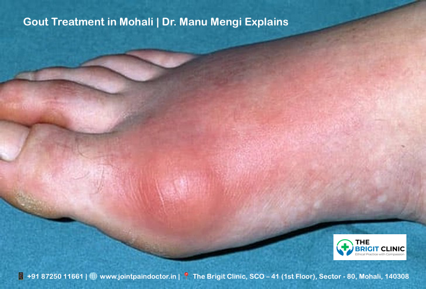
Gout Treatment in Mohali | Dr. Manu Mengi Explains

Have you ever woken up in the middle of the night with your big toe feeling like it’s on fire? This intense pain is often a hallmark of gout, a painful form of arthritis that affects about 1-2% of adults. Finding effective Gout Treatment in Mohali is the first step toward lasting relief. Historically known as “the disease of kings,” gout has actually become more common in recent decades.

Gout occurs when urate crystals accumulate in your joints, causing inflammation and intense pain during an attack. This condition most commonly affects your big toe joint, though it can target other joints as well. High uric acid levels in your body are the primary culprit, often resulting from eating or drinking foods high in purines. Additionally, certain risk factors increase your likelihood of developing gout, including being overweight, having high blood pressure, diabetes, kidney disease, or heart failure. Men are three times more likely to develop this condition than women, though anyone can be affected.

Fortunately, with early diagnosis, appropriate treatment, and lifestyle changes, gout is one of the most controllable forms of arthritis. In this comprehensive guide, Dr. Manu Mengi explains everything you need to know about managing and preventing gout flares.

> > > CTA: Is your big toe swollen and red? Don't ignore the signs. Consult with Dr. Manu Mengi, a leading Red Painful Toe Specialist in Mohali, for an accurate diagnosis. Book your appointment today: +91 87250 11661

Understanding Gout: What It Is and How It Starts

Gout represents a complex form of inflammatory arthritis that occurs when sharp, needle-shaped crystals form in and around your joints. These crystals cause intense inflammation, resulting in sudden attacks of severe pain, swelling, and tenderness.

What is gout and how it affect joints

Gout is characterised by the formation of monosodium urate (MSU) crystals in joints and surrounding tissues. These crystals trigger highly inflammatory responses in the affected areas. The inflammation doesn’t just cause pain—it can eventually lead to joint damage if left untreated. Furthermore, gout progresses through several stages, beginning with asymptomatic hyperuricemia, followed by acute gout flares, intervals between attacks, and eventually, in some cases, chronic gout with tophi (crystal deposits under the skin).

The role of uric acid and purines

At the heart of gout is uric acid, the final product of purine metabolism. Purines are chemicals naturally found in your body and certain foods. Normally, uric acid dissolves in your blood and passes through your kidneys into urine. However, problems arise in two scenarios: either your body produces too much uric acid, or your kidneys don’t excrete enough.

Consequently, uric acid can build up in your bloodstream—a condition called hyperuricemia. When levels exceed 6.8 mg/dL (the saturation threshold), uric acid can form those painful MSU crystals. Essentially, these crystals are what cause the inflammation and intense pain of a gout attack.

Why the big toe is often the first target

The first metatarsophalangeal joint (your big toe joint) is ground zero for approximately 50% of initial gout attacks, with about 90% of patients experiencing at least one attack in this location. This preference for the big toe isn’t random—there are scientific reasons behind it.

Temperature plays a significant role in crystal formation. A reduction of even 2°C is sufficient to lower the solubility point of urate from 6.8 to 6.0 mg/dL. The big toe, being farther from your body’s core and having less tissue coverage, naturally maintains a slightly lower temperature. Additionally, this joint experiences repetitive trauma from walking, creating an ideal environment for crystal formation.

Unlike other forms of arthritis, gout is largely controllable with proper management of uric acid levels. Understanding this connection between uric acid and gout symptoms is the first step toward effective treatment.

> > > CTA: Struggling with high uric acid? Get personalised advice from the Best Gout Doctor in Mohali. Dr. Mengi creates tailored management plans to prevent crystal formation and stop the pain. Call now: +91 87250 11661

Recognising the Signs: Symptoms and Triggers

The sudden, excruciating pain of a gout attack often strikes without warning, leaving you searching for relief in the middle of the night. Recognising the symptoms early can help you seek treatment promptly and potentially reduce the severity of flares.

Gout symptoms in the foot and other joints

While the big toe is gout’s favourite target, the condition doesn’t limit itself to just one location. Gout can also affect your ankles, knees, elbows, wrists, and fingers. The pain is typically most severe within the first 4-12 hours after it begins. As gout progresses, you might experience a limited range of motion in the affected joints.

Swollen big toe joint and severe tenderness

The joint at the base of your big toe often bears the brunt of a gout attack. During a flare, this joint becomes so tender that even the light touch of a bedsheet can cause excruciating pain. You’ll notice significant swelling along with this tenderness, making it difficult to wear shoes or walk normally.

Redness & heat in joints during a flare

Beyond pain and swelling, visual cues help identify a gout attack. The affected joint typically becomes noticeably red or discoloured. Moreover, you’ll feel warmth or even a sensation like the joint is “on fire”. This inflammation is your body’s response to the sharp urate crystals irritating the joint.

Gout attack signs and how long they last

Gout flares often begin suddenly at night, with pain intense enough to wake you from sleep. A typical gout attack lasts between one and two weeks. Between flares, you usually won’t experience any symptoms. Nevertheless, without proper treatment, attacks may become more frequent and last longer over time.

Alcohol binge and other common triggers

Research shows alcohol consumption is the most common trigger for gout attacks, responsible for 14% of flares in one study. Specifically, drinking more than one alcoholic beverage in a day raises your risk of experiencing a gout attack. Beer appears to pose a greater risk than spirits, due to its high purine content.

Other common triggers include:

  • Consuming red meat or seafood
  • Dehydration
  • Physical injury or excessive activity
  • Extremely warm or cold weather

For those with gout, experts recommend avoiding alcoholic beverages entirely, despite the potential benefits of moderate consumption for other health conditions.

> > > CTA: Woken up by a sudden, fiery joint pain? You don't have to suffer through a gout flare alone. Contact Dr. Manu Mengi's Clinic in Mohali for urgent consultation and effective pain relief strategies.

What Causes Gout: Risk Factors and Underlying Conditions

Understanding what causes gout begins with identifying its root mechanism – disrupted uric acid balance in your body. Let’s explore the factors that put you at risk.

High uric acid levels and hyperuricemia

Hyperuricemia occurs when uric acid levels exceed 6.8 mg/dL in your blood. This condition happens when your body either produces too much uric acid or your kidneys don’t excrete enough. Although one-quarter of people with hyperuricemia develop gout, most remain symptom-free.

Risk factors for gout – diet, alcohol, obesity

Your dietary choices significantly impact gout risk. Foods high in purines, such as red meat, organ meats, and certain seafood, can raise uric acid levels. Beer consumption poses a greater risk than other alcoholic beverages because of its high guanosine content. Weight gain is another major factor – obesity increases your risk of gout by 3.26 times compared to a normal weight.

Genetics & gout: family history matters

Gout runs in families, with heritability estimated at approximately 60%. About 20% of people with gout have a relative with the condition. Research shows that an additive genetic score of high-risk alleles can increase gout risk up to 41-fold.

Medications that trigger gout

Certain medications raise uric acid levels in your blood. Diuretics increase the relative risk of gout by almost 80%. Other culprits include low-dose aspirin, cyclosporine, and some transplant medications.

Hormonal changes in gout (e.g., menopause)

For women, menopause significantly increases gout risk. The hormone estrogen helps eliminate uric acid through urine. After menopause, estrogen production declines, causing uric acid levels to rise.

Gout & kidney disease: a two-way link

A bidirectional relationship exists between gout and kidney disease. Kidney disease impairs uric acid excretion, raising gout risk. Conversely, high uric acid can damage the kidneys by forming stones and causing scarring.

> > > CTA: Confused about how your diet or medications affect your gout? Get a personalised Uric Acid Diet Consultation in Mohali with Dr. Mengi to understand your triggers and build a prevention plan.

Diagnosis and Treatment Options Explained

Getting an accurate diagnosis is crucial for effective gout management. Modern diagnostic techniques have improved physicians’ ability to detect and monitor this painful condition.

Ultrasound for gout detection

Ultrasound has emerged as a valuable tool for diagnosing gout, offering high-resolution imaging without radiation. The characteristic “double contour sign” (an irregular hyperechoic line over cartilage) has 43.7% sensitivity but 99% specificity for gout. Ultrasound can also identify tophaceous deposits in soft tissues and joints that might not be evident during clinical examination.

X-ray changes in chronic gout

X-rays typically show normal findings early in the disease. In chronic gout, hallmark features include marginal erosions with overhanging edges and sclerotic margins. Soft-tissue nodules (tophi) may appear as dense masses with higher attenuation than surrounding tissues.

Corticosteroids in gout treatment

Corticosteroids effectively treat acute gout flares, especially for patients who cannot tolerate NSAIDs. Options include oral prednisone (30-40mg daily for 5 days), intravenous administration, or direct joint injections. For single-joint gout, intra-articular injections offer fewer systemic side effects.

Gout treatment without medications

Non-pharmaceutical approaches include applying ice to affected joints, resting the inflamed area, and drinking plenty of water. Maintaining a healthy weight through regular exercise helps prevent flares, with low-impact activities like walking or swimming being ideal.

Managing chronic gout vs acute gout

Acute gout requires rapid inflammation control within 24 hours of symptom onset. Chronic gout management focuses on lowering serum uric acid below 6 mg/dL through long-term therapy. This distinction is essential—treating inflammation during flares differs from preventing future attacks through urate-lowering strategies.

> > > CTA: Need a precise diagnosis? As a leading Uric Acid Treatment Doctor in Mohali, Dr. Mengi uses advanced ultrasound imaging to accurately detect gout crystals and create a targeted treatment strategy for both acute and chronic gout. Visit our clinic to know more.

Conclusion

Gout, while extremely painful, remains one of the most manageable forms of arthritis when properly addressed. Understanding the connection between uric acid levels and gout attacks enables you to take control of this condition. Certainly, the buildup of urate crystals causes those excruciating flares, but knowledge about triggers like alcohol consumption, purine-rich foods, and certain medications gives you the power to prevent attacks.

Therefore, working closely with healthcare providers becomes essential for proper diagnosis and treatment. Advanced diagnostic tools, such as ultrasound, can detect gout even before visible symptoms appear. After diagnosis, treatment options range from medications that reduce inflammation during acute attacks to long-term strategies for lowering uric acid levels below the crystal-forming threshold.

Besides medical interventions, lifestyle modifications play a crucial role in gout management. Weight loss, adequate hydration, and avoiding high-purine foods significantly reduce your risk of future flares. Additionally, limiting alcohol consumption, particularly beer, helps maintain lower uric acid levels.

Remember that gout management requires both immediate relief during attacks and ongoing prevention between episodes. Though historically called “the disease of kings,” gout does not have to rule your life. With early detection, appropriate treatment, and consistent lifestyle adjustments, you can effectively manage this condition and maintain joint health for years to come.

Key Takeaways

Understanding gout empowers you to manage this painful but controllable form of arthritis effectively through proper diagnosis, treatment, and lifestyle modifications.

• Gout occurs when uric acid crystals form in joints, most commonly affecting the big toe first due to lower temperature and repetitive trauma from walking.

• Alcohol consumption is the top trigger for gout attacks, with beer posing a higher risk than other alcoholic beverages due to its purine content.

• Early diagnosis using ultrasound and X-rays enables targeted treatment, distinguishing between acute flare management and chronic prevention strategies.

• Lifestyle changes like weight loss, hydration, and avoiding high-purine foods significantly reduce attack frequency and severity.

• Gout is highly manageable when uric acid levels stay below 6 mg/dL through medication, diet modifications, and consistent medical monitoring.

With proper understanding of triggers, symptoms, and treatment options, gout doesn’t have to control your life. The key lies in working with healthcare providers to develop a comprehensive management plan that addresses both immediate pain relief and long-term prevention strategies.

> > > CTA: Ready to take control of your gout? Partner with Dr. Manu Mengi Gout Specialist Mohali, for a comprehensive management plan that combines advanced treatment with sustainable lifestyle changes. Your journey to a pain-free life starts here.

FAQs

Q1. What exactly is gout, and how does it affect the body?

A1. Gout is a form of inflammatory arthritis caused by the buildup of uric acid crystals in joints, most commonly in the big toe. It results in sudden attacks of severe pain, swelling, and tenderness. Gout occurs when the body either produces too much uric acid or the kidneys don’t excrete enough, leading to crystal formation in joints.

Q2. What are the most common triggers for a gout attack?

A2. The most common trigger for gout attacks is alcohol consumption, particularly beer, due to its high purine content. Other triggers include consuming red meat or seafood, dehydration, physical injury, and extreme weather conditions. Avoiding these triggers, especially limiting alcohol intake, can help reduce the frequency of gout flares.

Q3. How can I quickly relieve gout symptoms at home?

A3. To quickly relieve gout symptoms at home, apply ice to the affected joint, rest the inflamed area, and drink plenty of water. Some people find cherry juice helpful in reducing uric acid levels. However, it’s important to consult with a healthcare provider for proper treatment, as these methods provide temporary relief but don’t address the underlying cause.

Q4. Are there any non-medication approaches to managing gout?

A4. Yes, there are several non-medication approaches to managing gout. These include maintaining a healthy weight through regular low-impact exercise like walking or swimming, staying well-hydrated, and avoiding foods high in purines. Lifestyle modifications play a crucial role in preventing gout flares and managing the condition long-term.

Q5. How is gout diagnosed, and what treatment options are available?

A5. Gout is typically diagnosed through a combination of clinical symptoms, blood tests to measure uric acid levels, and imaging techniques like ultrasound or X-rays. Treatment options include medications to reduce inflammation during acute attacks, such as NSAIDs or corticosteroids, and long-term therapies to lower uric acid levels. The goal is to bring serum uric acid below 6 mg/dL to prevent future attacks and complications.

Dr. Manu Mengi – Gout Specialist Mohali

If you are looking for expert and compassionate care for gout in Mohali, look no further than Dr. Manu Mengi. Renowned as one of the Best Orthopedic Doctors in Chandigarh Mohali, Dr. Manu Mengi specialises in the diagnosis and management of gout and other joint pain disorders. His patient-centric approach combines cutting-edge diagnostic technology with evidence-based treatment protocols to provide both immediate relief and long-term prevention strategies. Whether you’re experiencing your first painful flare or struggling with chronic gout, Dr. Mengi and his team are dedicated to helping you regain your mobility and live a pain-free life. Schedule a consultation at his Mohali clinic to begin your journey toward effective gout management.

Book Appointment - Call Us: +91 87250 11661 | Find Our Clinic: Get Directions


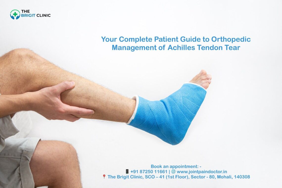
Your Complete Patient Guide to Orthopedic Management of Achilles Tendon Tear

Despite being the largest tendon in your body and capable of withstanding forces up to 10 times your body weight, the Achilles tendon is surprisingly vulnerable to complications. Understanding the Orthopedic Management of Achilles Tendon Tear is the first step toward a successful recovery. Achilles tendon ruptures are very common sports injuries, occurring most frequently in people ages 30 to 40 and affecting men more often than women.

If you’re among the “weekend warriors” who exercise intensely without regular training, you face a higher risk of tearing your Achilles than younger, well-trained athletes. Additionally, these injuries can be missed in up to 25% of cases, making proper diagnosis crucial. Whether you’re dealing with a partial or complete tear, understanding your treatment options is essential for recovery. This comprehensive guide will walk you through everything you need to know about Achilles tendon tears—from identifying symptoms and risk factors to exploring both conservative and surgical treatment approaches that can help you return to your normal activities.

For expert diagnosis and a personalised treatment plan, 
Consult the Best Orthopedic Doctor in Mohali or call +91 87250 11661

Understanding Achilles Tendon Tear

The Achilles tendon is a critical structure that plays a significant role in your daily movements. Understanding its function and how it can tear will help you better recognise and manage this injury.

What is the Achilles tendon?

The Achilles tendon, also called the calcaneal tendon, is the thickest and strongest tendon in your body. This fibrous band of tissue connects your calf muscles (gastrocnemius and soleus) to your heel bone (calcaneus). Located at the back of your lower leg, this powerful tendon spans approximately 6 to 10 inches in adults.

What makes the Achilles tendon remarkable is its incredible strength—it can support forces up to four times your body weight. This impressive capacity allows you to perform essential movements like walking, running, and jumping. When your calf muscles contract, they pull on the Achilles tendon, causing your foot to point downward (plantarflexion), which helps lift your heel off the ground during physical activities.

Partial vs complete Achilles tendon tear

Achilles tendon tears exist on a spectrum from minor damage to complete rupture. Understanding the difference between partial and complete tears is crucial for proper treatment.

In a partial tear, only a portion of the tendon fibres is damaged. Think of this like a frayed rope where some strands remain intact while others are torn. With a partial tear, you’ll experience:

  • Localised soreness around the tendon
  • Some swelling that gradually decreases
  • Full function of the ankle despite pain

A complete tear occurs when the tendon ruptures entirely, separating into two distinct parts. This severe injury results in:

  • A noticeable “pop” or snapping sound at the moment of injury
  • Complete loss of strength and function in the ankle
  • Extreme difficulty walking or exercising
  • Significant swelling around the ankle
  • In some cases, visible displacement of calf muscles

The Thompson test is commonly used by doctors to diagnose a complete rupture—when squeezing the calf fails to produce normal foot movement, it indicates a tear.

Common causes and overuse injuries

Most Achilles tendon tears happen during recreational sports or athletic activities. The tendon typically ruptures when exposed to sudden, powerful forces, especially during activities requiring quick stops, starts, and pivots—such as soccer, football, basketball, tennis, or squash.

Several specific scenarios commonly lead to tears:

  • Landing awkwardly from a jump
  • Cutting movements during sports
  • Sudden acceleration or forceful pushing off with the foot
  • Direct trauma to the tendon
  • Tripping or falling, particularly when the foot is forced upward[18]

Certain factors increase your risk of experiencing an Achilles tendon tear. As you age, the tendon naturally becomes stiffer and weaker. “Weekend warriors”—adults who exercise intensely without regular training—face a higher risk than consistently trained athletes.

Furthermore, medical conditions like inflammatory diseases, diabetes, obesity, and certain medications (including fluoroquinolone antibiotics and corticosteroids) can weaken the tendon structure. Mechanical issues such as tight calf muscles, improper footwear, and training errors also contribute to vulnerability.

Overall, Achilles tendon ruptures affect approximately 12 per 100,000 individuals, most commonly occurring between ages 40 and 50, with men experiencing them 2 to 12 times more frequently than women.

If you're active and experiencing heel pain, visit a Sports Injury Clinic Mohali for a risk evaluation. Book your appointment or call +91 87250 11661

Recognising Symptoms and Risk Factors

Recognising an Achilles tendon tear promptly can make a significant difference in your treatment outcomes. By understanding the tell-tale signs and knowing your risk profile, you might prevent a delayed diagnosis that occurs in up to 25% of cases.

Sudden pop in the back of the ankle

The most distinctive sign of an Achilles tendon rupture is experiencing (and sometimes hearing) a sudden “pop” or “snap” at the back of your ankle. This sensation is so pronounced that many mistake it for being struck from behind. The sound represents the actual moment when your tendon fibres separate.

After this characteristic pop, you’ll likely feel immediate, intense pain. The sensation resembles what would happen if someone kicked you forcefully in the lower leg. Unlike gradual onset injuries, this moment is unmistakable and marks a clear point when damage occurred.

Call your doctor immediately if you experience this sudden snap during physical activity. This symptom alone strongly suggests a complete tear rather than a minor strain, consequently requiring proper medical evaluation.

Heel pain & swelling

Following a tear, sharp, sudden pain typically develops near your heel. Initially, this pain might be unbearable, though it sometimes subsides slightly after the acute injury. The area around your Achilles tendon will swell noticeably, often accompanied by bruising along the back of the ankle.

The discomfort pattern differs from other foot conditions. With an Achilles tendon tear, pain tends to be localised specifically to the back of the ankle where the tendon attaches to your heel bone. Throughout the day, this pain might intensify with activity.

Morning stiffness presents another common symptom, where the affected area feels particularly tight and sore when you first get up. As you move around, this stiffness sometimes improves temporarily.

Calf pain after injury

Beyond the immediate heel area, pain often radiates upward into your calf muscle. This happens because the calf muscles connect directly to the Achilles tendon, creating a continuous pain pathway.

The discomfort in your calf typically worsens during activities that engage these muscles, such as climbing stairs or walking uphill. Furthermore, weakness in the leg becomes apparent when attempting to push off with the affected foot.

For partial tears, you might maintain some function despite the pain. However, with complete ruptures, standing on tiptoes or performing basic foot movements becomes nearly impossible.

Risk factors for Achilles tendon tear

Several factors increase your vulnerability to Achilles tendon tears:

Age and Gender: The peak age for ruptures occurs between 30-40 years, though some sources identify 40-50 as the highest risk period. Men experience these injuries four to five times more frequently than women.

Activity Patterns: “Weekend warriors” face significantly higher risk than regularly trained athletes. Sports involving quick stops, starts, and pivots present the greatest danger—particularly soccer, basketball, tennis, football, and racquet games.

Medical Factors: Certain conditions predispose you to tendon injuries:

  • Previous Achilles tendinopathy
  • Diabetes
  • High cholesterol or blood pressure
  • Psoriasis
  • End-stage kidney disease
  • Inflammatory arthritis

Medication Usage: Some medications weaken tendon structure, notably:

  • Fluoroquinolone antibiotics[28]
  • Corticosteroid injections
  • Oral bisphosphonates

Biomechanical Issues: Physical characteristics matter too. Having tight calf muscles, flat arches, overpronation (ankles rolling inward), or legs of different lengths all increase your risk[30].

Understanding these symptoms and risk factors helps you identify potential problems early and seek appropriate orthopaedic management for Achilles tendon tears before complications develop.

Experienced a pop or snap in your ankle? Seek immediate consultation with an Orthopedic Doctor in Mohali for Achilles Tear, call +91 87250 11661

How Achilles Tendon Tears Are Diagnosed

Getting an accurate diagnosis is essential for proper orthopaedic management of an Achilles tendon tear. Medical professionals use several examination techniques combined with imaging to confirm your injury.

Physical exam and Thompson test

Your doctor will begin by asking about your symptoms and medical history, followed by a thorough physical examination of your lower leg. During this examination, they’ll check for tenderness, swelling, and potentially feel for a gap in your tendon if it has ruptured completely.

The Thompson test (sometimes called the calf squeeze test) is a key diagnostic procedure with 96-100% sensitivity and 93-100% specificity for detecting Achilles ruptures. Here’s how it works:

  • You’ll lie face down with your feet hanging over the edge of the exam table
  • Your doctor will gently squeeze your calf muscle
  • In a healthy tendon, this causes your foot to point downward naturally
  • If your foot doesn’t move during the squeeze, it indicates a likely rupture

To confirm the diagnosis, doctors often use additional clinical signs like checking for a palpable gap in the tendon (typically 3-6cm above the heel) and assessing plantar flexion strength.

MRI vs Ultrasound in Achilles tendon tear

Both MRI and ultrasound provide valuable diagnostic information, albeit with different strengths:

Ultrasound shows the tendon in real-time and demonstrates how it responds to movement. It’s highly accurate with 95% sensitivity and 99% specificity for detecting full-thickness tears. Ultrasound is generally:

  • More cost-effective
  • Readily available
  • Excellent for detecting tendinopathy and complete ruptures

MRI creates detailed images of soft tissues and is particularly valuable for:

  • Detecting partial tears (superior to ultrasound)
  • Assessing the distance between torn tendon ends
  • Postoperative evaluation
  • Ruling out other injuries with similar symptoms

Most specialists recommend ultrasound over MRI for initial diagnosis and monitoring, though your doctor may order both depending on your specific situation.

When to see a doctor

Seek immediate medical attention if you experience:

  • A popping or snapping sound at the time of injury
  • Suddenly, severe pain in the back of your ankle
  • Difficulty walking or standing on tiptoes
  • Visible swelling around the heel area

Even if you can walk with a ruptured Achilles (which many people can), it’s crucial to see a healthcare provider promptly. Using your ankle and putting full weight on it before diagnosis can worsen the injury. Importantly, up to 20% of Achilles tendon ruptures are initially misdiagnosed, often confused with ankle sprains, making proper medical evaluation essential for effective treatment.

For advanced diagnostic imaging and expert interpretation, visit the Best Ortho Doctor in Mohali. Schedule your visit.

Treatment Options: Conservative and Surgical

Treatment decisions for Achilles tendon tears depend on several factors, including your age, activity level, and the severity of your injury. Both non-surgical and surgical approaches offer viable pathways to recovery, each with distinct advantages.

Achilles tendon tear – conservative management

Conservative treatment involves non-surgical approaches that typically include rest, immobilisation, and controlled rehabilitation. This option is often suitable for older patients, those with limited activity goals, or individuals with health conditions that increase surgical risks.

For partial tears with less than 5mm gap between ruptured tendon edges, conservative management can be particularly effective. The traditional approach involves wearing a below-knee cast in an equinus (pointed down) position for four weeks without weight-bearing, followed by a neutral position cast with weight-bearing for another four weeks.

Surgical treatment of Achilles tendon tear

Surgical intervention appears to be the preferred method for athletes and younger, active individuals. The primary benefit of surgery is a lower re-rupture rate compared to non-surgical treatment.

The procedure typically involves making an incision in the back of your leg and stitching the torn tendon together. In cases of severe degeneration, surgeons may remove damaged portions and repair the remaining healthy tendon.

Minimally invasive Achilles tendon tear surgery

This advanced technique involves a small 3-4cm incision instead of the traditional 10cm cut. Through this smaller opening, specialised instruments guide sutures into the tendon to complete the repair.

The minimally invasive approach offers several advantages:

  • Reduced wound healing issues
  • Lower infection rates
  • Less scar tissue formation
  • Faster return to normal activities

Immobilisation vs early mobilisation in Achilles tendon tear

Historically, rigid cast immobilisation for six weeks was standard practice. Nevertheless, recent research strongly supports early functional rehabilitation and mobilisation.

Studies demonstrate that early mobilisation doesn’t increase re-rupture rates. Moreover, it offers superior benefits:

  • Decreases excessive adhesion formation
  • Improves the biomechanical properties of healing tissue
  • Enhances tendon gliding function
  • Reduces joint stiffness and muscle atrophy

Medication for tendon inflammation

Pain management typically begins with over-the-counter options like ibuprofen or naproxen sodium. For persistent discomfort, prescription medications might include COX-2 inhibitors, which potentially cause fewer gastrointestinal side effects than traditional NSAIDs.

PRP Achilles tendon tear therapy

Platelet-rich plasma (PRP) therapy involves injecting a concentrated solution of your own platelets into the injured area. These platelets contain growth factors that may promote tissue repair and regeneration.

Currently, evidence regarding PRP effectiveness remains mixed. Some studies show improvements in ankle dorsiflexion angle and calf circumference, whereas others found no significant differences in patient-reported outcomes at two years post-injury.

Explore all treatment options, including Minimally Invasive Achilles Surgery in Mohali, with the Best Orthopedician in Mohali. Discuss your choices at https://jointpaindoctor.in/ or 
Call *+91 87250 11661* to learn more about the Achilles Tear Surgery Cost Mohali.

Recovery, Rehab, and Return to Activity

Full healing from an Achilles tendon tear requires a comprehensive rehabilitation approach tailored to your specific needs. The recovery journey typically spans four to six months, regardless of whether you underwent surgical or non-surgical treatment.

Physical therapy and strengthening

Physical therapy serves as the cornerstone of Achilles tendon rehabilitation. The duration varies based on injury severity—from a few weeks to several months. Your therapist will focus on three primary goals: pain relief through various modalities, restoring proper movement patterns, and rebuilding muscle strength and balance.

Eccentric exercises stand out as the most evidence-based intervention for Achilles rehabilitation. This approach, typically performed twice daily for at least 11 weeks, has been shown to reduce pain by an average of 60% across multiple clinical trials. The Alfredson protocol remains the gold standard, gradually progressing from bilateral to single-leg heel raises.

For optimal recovery, maintain a consistent exercise regimen alongside gradually increasing weight-bearing activities. Initially, you’ll use a walking boot with progressively decreasing heel wedges until reaching a neutral position, usually around 6-8 weeks post-injury.

Custom orthotics post Achilles repair

Bespoke orthotics play a valuable role in recovery by providing proper foot alignment, enhancing shock absorption, and correcting biomechanical issues that might stress your healing Achilles tendon. These devices primarily keep your heel raised, reducing the workload on the tendon while protecting against re-rupture.

Studies have demonstrated that custom foot orthoses can significantly improve symptoms in athletes with Achilles tendinopathy, with participants reporting an average 92% improvement when using high-density EVA orthotics.

Equinus contracture after Achilles tendon tear

Equinus contracture—excessive tightness limiting ankle dorsiflexion—often develops following Achilles injuries. Conservative management through physical therapy, stretching, and night splints should be attempted first. For refractory cases, surgical options include gastrocnemius lengthening, soleus fascial release, or Achilles tendon lengthening procedures.

Return to sports after Achilles tendon tear

Returning to sports requires patience—full athletic activities should be avoided for at least 6 months post-injury. The return process follows a carefully structured progression: controlled strengthening, followed by plyometric training, and finally sport-specific movements.

Before resuming competitive activities, you should achieve specific milestones: single-leg heel raise at 90% height compared to your uninjured side, normal gait mechanics, and pain-free performance of sport-specific movements. Even with optimal rehabilitation, expect some persistent strength deficits (10-30%) in the affected leg beyond the one-year mark.

Access comprehensive rehabilitation programs at our Orthopedic Hospital in Mohali for the Achilles Rupture facility. Start your recovery journey with the Best Achilles Tendon Surgeon in Mohali by calling +91 87250 11661

Conclusion

Achilles tendon tears represent serious injuries that require prompt diagnosis and appropriate treatment for optimal recovery. Throughout this guide, we’ve explored how these tears happen, their symptoms, and the available treatment approaches. Whether you choose conservative management or surgical intervention, your recovery journey demands patience and commitment to rehabilitation protocols.

Most patients can expect a full recovery period of four to six months, though some strength deficits might persist beyond the one-year mark. During this time, physical therapy will become your essential ally, particularly through eccentric strengthening exercises that have proven highly effective for tendon healing.

Remember that each case differs based on factors like age, activity level, and tear severity. Therefore, working closely with healthcare professionals remains crucial for developing a personalised treatment plan. Custom orthotics might benefit your recovery by improving foot alignment and reducing stress on your healing tendon.

Though returning to sports and normal activities takes time, a structured approach to rehabilitation significantly improves your outcomes. Above all, don’t rush this process. Your body needs adequate time to rebuild the strongest tendon in your body.

Armed with this knowledge about Achilles tendon tears, you can now make informed decisions about your care if faced with this injury. Early recognition of symptoms, prompt medical attention, and dedication to your rehabilitation program will ultimately determine your successful return to the activities you enjoy.

For a successful recovery under expert guidance, book your final consultation at https://jointpaindoctor.in/ or call +91 87250 11661

Key Takeaways

Understanding Achilles tendon tears and their proper management can significantly impact your recovery outcomes and help you make informed treatment decisions.

• Recognise the warning signs early: A sudden “pop” sound, severe heel pain, and inability to stand on tiptoes indicate a potential Achilles rupture requiring immediate medical attention.

• Both surgical and conservative treatments work: Your age, activity level, and tear severity determine the best approach—athletes often benefit from surgery while older patients may succeed with non-surgical management.

• Early mobilisation beats prolonged immobilisation: Modern rehabilitation emphasises controlled movement over extended casting, leading to better outcomes and faster functional recovery.

• Recovery takes 4-6 months minimum: Patience is crucial as rushing back to activities increases re-rupture risk—expect some strength deficits even after one year.

• Physical therapy is non-negotiable: Eccentric strengthening exercises, particularly the Alfredson protocol, form the foundation of successful rehabilitation regardless of treatment method chosen.

The key to successful Achilles tendon recovery lies in prompt diagnosis, appropriate treatment selection, and unwavering commitment to structured rehabilitation. Don’t underestimate this injury—proper management now prevents long-term complications and ensures your return to normal activities.

Ready to start your treatment? Contact the Best Orthopedic Doctor Mohali today 
or call +91 87250 11661

FAQs

Q1. What are the main symptoms of an Achilles tendon tear?

A1. The primary symptoms include a sudden “pop” or snapping sensation in the back of the ankle, intense heel pain, swelling around the affected area, and difficulty walking or standing on tiptoes.

Q2. How long does it typically take to recover from an Achilles tendon tear?

A2. Recovery usually takes 4-6 months, regardless of whether surgical or non-surgical treatment is chosen. However, some strength deficits may persist for over a year.

Q3. Is surgery always necessary for an Achilles tendon tear?

A3. Not always. The decision between surgical and conservative treatment depends on factors like age, activity level, and tear severity. Athletes often benefit from surgery, while older patients may succeed with non-surgical management.

Q4. What role does physical therapy play in Achilles tendon tear recovery?

A4. Physical therapy is crucial for recovery, focusing on pain relief, restoring proper movement, and rebuilding strength. Eccentric exercises, particularly the Alfredson protocol, are considered highly effective for rehabilitation.

Q5. When can I return to sports after an Achilles tendon tear?

A5. Full athletic activities should be avoided for at least 6 months post-injury. Before returning to competitive sports, you should achieve specific milestones like single-leg heel raises at 90% height compared to the uninjured side and pain-free performance of sport-specific movements.

About the Doctor – Dr. Manu Mengi

Dr. Manu Mengi is a highly skilled and renowned Orthopedic Surgeon in Mohali, specialising in the management and treatment of sports injuries, particularly complex Achilles tendon tears. With extensive experience and a commitment to adopting the latest surgical techniques, including minimally invasive procedures, Dr. Mengi provides personalised care to each patient. He leads a state-of-the-art Ortho Clinic in Mohali that is equipped with advanced diagnostic technology to ensure accurate assessments and the most effective treatment plans. Dedicated to helping patients return to their active lifestyles, Dr. Mengi is considered one of the best orthopedic doctors in the region for Achilles tendon repair and rehabilitation.


Book an Appointment today - 
📱 +91 87250 11661 | 🌐 Read Reviews | 📍 Get Directions

Orthopedic Complications in Diabetic Patients

Diabetic Bone & Joint Problems: Orthopedic Risks & Solutions

Diabetes & bone health concerns will significantly affect more people in the coming years, with projections showing diabetes cases increasing from 285 million in 2010 to a staggering 439 million by 2030. This growing health crisis not only impacts blood sugar management but also creates serious orthopaedic complications that can dramatically reduce your quality of life. If you’re seeking expert care, Dr. Manu Mengi, renowned as the Best Orthopedic Doctor in Mohali, emphasises early intervention for diabetic patients at his Ortho Clinic in Mohali.

If you’re living with diabetes, your risk for developing musculoskeletal disorders increases substantially. Diabetic patients face a seven times higher likelihood of surgical site infections, experience delayed fracture healing, and account for 85% of all non-traumatic amputations. Furthermore, your chances of developing hip fractures increase 1.4-1.8 fold with type 2 diabetes and 6-7 fold with type 1 diabetes. The financial burden is equally concerning—diabetes-related healthcare expenditures reached $376 billion in 2010 and are expected to exceed $490 billion by 2030.

Throughout this article, you’ll discover the complex relationship between diabetes and orthopedic health, common complications like Charcot foot and Dupuytren’s contracture, and essential strategies to protect your musculoskeletal system while managing your diabetes.

Understanding How Diabetes Affects the Musculoskeletal System

The relationship between diabetes and musculoskeletal issues stems from complex biochemical processes that affect your entire body. Diabetes doesn’t just impact your blood sugar—it fundamentally alters how your bones, muscles, joints, and connective tissues develop, maintain, and repair themselves.

Types of Diabetes and their Orthopedic Implications

Type 1 and Type 2 diabetes affect your skeletal system in markedly different ways. With Type 1 diabetes (T1DM), the absolute lack of insulin and low insulin-like growth factor 1 (IGF1) levels suppress the development of bone-forming osteoblasts. This inhibits proper skeletal growth during youth, preventing adequate bone mass accumulation. Consequently, people with T1DM face a staggering 6-7 fold increase in hip fracture risk compared to non-diabetic individuals.

In contrast, Type 2 diabetes (T2DM) presents a paradoxical situation. Despite typically having higher bone mineral density (BMD) than non-diabetics, T2DM patients still experience a 1.4-1.8 fold increased risk of hip fractures. This occurs because T2DM negatively affects bone quality rather than quantity. Advanced stages of T2DM combine multiple detrimental factors—insulinopenia, hyperglycemia, advanced glycation end products (AGEs), chronic inflammation, and microvascular disease—that collectively compromise bone architecture and strength.

Notably, certain musculoskeletal complications appear more frequently in specific diabetes types. For instance, diffuse idiopathic skeletal hyperostosis (DISH) occurs more commonly in Type 2 diabetes, particularly in obese patients, with prevalence rates of 13-49% compared to just 1.6-13% in the general population.

Role of Insulin Resistance in Bone and Joint Health

Insulin resistance—the key pathology underlying Type 2 diabetes—significantly impacts your bone strength. Research shows that greater insulin resistance correlates with lower femoral neck strength relative to load, with every doubling of insulin resistance associated with a 0.34 to 0.40 standard deviation decrease in bone strength indices.

Interestingly, it’s hyperinsulinemia (high insulin levels) rather than hyperglycemia (high blood sugar) that primarily drives this negative effect on bone quality. Although insulin typically increases bone mineral density through skeletal loading effects, insulin resistance prevents this beneficial adaptation. Therefore, despite having a higher body weight that should strengthen bones, people with insulin resistance don’t gain the expected skeletal benefits from their increased mass.

The relationship extends beyond bone density—insulin resistance also impacts joint health. Eight out of ten Type 2 diabetes patients likely experience musculoskeletal complications, with obesity being a common denominator for both conditions.

Impact of Hyperglycemia on Immune and Healing Response

Chronic high blood sugar profoundly disrupts your body’s healing and immune functions through several mechanisms:

  • Impaired inflammation control: Hyperglycemia increases pro-inflammatory cytokines like TNF-α, interleukin-1β, and interleukin-6, creating a state of chronic low-grade inflammation.
  • Compromised immune function: High blood sugar inhibits interleukin-1 release from macrophages, impairs phagocytosis, and diminishes oxygen radical production from neutrophils, collectively weakening your immune response.
  • Delayed tissue repair: Hyperglycemia reduces important cytokine production and decreases new blood vessel formation at fracture sites, slowing healing.

Moreover, hyperglycemia leads to the formation of advanced glycation end products (AGEs) that cross-link with collagen fibres in both trabecular and cortical bone. This makes bones more brittle and deteriorates their post-yield properties without necessarily changing bone mineral density. Additionally, AGEs directly interfere with osteoblast function, inhibiting proper bone formation.

These metabolic disruptions help explain why diabetic patients show higher rates of delayed union and non-union during fracture healing, with healing time potentially prolonged by up to 87%.

Common Orthopedic Complications in Diabetic Patients

Living with diabetes involves managing numerous musculoskeletal complications beyond blood sugar control. Prolonged hyperglycemia damages tissues throughout your body, leading to several distinct orthopedic conditions that can severely impact mobility and quality of life.

Charcot joint and Diabetic Foot Ulcers

Charcot neuroarthropathy, often called Charcot foot, represents one of the most serious diabetes-related orthopedic emergencies. This condition occurs when undetected injuries to foot bones lead to joint deterioration due to nerve damage, a common diabetes complication primarily affecting the feet.

The earliest sign is typically swelling, which can occur without obvious injury. Redness and warmth frequently follow, often mistaken for infection. As Charcot foot progresses, joints collapse and deform, creating a characteristic “rocker-bottom” foot shape.

What makes Charcot foot particularly dangerous is that 50% of patients who develop ulcers face amputation risk, versus only 10% without ulcers. Indeed, 85% of non-traumatic amputations stem from diabetes. Early detection and limiting weight-bearing activities are crucial for slowing progression.

Osteoarthritis and Joint Stiffness

Type 2 diabetes increases osteoarthritis (OA) risk beyond what would be expected from obesity alone. The link stems primarily from hyperglycemia-induced advanced glycation end products (AGEs), which accumulate in cartilage, trigger inflammation, and accelerate joint degradation.

Specifically, AGEs activate chondrocytes and synoviocytes to produce pro-inflammatory mediators. Studies show diabetic OA patients have higher levels of matrix metalloproteinases and experience stronger local inflammation with pronounced synovitis.

Joint pain is typically worse in diabetic patients with OA, especially in knee and hand joints. A recent large cohort study confirmed that increased OA pain is a specific feature of diabetes, not merely a result of increased OA progression or higher BMI.

Frozen Shoulder and Limited Joint Mobility

Frozen shoulder (adhesive capsulitis) occurs seven times more frequently in diabetic patients than in the general population. This painful condition limits shoulder motion and progresses through three distinct phases: freezing (painful with decreasing motion), frozen (less painful but stiffer), and thawing (gradual improvement).

Limited joint mobility syndrome (LJMS), another common complication, initially affects small joints in hands and feet but can eventually impact all joints, including shoulders, hips, and spine. The “prayer sign” and “tabletop sign” are clinical tests strongly supporting diagnosis.

Research using three-dimensional kinematic analysis found diabetic patients show 11-16° decreased glenohumeral external rotation and 10-14° decreased humerothoracic elevation. Further, external rotation with the arm abducted was reduced by 20-22° in diabetic patients compared to controls.

Dupuytren’s Contracture and Hand Deformities

Dupuytren’s contracture creates a progressive deformity where fingers bend toward the palm due to thickening and scarring of connective tissue. Notably, patients with Type 2 diabetes have a 1.641 times higher prevalence of this condition compared to those with Type 1 diabetes.

This difference is likely related to glycosylation—a process where glucose molecules attach to proteins, making collagen sticky and restricting movement. Among diabetic patients, those using insulin show higher Dupuytren’s prevalence than those taking metformin.

DISH and Spinal Complications

Diffuse idiopathic skeletal hyperostosis (DISH) involves abnormal bone growth along the spine’s anterolateral aspect, affecting at least four contiguous vertebrae. This condition occurs disproportionately in diabetic patients, with a 40% prevalence in obese type 2 diabetics compared to only 13% in the general population.

DISH causes spinal stiffness and can lead to unstable spine fractures even with minimal trauma. Meta-analysis confirms a significant association between DISH and carbohydrate metabolism disorders, potentially related to lower serum levels of natural osteogenesis inhibitor Dickkopf-1 (DKK-1).

Bone and Tendon Healing Challenges in Diabetes

Beyond common complications, diabetes creates profound challenges in healing processes that can significantly extend your recovery time following injuries. This section explores how diabetes alters your body’s natural repair mechanisms.

Delayed Bone Healing in Diabetics

Your fracture risk increases markedly with diabetes, 6-7 fold with Type 1 diabetes and 1.4-1.7 fold with Type 2 diabetes. Subsequently, these fractures heal more slowly. Research shows healing time in diabetic patients is prolonged by a staggering 87%, often resulting in prolonged disability and reduced mobility.

The mechanisms behind this delayed healing include insulin insufficiency, hyperglycemia, and oxidative stress—all of which reduce osteoblast differentiation, increase osteoclast activity, and alter chondrocyte apoptosis. Typically, these disruptions interfere with the normal transition from cartilage to bone during the healing process.

Fracture Healing in Diabetes: What Research Shows

Clinical studies reveal diabetic patients face a 3.4-fold higher risk of fracture complications, including delayed union, non-union, and redislocation. To clarify, diabetes damages multiple phases of the healing process. The inflammatory phase becomes dysregulated, decreasing the availability of cytokines and growth factors necessary for repair.

Animal studies demonstrate that diabetic fracture healing shows decreased bone apposition and mineralisation. Likewise, researchers observed diabetic mice exhibit increased callus adiposity and hampered biomechanical properties. First thing to remember is that these changes reflect an altered balance of stem cell differentiation, favouring adipocytes over osteoblasts in the fracture callus.

Tendon Abnormality in Diabetic Patients

As a result of metabolic disturbances, your risk for tendon problems increases threefold with diabetes. Unfortunately, these conditions prove more treatment-resistant than in non-diabetic patients. Common manifestations include:

  • Achilles tendinopathy
  • Rotator cuff syndrome
  • Lateral epicondylitis (tennis elbow)
  • Trigger finger

Approximately 5% of diabetic patients develop tendinopathy annually, with risk increasing for those on insulin and with longer disease duration. Diabetes-induced hyperglycemia increases the expression of inflammatory factors that impair tendon healing through elevated reactive oxygen species and apoptotic cells.

Impact of Glycemic Control on Bone Healing

Important to realise, glycemic control significantly influences healing outcomes. Studies consistently show that longer disease duration and worse glycemic control correlate with higher fracture risks. Up to the present time, research demonstrates elevated haemoglobin A1c levels 3-6 months after rotator cuff repair surgery are directly associated with increased re-tear rates.

Conversely, diabetic patients with effective perioperative glycemic control show re-tear rates comparable to non-diabetic patients, highlighting the critical importance of blood sugar management. Treatment with systemic insulin can reverse impaired fracture healing, suggesting insulin signalling plays a necessary role in repair. Nevertheless, local insulin application to fracture sites shows even more promise by directly stimulating cell proliferation.

Surgical Risks and Outcomes in Diabetic Patients

Undergoing orthopaedic surgery with diabetes presents unique challenges that extend far beyond standard surgical risks. Understanding these factors can help you prepare for procedures and improve your chances of successful outcomes.

Diabetes & Orthopedic Surgery Risks

Orthopedic surgery involving diabetes carries substantial complications across all subspecialties, including foot and ankle, upper extremity, adult reconstructive, paediatrics, spine surgery, and sports medicine. Patients with diabetes utilise approximately 45% more hospital bed days compared to non-diabetic patients. Furthermore, non-cardiac surgeries show significantly higher perioperative mortality rates among diabetic patients. The risk of cardiovascular complications such as coronary artery disease, hypertension, and cerebrovascular accidents increases 100%-300% in patients with diabetes.

Surgical Outcomes in Diabetic Orthopedic Patients

Surgical outcomes differ remarkably between diabetic types. Type 1 diabetes patients experience longer hospital stays and higher costs than those with Type 2 after hip and knee arthroplasty. Both surgical complications (haemorrhage and wound infection) and non-surgical complications (heart attack, pneumonia, urinary infections, and death) occur more frequently in Type 1 diabetic patients. Interestingly, well-controlled diabetic patients without comorbidities demonstrate similar outcomes to non-diabetic patients.

Peripheral Vascular Disease & Orthopedic Surgery

Peripheral vascular disease (PVD) dramatically complicates orthopedic surgery. Macrovascular disease often results in atherosclerosis that impedes wound healing. Once diabetic foot ulcers develop (in 15%-25% of diabetic patients during their lifetime), infection risk rises dramatically. Severe diabetic foot infections requiring transtibial amputation occur in nearly 20% of cases. Atherosclerosis affecting osseous structures can weaken bones, especially among patients with diabetic nephropathy and vitamin D deficiency.

Infection and Implant Failure Risks

Infection rates increase substantially with hyperglycemia. Patients showing serum glucose above 220 mg/dL experienced a 25% overall infection rate. Perioperative glucose exceeding 220 mg/dL increases infection likelihood sevenfold in orthopedic trauma patients. Notably, patients with complicated diabetes (neuropathy, PAD or nephropathy) face seven times higher surgical site infections compared to non-diabetic patients. Hyperglycemia simultaneously increases pulmonary embolism risk by 200% after joint replacement procedures.

Strategies for Prevention and Management

Managing orthopedic complications requires proactive strategies that extend beyond basic diabetes care. Proper interventions can significantly reduce your risk of serious musculoskeletal issues and improve surgical outcomes.

Importance of Glycemic Control Before Surgery

Preoperative glycemic control is essential for minimising surgical complications. Studies show that HbA1c levels above 7.0% are associated with higher rates of surgical site infections in spinal surgery (35.3% compared with 0.0%). Most orthopedic surgeons recommend an HbA1c target below 8% for elective procedures. For trauma patients where surgery cannot be delayed, comanagement with internal medicine can help rapidly reduce blood glucose levels.

Beyond preoperative optimisation, maintaining glycemic control throughout hospitalisation is crucial. Current guidelines recommend pre-prandial glucose levels below 140 mg/dL and random glucose below 180 mg/dL for non-ICU patients. In the ICU setting, serum glucose levels should be maintained below 150 mg/dL.

Physical Therapy and Lifestyle Interventions

Physical therapy plays a vital role in managing diabetic musculoskeletal complications. Recommended exercises include:

  • Strength training to improve muscle function and reduce neuropathic pain
  • Aerobic activity for cardiovascular health and glycemic control
  • Balance and posture training for falls prevention (particularly important as diabetes increases fall risk by 200-300%)
  • Flexibility exercises to improve joint mobility

Regular physical activity reduces diabetes risk by approximately 30%. In fact, brisk walking for at least 2.5 hours weekly is associated with reduced type 2 diabetes risk regardless of body mass index.

Monitoring for Neuropathy and Vascular Issues

Regular foot examinations are crucial since 85% of non-traumatic amputations among diabetic patients begin with chronic foot ulcers. Screening should include thorough visual inspection for calluses, blisters, and redness—all precursors to skin breakdown.

Additionally, vascular assessment should evaluate peripheral artery disease, a component cause in approximately one-third of foot ulcers. Palpate the dorsalis pedis and tibialis posterior arteries, and check capillary refill (refill exceeding 4.5 seconds indicates compromised circulation).

When to consider surgical intervention

Surgical intervention becomes necessary when conservative measures fail or complications threaten limb viability. Prior to orthopedic surgery, vascular assessment is essential—revascularisation surgery should systematically precede orthopedic procedures in cases of critical vascular insufficiency.

For diabetic foot complications, prophylactic surgery to limit ulceration risks should be approached cautiously and only when justified. Acute diabetic foot is considered a medical emergency requiring immediate empiric antibiotics.

Conclusion

Living with diabetes undoubtedly presents significant orthopedic challenges that extend far beyond blood sugar management. Throughout this article, we’ve examined how diabetes fundamentally alters your musculoskeletal system through complex biochemical mechanisms. Certainly, the statistics paint a concerning picture—diabetes increases surgical site infection risk sevenfold, delays fracture healing by up to 87%, and accounts for 85% of all non-traumatic amputations.

Effective management of orthopedic complications requires a comprehensive approach. First and foremost, maintaining tight glycemic control stands as your primary defence against serious complications. Research conclusively demonstrates that well-controlled diabetic patients without comorbidities show surgical outcomes similar to non-diabetic individuals. Additionally, regular physical activity not only improves your overall diabetic control but also specifically strengthens bones and muscles while reducing fall risk.

The relationship between diabetes and orthopedic health highlights the importance of preventive care. Regular screening for neuropathy and vascular issues allows early intervention before complications become severe. Though orthopedic challenges associated with diabetes may seem overwhelming, understanding these risks empowers you to take proactive steps. Remember that early detection, consistent monitoring, and appropriate lifestyle modifications significantly reduce your risk of developing serious musculoskeletal complications that could permanently affect your mobility and quality of life.

FAQs

Q1. What are the most common orthopedic complications associated with diabetes?

A1. Common orthopedic complications in diabetic patients include Charcot foot, diabetic foot ulcers, osteoarthritis, frozen shoulder, Dupuytren’s contracture, and diffuse idiopathic skeletal hyperostosis (DISH). These conditions can significantly impact mobility and quality of life.

Q2. How does diabetes affect bone healing?

A2. Diabetes can delay bone healing by up to 87%. It impairs the inflammatory phase, decreases bone formation, and alters stem cell differentiation in fracture sites. This results in a higher risk of complications such as delayed union, non-union, and redislocation of fractures.

Q3. What are the risks of orthopedic surgery for diabetic patients?

A3. Diabetic patients face increased risks during orthopedic surgery, including higher rates of surgical site infections, longer hospital stays, and greater chances of cardiovascular complications. Well-controlled diabetes, however, can lead to outcomes similar to those of non-diabetic patients.

Q4. How can diabetic patients prevent orthopedic complications?

A4. Prevention strategies include maintaining good glycemic control, engaging in regular physical activity (including strength training and balance exercises), monitoring for neuropathy and vascular issues, and undergoing regular foot examinations to detect early signs of complications.

Q5. When should a diabetic patient consider surgical intervention for orthopedic issues?

A5. Surgical intervention should be considered when conservative measures fail or when complications threaten limb viability. However, it’s crucial to optimise glycemic control before elective procedures and to assess vascular health, as revascularisation may be necessary before orthopedic surgery in some cases.

About the Doctor

Dr. Manu Mengi is a leading Ortho Doctor in Mohali, specialising in diabetic orthopaedic complications at his state-of-the-art Orthopedic Clinics in Mohali. With decades of experience, he has helped countless patients manage conditions like Charcot foot, diabetic fractures, and joint disorders through evidence-based Orthopedic Treatment in Mohali. Recognised as the Best Orthopedician in Mohali, Dr. Mengi combines advanced surgical expertise with personalised rehabilitation programs to restore mobility and quality of life.

Your Essential Guide to Bone & Joint Health During Pregnancy: From First Trimester to Delivery & Recovery.

Bone & Joint Health During Pregnancy: Expert Guide by Dr. Manu Mengi 

During pregnancy, your body undergoes remarkable changes in bone and joint health, with studies showing that 3 to 5 percent of bone mass can fluctuate throughout this journey. The hormone relaxin loosens your ligaments, while increased estrogen levels help build stronger bones.

However, these changes often bring challenges, especially during the second and third trimesters. Your expanding uterus shifts your centre of gravity, leading to lower back pain, while extra weight creates pressure on your knees and other joints. The good news is that your body becomes more efficient at absorbing calcium during pregnancy, specifically in the later months.

This comprehensive guide, reviewed by Dr. Manu Mengi (Leading Orthopedic Specialist in Mohali), covers everything you need to know about maintaining strong bones and healthy joints throughout your pregnancy. You’ll learn about essential nutrients, safe exercises, and practical strategies to manage common discomforts from your first trimester through recovery.

Understanding Bone and Joint Changes in Early Pregnancy

Your body begins preparing for your baby’s development from the moment of conception. The first trimester marks the beginning of significant bone and joint changes that will continue throughout your pregnancy journey.

How hormones affect your skeletal system

The early weeks of pregnancy trigger a cascade of hormonal changes that directly impact your skeletal system. By 8-12 weeks, you experience the first peak of relaxin, making this an especially vulnerable period for your joints and supporting ligaments. This hormone works by breaking down collagen in targeted tissues and replacing it with a modified form containing higher water content.

Relaxin isn’t working alone, though. Alongside progesterone, it increases connective tissue pliability and extensibility, resulting in reduced passive joint stability. This loosening process begins around 10 weeks of pregnancy, preparing your body for the eventual accommodation of your growing baby.

In contrast to these loosening effects, estrogen plays a protective role for your bones. High estrogen levels during pregnancy are beneficial for bone building, helping to counter some of the potential calcium loss. At the same time, your body begins producing parathyroid hormone-related protein (PTHrP), which increases from the first trimester until labor. This hormone works to suppress and replace regular parathyroid hormone, preventing hyperparathyroidism.

First signs of joint sensitivity

As these hormonal shifts occur, you might begin noticing subtle changes in how your body feels. Early pregnancy often brings minor joint sensitivity that many women dismiss as normal body aches. Nevertheless, the combination of ligament relaxation due to relaxin, postural shifts, and beginning weight changes can trigger initial discomfort.

You may first notice these changes when performing daily activities, as some joints begin losing their normal stability. At the microstructural level, bone biopsies suggest increased bone resorption occurs as early as 8-10 weeks of pregnancy, though this process typically normalizes by term.

Many women report slight discomfort in the pelvic region, particularly around the sacroiliac joints. Furthermore, the symphysis pubis experiences measurable changes, with the normal gap of 4-5 mm showing an average increase of 3 mm during pregnancy.

Preparing your body for changes ahead

The first trimester provides a critical window to prepare for the more pronounced joint challenges of later pregnancy. According to pregnancy fitness experts, this is an ideal time to learn proper pelvic bracing techniques that you’ll use throughout your pregnancy.

To protect your joints as they become more vulnerable:

  • Begin strengthening your deep core and pelvic floor muscles immediately
  • Perform cardio and strength exercises in a controlled manner to avoid hyperextending joints
  • Increase your calcium intake through food sources rather than supplements for better absorption
  • Be mindful of your posture as your center of gravity begins to shift

Although your joints are becoming more relaxed, the good news is that bone resorption appears to be regained through the addition of new trabeculae by late pregnancy. Additionally, your intestinal calcium absorption improves during early pregnancy to help meet your baby’s growing calcium requirements.

Consider speaking with your healthcare provider about appropriate prenatal exercises that can strengthen the muscles surrounding your joints. Consequently, this additional support becomes vital as your ligaments continue to loosen throughout pregnancy. With proper preparation now, you can significantly reduce discomfort in the coming months as your body continues its remarkable adaptation process.

Second Trimester: Managing Increasing Joint Pressure

As your pregnancy progresses into the second trimester, the subtle joint changes you experienced earlier become more pronounced. The growing weight of your baby, combined with hormonal shifts, creates new challenges for your skeletal system, particularly affecting your back, pelvis, and knees.

Why does back pain typically begin now?

Most pregnant women experience back pain starting in the second half of pregnancy. This timing isn’t random – it corresponds with several significant bodily changes. As your uterus expands, it puts pressure on the blood vessels and nerves in your pelvis and back, creating discomfort that wasn’t present before.

Moreover, your centre of gravity shifts noticeably during this period. Your body gradually begins to adjust posture and movement patterns, often without you even realising it. The pelvis also tilts forward as pregnancy progresses, creating a chain reaction through your spine.

Perhaps most importantly, the hormone relaxin continues its work, allowing ligaments supporting the spine to loosen. This increased flexibility, while essential for childbirth preparation, leads to greater instability and potential pain throughout your back.

The science behind pelvic expansion

The second trimester marks a critical period for pelvic joint changes. Your pelvis begins a measurable process of expansion, with both the anterior and posterior pelvic joints continually opening to accommodate your growing baby. This expansion is directly facilitated by relaxin, which helps your pelvic joints and ligaments relax.

From a biomechanical perspective, your pelvis serves a crucial function – transferring loads generated by body weight and gravity during daily activities. This function becomes even more vital now as your body weight increases substantially over a relatively short period.

Research shows that increased pelvic asymmetry during pregnancy is a risk factor for pregnancy-related sacroiliac joint pain. The relatively small and flat sacroiliac joint of women compared to men, combined with hormonal weakening of ligaments, often leads to sacroiliac joint instability.

Protecting your knees as weight increases

In the second trimester, more structural changes begin to affect your knees. The forward tilting pelvis realigns your center of gravity, subtly changing your posture and how exercise puts stress on your knees.

Increased weight puts direct pressure on your knees and may intensify any existing knee discomfort. Additionally, gaining water weight during this period can increase stiffness in your feet, hips, knees, and ankles.

To protect your knees:

  • Wear supportive, comfortable footwear with good arch support instead of high heels
  • Try core stability exercises like side planks, clam shells, and leg raises
  • Perform foot strengthening exercises such as toe-stands and foot arch lifts
  • Take time to rest, giving your joints regular breaks

Safe exercises for maintaining joint stability

Building off first trimester core strengthening, you can now add functional movements that support joint stability. Regular exercise during this period improves range of motion and keeps your joints flexible.

For optimal joint protection, focus on:

  1. Low-impact activities: Walking and swimming are gentler on your joints while still providing excellent conditioning.
  2. Core strengthening: The bird dog exercise increases stability, promotes a neutral spine, and alleviates low back pain. It improves posture while strengthening your core, hips, and back muscles.
  3. Lower body support: Squats strengthen your abdominal and leg muscles, helping prepare you for labour while reducing the risk of knee and ankle injuries.
  4. Pelvic stability: Standing pelvic tilts help alleviate back pain by relieving lower back stiffness and gently stretching back muscles.

If you experience joint discomfort, applying warm compresses to painful areas and wearing low-heeled, supportive shoes can provide significant relief. Likewise, avoid standing on one leg, and climb stairs one at a time to minimise pelvic stress.

Third Trimester: Navigating Peak Joint Stress

The final stretch of pregnancy brings the heaviest joint burden as your body reaches maximum physical adaptation. The third trimester introduces peak joint stress, with many women experiencing their most significant discomfort during these crucial weeks.

Why do joints hurt more in the final weeks?

The third trimester triggers relaxin’s second peak, creating even greater joint instability at precisely the time your body carries maximum weight. This hormonal surge increases the shearing forces at your pubic symphysis joint, often causing deeper discomfort than in previous months. First of all, your centre of gravity shifts dramatically as your baby grows, forcing your body to compensate through posture adjustments that put additional pressure on your lower back and hips.

Meanwhile, the extra pregnancy weight now places maximum strain on your joints. Your abdominal muscles, increasingly stretched to accommodate your growing uterus, weaken further, providing less support for your spine and contributing to joint discomfort. Above all, the continued loosening of ligaments supporting your pelvis reaches its peak effectiveness in preparation for childbirth.

Managing hip and sacroiliac pain

Wearing a belly band starting in mid-to-late third trimester provides critical support for your abdomen, notably relieving both lower back pain and sacroiliac joint discomfort. To protect vulnerable joints, avoid activities that place uneven weight distribution across your pelvis – no leaning on one leg, no hip jutting, and no crossing your legs when sitting.

For sacroiliac pain management:

  • Rest frequently to take the weight off your pelvis
  • Take smaller, slower steps when walking
  • Climb stairs one at a time to minimise pelvic stress
  • Change positions regularly to avoid prolonged sitting or standing

Wearing supportive footwear becomes increasingly important, as does using hot and cold therapy. Thereafter, consider physical therapy if pain persists, since strengthening your deep core and glutes often helps stabilise the sacroiliac joint.

Sleeping positions that reduce joint pressure

Left-side sleeping offers optimal circulation and reduces swelling, primarily benefiting women with high blood pressure or other complications. Place a pillow between your legs to improve alignment and decrease hip pressure. For lower back discomfort, try side sleeping with a pillow supporting both your legs and under your belly.

If experiencing hip pain, adding a pillow underneath your hips helps improve alignment. Alternatively, if you find yourself on your back, try elevating your upper body at a 45-degree angle using pillows to prevent compression of the vena cava while taking pressure off your spine.

When to be concerned about severe pain

Contact Dr. Manu Mengi, Mohali’s Best Orthopedic Doctor, if joint pain intensifies, prevents normal daily activities, or leads to additional symptoms. Be particularly alert to unrelenting pain, deep, sharp sensations, or sudden inability to walk or get up. Importantly, rhythmic low back pain occurring every few minutes might indicate contractions, which sometimes present as back pain rather than abdominal cramping.

Even if pain seems manageable, mention any joint discomfort at your next appointment. Your provider can help develop strategies to decrease pain and potentially prevent it from worsening as you approach delivery.

Essential Nutrition for Bone Health Throughout Pregnancy

Proper nutrition plays a pivotal role in supporting your changing skeletal system throughout pregnancy. Your nutritional needs shift dramatically as your body works to maintain your bone health while building your baby’s developing skeleton.

Calcium requirements and absorption changes

During pregnancy, your daily calcium needs range from 1200-1500mg depending on your age. Remarkably, your body becomes more efficient at absorbing this vital mineral, with intestinal calcium absorption doubling as early as 12 weeks into pregnancy. This increased absorption appears to be your primary maternal adaptation to meet fetal calcium demands.

Your calcium absorption rate increases from 27% (non-pregnant) to 54% by the fifth or sixth month of pregnancy. This efficiency is crucial because, without adequate intake, your body will extract calcium from your own bones to supply your baby. Studies indicate that high maternal calcium intake positively affects bone mineral density in offspring even at age 16.

Beyond calcium: other minerals your bones need

Calcium doesn’t work alone. Research shows that magnesium and phosphorus also play essential roles in maternal and fetal bone development. Your magnesium needs increase during pregnancy to support proper muscle function and bone formation.

Vitamin D is equally important as it helps your body absorb calcium effectively. Adults need 10 micrograms (400 IU) daily, primarily obtained through sunlight exposure and diet. Without sufficient vitamin D, calcium absorption diminishes significantly – studies demonstrate that maternal vitamin D deficiency correlates with declining bone mineral density.

Foods that fight inflammation and joint pain

Incorporating anti-inflammatory foods can help manage pregnancy-related joint discomfort. Focus on consuming:

  • Fruits rich in antioxidants (blueberries, apples)
  • Leafy greens containing polyphenols and calcium
  • Nuts with protective compounds that fight inflammation
  • Fatty fish providing omega-3 fatty acids and vitamin D
  • Whole grains that help produce anti-inflammatory fatty acids

In fact, studies indicate that polyphenols in foods like coffee may provide additional anti-inflammatory benefits. Garlic, turmeric, and ginger contain potential anti-inflammatory properties that support joint health. Overall, maintaining a Mediterranean-style diet high in fruits, vegetables, and healthy fats offers the best approach for reducing inflammation while supporting optimal bone development.

Practical Daily Movements to Protect Your Joints

Daily movement patterns significantly impact how your joints respond to pregnancy’s changing demands. Small adjustments to your routine activities can make a substantial difference in preventing discomfort and protecting your skeletal system.

Proper sitting and standing postures

For sitting: Keep your back straight with shoulders pulled back, allowing your buttocks to touch the chair back. Place a small pregnancy pillow behind your lower back for added support. Maintain your knees and hips at a 90-degree angle with feet flat on the floor—never cross your legs. Take breaks every 30 minutes, as prolonged sitting increases joint strain.

For standing: Hold your head straight with chin in and shoulders back. Keep knees straight but not locked while maintaining weight balanced evenly on both feet. Avoid standing in the same position for extended periods; elevate one foot on a small stool when standing for longer durations, switching feet every 5-15 minutes to relieve pressure.

Safe lifting techniques when pregnant

First of all, avoid lifting objects heavier than 20 pounds during pregnancy. When lifting is necessary, stand with feet shoulder-width apart and bend at your knees—never at your waist. Keep your back straight while tightening your stomach and pelvic floor muscles before lifting. Hold objects close to your body with your arms bent, taking small steps as you move.

Core stability exercises for back protection

Strengthening your core muscles helps prevent back pain and improves stability. Try these exercises:

  • Pelvic tilts: While lying with knees bent, flatten your lower back against the floor to engage abdominals
  • Bird dog: From hands and knees position, extend opposite arm and leg while maintaining a stable core
  • Wall squats: With back against wall, slide down into a sitting position, engaging thighs and abdominals

Hold strengthening poses for 3-10 seconds, repeating 10-30 times. Remember to breathe out during exertion and in during relaxation.

Using supportive devices effectively

Supportive devices can significantly reduce joint stress. Consider wearing a belly band in later pregnancy to provide abdominal support and relieve lower back pressure. Importantly, research shows seven out of ten pregnant women are willing to change behaviors based on personalized recommendations from wearable monitoring devices.

Conclusion

Pregnancy brings remarkable changes to your bones and joints.

Pregnancy brings remarkable changes to your bones and joints, though understanding these shifts helps you navigate them successfully, for expert guidance, consult Dr. Manu Mengi, the Best Orthopedic Doctor in Mohali, at his clinic for personalised care. Book an appointment today!”. Your body becomes more efficient at absorbing nutrients while hormones prepare your skeletal system for childbirth. This natural wisdom of your body, paired with proper care, supports both your health and your baby’s development.

Maintaining strong bones and stable joints throughout pregnancy requires a balanced approach. Regular gentle exercise, especially activities like swimming and walking, strengthens supporting muscles without overtaxing your joints. Meanwhile, calcium-rich foods and anti-inflammatory nutrients provide essential building blocks for skeletal health.

Small daily adjustments make significant differences in joint comfort. Proper posture, careful movements, and supportive devices reduce unnecessary strain on your changing body. Remember that mild discomfort is normal, though sharp or persistent pain warrants a conversation with your healthcare provider.

After delivery, your joints gradually return to their pre-pregnancy state as hormone levels normalize. This recovery period deserves the same careful attention you gave your body during pregnancy. Stay patient with yourself and maintain the healthy habits you’ve developed – they’ll serve you well beyond pregnancy.

FAQs

Q1. How can I manage joint pain during pregnancy?

A1. To manage joint pain, engage in gentle exercises like swimming or walking, apply warm compresses or ice packs, get adequate rest, and wear supportive gear such as pregnancy belts and cushioned shoes. Also, maintain good posture and consider prenatal massage for relief.

Q2. Which trimester is typically the most challenging for joint health?

A2. The third trimester often presents the greatest challenges for joint health. As your baby grows, there’s increased pressure on your joints, particularly in your lower back, hips, and pelvis. The hormone relaxin peaks during this time, leading to greater joint instability.

Q3. What nutritional considerations are important for bone health during pregnancy?

A3. Focus on calcium-rich foods, as your body becomes more efficient at absorbing calcium during pregnancy. Include other essential minerals like magnesium and phosphorus, and ensure adequate vitamin D intake. Consume anti-inflammatory foods such as leafy greens, fatty fish, and whole grains to support joint health.

Q4. How should I modify my daily movements to protect my joints?

A4. Maintain proper posture when sitting and standing, taking regular breaks to move around. When lifting, bend at your knees, keep your back straight, and avoid heavy objects. Incorporate core stability exercises like pelvic tilts and wall squats to strengthen supporting muscles.

Q5. When should I be concerned about joint pain during pregnancy?

A5. While some discomfort is normal, contact your healthcare provider if you experience severe, persistent pain that interferes with daily activities or if you have sudden difficulty walking or getting up. Also, be aware that rhythmic low back pain could potentially indicate contractions.

For persistent pain, schedule a consultation at Dr. Manu Mengi’s Orthopaedic Clinic in Mohali for expert pregnancy joint care.