Weight Loss and Knee Pain: Why 5-10% Makes a Bigger Difference Than You Think – Dr. Manu Mengi

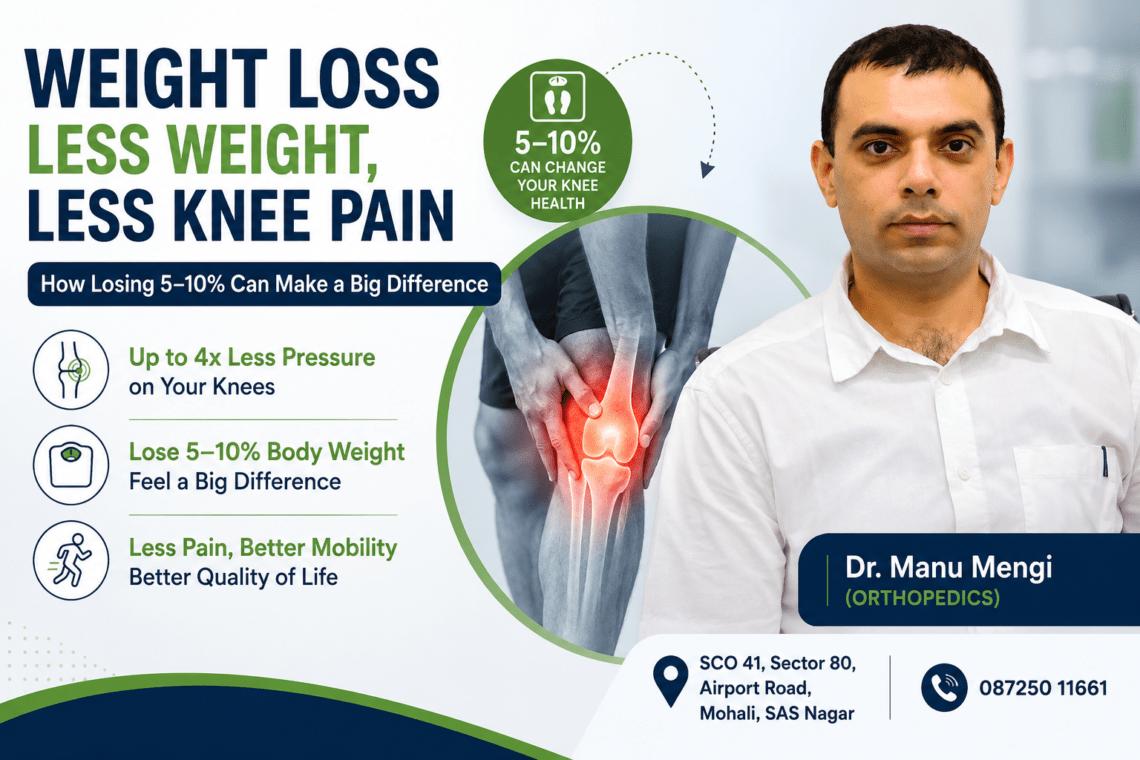
Can losing just 5-10% of your body weight reduce knee pain? The science says yes, and the numbers are more dramatic than you’d expect. Research shows that for every pound you lose, your knees experience four pounds less pressure with each step. That means dropping just 10 pounds removes 40 pounds of load from your knee joints. Studies confirm that people with a healthy BMI have only a 3.7% prevalence of knee osteoarthritis, while those with grade 2 obesity face a 19.5% rate. In this article, we’ll explore why even modest weight loss creates measurable improvements in knee function, pain levels, and long-term joint health.

The Science Behind Weight and Knee Pain

Your knees function as shock absorbers that handle forces far exceeding what most people realize. In reality, these weight-bearing joints don’t simply support your body mass. They absorb and distribute loads that multiply with every movement you make.

How Extra Weight Affects Your Knee Joints

When you carry extra weight, your knee joints bear significantly more pressure than they’re designed to handle. Each additional kilogram of body weight adds 3-4 kg of pressure on the knees. This isn’t a one-to-one ratio because your joints act as dynamic hinges framed by powerful lever arms.

The reaction forces from the ground, bone levers, and quadriceps muscle action amplify the joint load with each step. Over time, this sustained pressure causes knee cartilage to break down faster than normal. The protective cushioning between your bones wears away, eventually leading to bone-on-bone contact that creates chronic knee pain and stiffness.

Fat cells contribute another layer of damage beyond mechanical stress. These cells release chemicals that trigger inflammation throughout your body. The more fat present, the higher the concentration of inflammatory factors circulating in your system. This metabolic inflammation accelerates joint deterioration and increases the severity of osteoarthritis symptoms.

The 4:1 Pressure Rule You Need to Know

The pressure on your knees changes dramatically based on what you’re doing. Walking across level ground generates forces reaching 2.5 to 2.8 times your body weight on each knee. A 200-pound person puts 500-560 pounds of pressure on their knees with every step.

Going down stairs increases the load to 3.1 times body weight. Running pushes it even higher to 3.6 times body weight. Some activities like squatting to pick up an object create forces of four to five times your body weight.

Given that these multiplier effects, being just 10 pounds overweight translates to 30-60 pounds of additional force on your knee joints with each step. The math works in reverse too. Losing one kilogram removes approximately 4 kg of pressure with every step. Over the course of a day’s walking, that single kilogram of weight loss reduces knee stress by more than 20 tons.

Understanding Knee Osteoarthritis and Body Weight

Osteoarthritis develops when cartilage on the ends of bones deteriorates, leaving bare joints to rub against each other. Excess weight is a clear risk factor for this degenerative condition. Overweight women face nearly 4 times the risk of knee osteoarthritis compared to women at a healthy weight, while overweight men have a 5 times greater risk.

The numbers become even more striking when looking at obesity. People who are overweight are approximately 2.5 times more likely to develop knee osteoarthritis than those with normal weight. Obese individuals face 4.6 times the likelihood of developing this condition.

Body weight also influences how severe osteoarthritis becomes once it develops. Obese individuals experience significantly more severe joint degeneration in their knees compared to people at normal weight. Research shows that persons in the highest quintile of body weight have up to 10 times the risk of knee osteoarthritis compared to those in the lowest quintile.

Why 5% Body Weight Loss Makes a Measurable Difference

Trials have consistently demonstrated that a 5% or greater weight loss results in significantly reduced pain and improved function. This threshold isn’t arbitrary. Research shows that reaching this percentage triggers measurable changes in joint mechanics, inflammation levels, and overall knee health that translate to real relief.

Reduced Mechanical Load on Weight-Bearing Joints

When you shed 5% of your body weight, the reduction in mechanical stress on your knees compounds with every step. A 200-pound person losing 10 pounds removes 40 pounds of pressure from their knee joints during walking. Over a full day of typical movement, this single change reduces cumulative knee stress by more than 20 tons.

Note that the benefits extend beyond simple arithmetic. Studies examining patients with obesity who lost considerable body fat found that both knee pain severity and movement limitation decreased. The weight-bearing joints experience less wear and tear, which slows the progression of cartilage damage. This mechanical unloading gives your knees a chance to function without constant overload.

Lower Inflammation Levels in Your Body

Weight loss acts as a powerful anti-inflammatory intervention. Adipose tissue produces and releases pro-inflammatory chemicals throughout your body. Reducing fat stores correspondingly lowers your overall inflammation levels.

Research tracking inflammatory markers shows that weight loss causes significant reductions in plasma levels of inflammatory cytokines. C-reactive protein (CRP) and alpha-1-acid glycoprotein levels drop substantially. One study documented a 73% reduction in visceral fat mass after weight loss, accompanied by decreased low-grade inflammation markers.

Fat cells infiltrate joints and produce adipokines that amplify arthritis symptoms. By the same token, removing excess fat reduces the concentration of these inflammatory signals. The diet groups in clinical trials showed lower concentrations of pro-inflammatory cytokine interleukin 6. This systemic inflammation reduction affects not just your knees but your entire musculoskeletal system.

Improved Knee Function and Mobility

The combination of reduced mechanical load and lower inflammation produces significant functional improvements. Studies document enhanced pain relief, function, mobility, and health-related quality of life. Patients report being able to perform daily activities with less discomfort and greater ease.

Research reveals a dose-response relationship between weight loss percentage and symptom improvement. In other words, the percentage of weight you lose determines how much better you feel, regardless of your starting BMI. One finding stands out: patients with worse baseline knee pain experienced nearly double the benefit across every weight loss bracket.

Weight loss ranging from 7.3% to 7.9% produces similar improvements across all BMI categories. Even patients starting at lower BMI levels see clinically relevant pain reduction. Some studies show that as little as 2.5% weight loss creates meaningful improvement in all baseline BMI categories. For people struggling with mobility decline related to aging, 5-10% weight loss attenuates this decline.

Real Numbers: What 5% Weight Loss Means for You

Here’s what 5% weight loss looks like in practice:

  • At 150 pounds: Lose 7.5 pounds, remove 30 pounds of knee pressure per step
  • At 180 pounds: Lose 9 pounds, remove 36 pounds of knee pressure per step
  • At 200 pounds: Lose 10 pounds, remove 40 pounds of knee pressure per step
  • At 250 pounds: Lose 12.5 pounds, remove 50 pounds of knee pressure per step

Washington University research found that 5% weight loss markedly lowered diabetes and cardiovascular disease risk while improving metabolic function in liver, fat, and muscle tissue. Insulin-secreting beta cell function improved, along with insulin sensitivity across multiple tissue types. Total body fat decreased, with substantially less fat accumulation in the liver.

The 10% Weight Loss Threshold: A Game Changer for Knee Health

Reaching the 10% weight loss threshold represents a critical turning point for people struggling with knee osteoarthritis. While 5% produces measurable benefits, doubling that effort unlocks substantially greater improvements across every metric that matters.

Enhanced Pain Relief and Quality of Life

A 10% weight loss produces improved pain, function, and quality of life while reducing joint loads and inflammation compared to no weight loss or less than 10% loss. This threshold aligns with general weight loss recommendations among obesity experts for improving health outcomes.

Research documents a clear dose-response relationship. Greater weight loss produces larger effect sizes for improvements in pain, function, and quality of life. One trial found that 10% weight loss decreased pain by 50% over an 18-month period. Patients who achieved this level could perform daily activities with significantly less discomfort.

The American College of Rheumatology strongly recommends at least 5% weight loss, noting that clinically important benefits continue to increase with weight loss of 5-10%, 10-20%, and greater than 20% of body weight. In essence, each increment delivers compounding returns. Studies show that losing 10-20% of starting body weight improved pain, function, and quality of life better than losing just 5%.

Slowing Knee Cartilage Degeneration

Weight loss protects knee cartilage and menisci from further damage. Research tracking 640 overweight or obese participants over 48 months found that those who lost 5-10% of their body weight showed lower rates of cartilage degeneration compared to people who didn’t lose weight. Even more striking, those who lost more than 10% retained more healthy cartilage and experienced less menisci damage.

MRI analysis confirmed these protective effects. People who lost 5-10% of their weight over this period significantly decreased knee cartilage degradation. In the group that lost more than 10%, cartilage degradation was much slower. This structural preservation matters because damaged menisci speed up degeneration of the knee joint overall.

Reducing Your Risk of Needing Surgery

Weight loss directly influences whether you’ll need knee replacement surgery. For every 1% weight loss, there’s a 2% reduced risk of knee replacement. A weight loss target greater than 7.5% is required to reduce the risk of total knee replacement in adults with overweight or obesity.

Given that 80-95% of people needing total joint replacement also had overweight or obesity, losing weight becomes critical for surgical avoidance. Adults with obesity face a 60% lifetime risk of developing symptomatic knee osteoarthritis.

Study Results: 10-20% vs 5% Weight Loss Outcomes

Doubling weight loss from 10% to 20% cuts pain and improves function by another 25%. At 18 months, participants who lost 20% or more could walk 10% farther in 6 minutes than those who lost less than 5%, covering 559 meters versus 508 meters. By comparison, moderate outcomes emerged at lower thresholds, but the greatest improvements consistently appeared in groups achieving substantial weight loss.

How Weight Loss Reduces Knee Pain Beyond the Scale

Obesity triggers biological changes that extend far beyond what bathroom scales reveal. Adults with obesity are almost twice as likely to report having arthritis than adults at healthy weights (32% and 17.6%, respectively). The connection stems from systemic inflammation and metabolic dysfunction that weight loss can reverse.

Decreasing Metabolic Inflammation

Obesity creates a state of low-grade systemic inflammation that researchers call “metaflammation”. Adipose tissue secretes pro-inflammatory cytokines and adipokines that circulate throughout your body. As fat cells increase in size, cell membranes become tense, triggering stress responses that initiate inflammation. Insufficient blood supply to expanding adipose tissue causes cell death, further amplifying inflammatory reactions.

Weight loss activates the reduction in plasma levels of inflammatory markers. The greatest improvements in inflammatory markers appear in studies achieving at least 10% weight loss. Specifically, C-reactive protein, TNF-alpha, IL-6, and leptin levels decrease, while anti-inflammatory adiponectin increases. Studies show weight loss is a critical factor to reduce inflammation, with hypocaloric diets producing anti-inflammatory effects independent of diet composition.

Improving Insulin Resistance and Joint Health

High insulin resistance associates strongly with knee osteoarthritis, particularly in individuals with metabolic syndrome and type 2 diabetes. Insulin resistance leads to increased expression of pro-inflammatory cytokines and downregulated synthesis of structural proteins in articular cartilage. Insulin normally binds to receptors on chondrocytes and promotes production of type 2 collagen and proteoglycans, preventing cartilage wear.

Research shows that TyG-related indices significantly associate with knee osteoarthritis prevalence. Weight loss-related improvement in insulin sensitivity occurs through changes in adipose cell size and central distribution of body fat. This metabolic correction protects joint cartilage and reduces inflammation-driven degeneration.

Reducing Fat Pad Inflammation

The infrapatellar fat pad (IPFP) represents intra-articular adipose tissue that contributes to inflammation and pain by secreting pro-inflammatory cytokines. As adipose tissue, the IPFP secretes fatty acids with pro-inflammatory effects and stores immune cells that produce inflammatory mediators. High BMI and obesity substantially impact fat pad syndrome.

Weight loss interventions produce significant reductions in IPFP volume. Exercise alone reduced volume by 2.1%, diet by 4.0%, and combined diet plus exercise by 5.2%. Across intervention groups, significant correlations existed between IPFP volume change and individual weight loss (r = 0.40) and change in total body fat mass (r = 0.44).

Lowering Disease Activity in Knee Osteoarthritis

Weight loss plays a role in both primary prevention (intervening before osteoarthritis occurs) and secondary prevention (intervening during early osteoarthritis). In the Framingham Study, a weight loss of 5.1 kg reduced women’s risk of developing knee osteoarthritis by 50% over 10 years. For adults with obesity, the risk of developing symptomatic knee osteoarthritis reaches 60%. Achieving and maintaining a healthy weight interrupts inflammatory processes and altered joint mechanics that degrade structural integrity and contribute to increased pain.

Practical Steps to Lose Weight and Protect Your Knees

Addressing weight loss and knee pain requires a strategic approach combining movement and nutrition. Research provides clear guidance on effective interventions.

Low-Impact Exercises for Knee Arthritis

Aerobic activities like walking, cycling, and swimming show the highest probability of being the best treatment for improving pain, function, gait performance, and quality of life. Swimming and water aerobics reduce joint stress through buoyancy while strengthening muscles. Regular swimming reduced joint pain and stiffness while improving muscle strength and functional capacity. Cycling works quadriceps and hamstrings while improving knee range of motion.

Strength Training Without Stressing Your Joints

Strengthening muscles around your knees reduces joint stress. Target your quadriceps, hamstrings, abductors, adductors, and gluteal muscles. Quadriceps strengthening performed over eight weeks proved as effective as anti-inflammatory medications for function improvement. Strong leg muscles provide crucial knee support and shock absorption.

Walking Programs for Knee Pain Relief

Walking at least 6000 steps daily offers the ideal threshold for preventing functional limitation. People with knee osteoarthritis who walk for exercise are significantly less likely to develop worse pain.

Anti-Inflammatory Diet Changes

Small pilot studies show positive effects of anti-inflammatory diets on knee osteoarthritis symptoms. The Mediterranean diet reduces inflammation by emphasizing fruits, vegetables, fish, nuts, beans, and olive oil while limiting processed foods and saturated fat. Some patients discontinue NSAIDs after transitioning to this eating pattern.

Setting Realistic Weight Loss Goals

Suggest an initial weight loss goal of 10%. A recommended rate is 1-2 pounds per week. Successful strategies include calorie reduction, increased physical activity, and behavior therapy.

Conclusion

Weight loss doesn’t need to be dramatic to transform your knee health. In fact, losing just 5-10% of your body weight removes significant pressure from your joints, lowers inflammation throughout your system, and slows cartilage degeneration. The 4:1 pressure rule means every pound you lose translates to four pounds less stress on your knees with each step.

Start with realistic goals and combine low-impact exercise with anti-inflammatory eating patterns. Whether you need to lose 10 pounds or 50, your knees will thank you for every single pound. The science is clear: modest weight loss delivers measurable results faster than you might expect.

Key Takeaways

Losing just 5-10% of your body weight can dramatically reduce knee pain and slow joint degeneration. Here’s what the research reveals about this powerful connection:

• Every pound lost removes 4 pounds of pressure from your knees during walking, meaning a 10-pound weight loss eliminates 40 pounds of force with each step.

• 5% weight loss triggers measurable improvements in pain levels, mobility, and inflammation markers, while 10% weight loss can reduce pain by 50% and significantly slow cartilage degeneration.

• Weight loss reduces knee pain through multiple pathways beyond mechanical relief—it decreases systemic inflammation, improves insulin resistance, and reduces inflammatory fat pad volume around joints.

• Walking 6,000+ steps daily combined with strength training and an anti-inflammatory Mediterranean diet creates the optimal approach for protecting knees while achieving sustainable weight loss.

• Overweight individuals face 2.5 times higher risk of developing knee osteoarthritis, while obese individuals face 4.6 times the risk—making weight management critical for prevention.

The dose-response relationship is clear: greater weight loss produces larger improvements in pain, function, and quality of life. Even modest reductions of 7-10 pounds can transform your knee health and reduce your lifetime risk of needing joint replacement surgery.

FAQs

1. Can losing just 5-10% of my body weight really reduce knee pain?

Yes. Research consistently shows that losing 5-10% of your body weight can significantly reduce knee pain, especially in people with knee osteoarthritis. Even modest weight loss decreases the pressure on the knee joint, reduces inflammation, and improves mobility. Many people notice less pain while walking, climbing stairs, and standing for long periods.

2. How does excess body weight increase knee pain?

Every extra kilogram of body weight increases the load on your knees by approximately 3-4 kilograms during walking. Over time, this extra stress accelerates cartilage wear, increases inflammation, and causes pain, stiffness, and reduced joint function.

3. How much weight should I lose to see improvement in knee pain?

Most experts recommend aiming for an initial weight loss of 5-10% of your current body weight. For example, someone weighing 90 kg may experience noticeable improvement after losing just 4.5-9 kg.

4. Is weight loss more effective than painkillers for knee osteoarthritis?

Weight loss doesn’t replace pain medications when they are needed, but it addresses one of the root causes of knee pain. Unlike painkillers that provide temporary relief, maintaining a healthy weight can reduce joint stress, improve function, and slow disease progression.

5. Why does losing weight reduce knee inflammation?

Body fat is metabolically active and releases inflammatory chemicals called cytokines. Excess fat increases chronic low-grade inflammation throughout the body, including the joints. Losing weight lowers these inflammatory substances, helping reduce pain and stiffness.

6. Can weight loss prevent knee replacement surgery?

Weight loss cannot reverse severe arthritis, but it may delay the progression of knee osteoarthritis, improve symptoms, and in many individuals postpone or even avoid knee replacement surgery by reducing stress on the joint.

7. Will my knee cartilage grow back after losing weight?

No. Damaged cartilage usually does not regenerate completely. However, weight loss reduces further cartilage damage, decreases inflammation, improves joint function, and slows the progression of osteoarthritis.

8. Which type of exercise is best for losing weight without worsening knee pain?

Low-impact exercises are usually the safest choices, including:

  • Walking on level ground
  • Cycling
  • Swimming
  • Water aerobics
  • Elliptical training
  • Strength training under guidance

These exercises help burn calories while placing less stress on painful knees.

9. Can diet alone improve knee pain?

A healthy calorie-controlled diet can promote weight loss, which often reduces knee pain. Combining proper nutrition with regular exercise generally produces better and longer-lasting results than diet alone.

10. How long does it take to notice less knee pain after losing weight?

Many people begin noticing improvement within 8-12 weeks of consistent weight loss and exercise. The degree of improvement depends on the severity of arthritis, muscle strength, and adherence to lifestyle changes.

11. Does belly fat affect knee pain more than overall body weight?

Yes. Excess abdominal fat contributes to chronic inflammation by releasing inflammatory hormones and cytokines. This can worsen osteoarthritis symptoms beyond the extra mechanical load placed on the knees.

12. Can younger people with knee pain also benefit from weight loss?

Absolutely. Even younger adults with obesity-related knee pain can experience reduced pain, improved mobility, and a lower risk of developing early osteoarthritis by achieving a healthier body weight.

13. Is walking safe if I already have knee osteoarthritis?

In most cases, yes. Walking at a comfortable pace is beneficial for joint health and weight management. However, severe pain, swelling, or instability should be evaluated by an orthopaedic specialist before starting an exercise program.

14. Besides weight loss, what else helps reduce knee pain?

A comprehensive treatment plan may include:

  • Quadriceps and hip muscle strengthening
  • Flexibility exercises
  • Physical therapy
  • Appropriate footwear
  • Activity modification
  • Adequate Vitamin D and nutrition
  • Medical treatment when necessary

Combining these measures with weight loss often provides the best outcomes.

15. Should I consult a doctor before starting a weight-loss program for knee pain?

Yes. If you have persistent knee pain, swelling, severe arthritis, diabetes, heart disease, or other medical conditions, consult your doctor before beginning a weight-loss or exercise program. An individualized plan can maximize benefits while minimizing the risk of injury.

16. How does losing 5-10% of body weight affect knee pain?

Losing 5-10% of body weight significantly reduces knee pain by decreasing mechanical pressure on joints, lowering inflammation levels, and improving mobility. Research shows that 5% weight loss can reduce joint pain by up to 50%, while 10% weight loss may decrease pain by 50% over 18 months and slow cartilage degeneration.

17. What is the 4:1 pressure rule for knees and weight loss?

The 4:1 pressure rule means that for every pound of body weight you lose, you remove approximately four pounds of pressure from your knee joints with each step. For example, losing 10 pounds eliminates 40 pounds of force on your knees during walking, which adds up to more than 20 tons of reduced stress over a full day.

18. Can weight loss improve cholesterol levels along with knee health?

Yes, a 5-10% weight loss can increase HDL (good) cholesterol by about five points, which helps protect against heart disease. For men, HDL levels above 40 mg/dl and for women above 50 mg/dl are considered protective, making weight loss beneficial for both joint health and cardiovascular wellness.

19. How much weight loss is needed to reduce the risk of knee replacement surgery?

For every 1% of body weight lost, there’s a 2% reduced risk of needing knee replacement surgery. Research indicates that losing more than 7.5% of body weight is required to significantly reduce the risk of total knee replacement in adults with overweight or obesity.

20. Does weight loss help with arthritis pain beyond just knee joints?

Yes, weight loss reduces arthritis pain throughout the body by decreasing systemic inflammation, improving insulin resistance, and lowering inflammatory markers like C-reactive protein and IL-6. This occurs because fat tissue produces inflammatory chemicals, so reducing body fat correspondingly lowers overall inflammation levels affecting all joints.
Patellofemoral Pain Syndrome: The Hidden Reason Your Knees Hurt While Running

Runner’s Knee Treatment in Mohali – Causes & Prevention Tips by Dr. Manu Mengi

One-third of people visiting healthcare providers with knee pain are diagnosed with patellofemoral syndrome—runner’s Knee, making it one of the most common knee conditions we treat at our Sports Injury Clinic in Mohali. This prevalent condition particularly affects athletes and physically active individuals who regularly run and jump.

As an athlete or regular runner, you might be familiar with that nagging, dull ache at the front of your knee that worsens when climbing stairs or during squats. This pain can affect one or both knees and typically appears during or after intense physical activities. Young female patients and adults aged 20 to 40 are particularly vulnerable to this condition, especially those who suddenly increase their activity levels.

In this comprehensive guide, we’ll explore the causes, symptoms, and effective treatments for patellofemoral pain syndrome, including the RICE method and targeted physical therapy exercises. We’ll also share practical strategies to prevent this condition from affecting your running routine.

What is Patellofemoral Pain Syndrome (Runner’s Knee)?

Patellofemoral Pain Syndrome (PFPS) refers to pain around or behind the kneecap (patella) where it meets the thigh bone (femur). This condition got its nickname “Runner’s Knee” because it frequently affects people who run or participate in activities that put repeated stress on the knee joint. However, unlike what the name suggests, this condition isn’t limited to runners—it can affect anyone who engages in activities that stress the knee joint.

Understanding the patellofemoral joint

The patellofemoral joint consists of your kneecap (patella) and the groove in your thigh bone (femur) where the patella slides. This joint plays a crucial role in your daily movements. The patella acts as a pulley, increasing the leverage of the quadriceps muscles and providing protection to the knee joint.

The kneecap is normally held in place by various structures:

  • Quadriceps muscles (front thigh)
  • Hamstring muscles (back thigh)
  • Medial and lateral retinaculum (tissue along the sides)
  • Patellar tendon (connecting to the shin bone)

In a healthy knee, the patella glides smoothly up and down in this femoral groove as you bend and straighten your leg. This movement should be pain-free and efficient. Nevertheless, if any element of this mechanical system is off, whether it’s muscle imbalance, abnormal anatomy, or poor movement patterns, problems begin to develop.

What happens in PFPS is that the patella doesn’t track properly in its groove. Instead, it may shift laterally (outward) or tilt, creating excessive pressure on certain areas of the cartilage. This improper tracking leads to irritation of the soft tissues and inflammation, consequently causing pain and discomfort.

Why runners are more prone to PFPS

Runners face a higher risk of developing patellofemoral pain syndrome, which is why many seek Runner’s Knee Treatment in Mohali for relief. First and foremost, running creates repetitive stress on the knee joint. With each stride, your knee absorbs a force equivalent to several times your body weight. Over thousands of strides during a typical run, this adds up to significant cumulative stress on the patellofemoral joint.

Furthermore, biomechanical factors make runners particularly vulnerable:

  1. Muscle imbalances – Tight quadriceps combined with weak hip abductors (particularly the gluteus medius) can pull the patella out of alignment during running.
  2. Running form issues – Overpronation (excessive inward rolling of the foot), crossover gait (feet crossing over the midline), or overstriding can all contribute to abnormal knee mechanics.
  3. Training errors – Sudden increases in mileage, intensity, or hill work without proper adaptation time put excessive stress on the knee structures.

The repetitive nature of running means that even minor misalignments or muscle imbalances get magnified over time. As a runner logs more miles, these small issues compound until they reach a threshold where pain develops.

Moreover, running on hard surfaces or uneven terrain increases impact forces and can exacerbate existing biomechanical issues. Similarly, worn-out running shoes that no longer provide proper support can alter your gait and contribute to patellofemoral issues.

Unlike traumatic knee injuries, PFPS typically develops gradually. Many runners ignore early warning signs—such as mild discomfort that disappears after warming up—until the pain becomes persistent enough to interfere with training. This is why understanding the underlying mechanics and recognising early symptoms is vital for preventing long-term problems.

Common Causes of Knee Pain While Running

Running places significant demands on your knees, which is why Knee Pain When Running Treatment in Mohali is a common request among athletes. Understanding what causes knee pain while running can help you prevent patellofemoral pain syndrome or address it before it worsens.

Overuse and repetitive stress

Approximately 80% of running-related injuries are associated with overload. Unlike acute injuries, overuse problems develop gradually through repeated microtrauma that exceeds your body’s natural repair capacity.

When you run, various tissues including muscles, tendons, bones, and ligaments experience physiological stress. These structures adapt differently to training loads—tendons and ligaments adapt more slowly than muscles, making them particularly vulnerable to injury.

The problem arises when the cumulative damage surpasses your tissue’s repair threshold. This typically happens when you don’t allow adequate recovery time between training sessions. For overzealous runners who push too hard too often, the damage accumulates until pain and dysfunction appear.

What makes overuse injuries challenging is their gradual onset. There’s rarely a single, identifiable traumatic cause. Instead, the damage builds up silently until symptoms become noticeable, at which point significant tissue stress has already occurred.

Muscle imbalances and poor biomechanics

Your running form directly impacts how effectively your body absorbs and distributes forces. Poor biomechanics significantly increase your risk of developing patellofemoral pain syndrome.

Muscle imbalances are among the most common culprits. About 25% of adults experience knee pain, representing a 65% increase over the last 20 years. Often, this pain stems from muscle imbalances around the joint:

  • Weak gluteal muscles (particularly gluteus medius and maximus) fail to properly control hip and knee alignment during running
  • Quadriceps weakness reduces support across the knee joint and strongly correlates with the advancement of knee problems
  • Tight hamstrings or calves alter how your knee moves through its normal range
  • An imbalance between inner and outer thigh muscles can pull the kneecap out of alignment

These imbalances change how your patella tracks in its groove, creating excessive pressure on certain areas of the cartilage. Notably, runners with anterior knee pain have weaker knee extensors and flexors than those without pain.

Inefficient running biomechanics, such as excessive knee valgus (“knocked knee”) or knee varus (“knee out”), create abnormal stress patterns that your knee structures weren’t designed to handle.

Sudden changes in training or surface

Making abrupt modifications to your running routine often triggers knee pain. Runners who suddenly increase their mileage, intensity, or hill work without proper adaptation time put excessive stress on their knees.

Contrary to popular belief, changing to a softer running surface doesn’t necessarily reduce injury risk. Different surfaces simply alter the biomechanical demands on your body. While softer surfaces may reduce impact loading rates, they increase demands on muscles and tendons. A sudden change in running surface may decrease stress in one area but increase injury risk in others.

Training errors account for many cases of runner’s knee. Adding too much distance too quickly or not allowing sufficient recovery between runs prevents proper adaptation. This explains why many patients develop patellofemoral pain syndrome after they start running at an incline or increase their miles or speed.

Recognising the Symptoms Before It Gets Worse

Early detection of Patellofemoral Pain Syndrome can prevent progression to more severe conditions, which is why visiting a Knee Pain Doctor in Mohali at the first sign of discomfort is crucial. Recognising the warning signs promptly allows for faster intervention and better outcomes. When detected early, most cases resolve with conservative treatment rather than requiring invasive procedures.

Pain patterns and when they appear

The hallmark of Patellofemoral Pain Syndrome is a dull, aching pain around or behind the kneecap, particularly where it meets the lower part of the thighbone. Unlike acute injuries, this pain typically develops gradually over time.

You’ll likely notice pain intensifying when:

  • Walking up or down stairs
  • Squatting or kneeling
  • Running, especially downhill
  • Sitting with knees bent for extended periods (sometimes called “theatre sign”)
  • Standing up after prolonged sitting

Pain is usually diffuse rather than pinpointed to a specific location. Many runners report that symptoms peak when the knee is bent at 60° to 90°, such as during deep squats, and subside when straightening the leg. Initially, discomfort might only appear during or after intense activities, but as the condition progresses, even daily movements can trigger pain.

Clicking or popping sounds in the knee

That strange clicking or popping sound in your knee has a medical name: crepitus. While often alarming, knee crepitus by itself isn’t necessarily a cause for concern. Scientists have studied this phenomenon since 1947, with real-time medical imaging finally revealing in 2015 that these sounds typically occur when tiny nitrogen bubbles in joint fluid collapse due to pressure changes.

Nevertheless, when clicking or popping accompanies pain, attention is warranted. In Patellofemoral Pain Syndrome, you might experience:

  • Rubbing, grinding, or clicking sensations when bending and straightening the knee
  • Popping sounds that worsen after periods of rest
  • Crunching sensations when climbing stairs or kneeling

Clicking at the front of the knee can result from an anatomical structure called a plica. While usually painless, when inflamed it causes “Plica syndrome,” another condition that frequently affects runners.

Swelling around the kneecap after activity

Swelling indicates inflammation in the knee joint and serves as an important diagnostic clue. Following intense activities, you might notice mild swelling around the front of your knee or kneecap, representing fluid buildup in or around the joint.

This swelling typically develops gradually rather than suddenly. Sudden swelling (within four hours of activity) is more concerning, as it may indicate bleeding within the knee (hemarthrosis) and possible structural damage. In contrast, the swelling associated with Patellofemoral Pain Syndrome usually appears more gradually and is milder in nature.

Persistent swelling that doesn’t improve with rest should prompt medical evaluation. Although mild swelling might resolve with home treatment, recurrent episodes indicate that the underlying issues causing your runner’s knee remain unaddressed. In essence, your body is signalling that something isn’t right with your knee mechanics.

PFPS typically worsens over time if left untreated, potentially increasing your risk of injuries beyond just pain and discomfort. Hence, promptly recognising these symptoms can help prevent progression to more serious conditions.

Effective Treatments for Patellofemoral Pain Syndrome

Treating Patellofemoral Pain Syndrome effectively requires a multi-faceted approach, which is why many patients opt for a Knee Pain Doctor in Mohali as part of their recovery. Most patients recover completely with conservative treatments, though recovery timeframes vary depending on severity and adherence to treatment protocols.

RICE method and pain relief options

First and foremost, the RICE method provides immediate relief for acute flare-ups:

  • Rest: Temporarily avoid activities that trigger pain
  • Ice: Apply cold packs for 15-20 minutes every 2-3 hours
  • Compression: Use an elastic bandage around the knee to reduce swelling
  • Elevation: Keep your knee above heart level when possible

Over-the-counter pain relievers can complement this approach. NSAIDs like ibuprofen and naproxen reduce both pain and inflammation, while acetaminophen (Tylenol) addresses pain without anti-inflammatory effects. Generally, these medications should not be taken for more than 10 days without medical supervision.

Physiotherapy and strengthening exercises

Physiotherapy forms the cornerstone of effective treatment. Studies show that performing knee- and hip-strengthening exercises three times weekly for six weeks significantly decreases knee pain and improves physical activity.

Key exercises often include:

  • Straight leg raises for quadriceps strengthening
  • Side-lying clam exercises for hip abductors
  • Wall sits for thigh muscle development
  • Step-ups for gluteal and quadriceps activation

Primarily, these exercises aim to correct muscle imbalances and faulty movement patterns that contribute to improper tracking of the kneecap.

Knee braces and orthotics for support

Supportive devices provide additional stability during recovery. Patellofemoral braces help track the kneecap properly, reducing pressure on affected areas. Unloader braces, the most commonly recommended type, redistribute weight-bearing forces away from painful areas.

Orthotic inserts, either over-the-counter or custom-made, can correct foot mechanics that contribute to knee misalignment. These interventions work best when combined with appropriate exercises.

When to consider surgery

Surgery represents a last resort, typically considered only after 6-12 months of consistent conservative treatment failure. Arthroscopic procedures allow surgeons to examine the knee joint and address specific issues like removing damaged cartilage or releasing tight lateral structures.

For severe cases with significant misalignment, realignment surgery may be necessary to adjust the position of the kneecap. Fortunately, most patients respond well to non-surgical approaches, making surgery rarely necessary for this condition.

Preventing PFPS and Staying Pain-Free

Preventing Patellofemoral Pain Syndrome demands a proactive approach, including Proper Running Form and Knee Pain Treatment in Mohali if symptoms arise.  As the saying goes, an ounce of prevention is worth a pound of cure—nowhere is this more applicable than with runner’s knee.

Stretching and warm-up routines

Before hitting the pavement, engage in dynamic stretching that mimics running movements to increase your heart rate and warm up muscles. Subsequently, save static stretching for after your run when muscles are warm. Many experts recommend a 5-10 minute walking warm-up to get blood flowing properly through your body. For optimal results, focus on stretching your quadriceps, hamstrings, calves, and hip flexors—all crucial muscle groups that impact knee alignment.

Choosing the right footwear

Your running shoes play a vital role in preventing knee pain. According to podiatrists, proper footwear should provide appropriate cushioning without raised heels, which can shift your center of gravity and place additional pressure on the knee joint. Typically, shoes with lower heel drops (under 8mm) help reduce forward knee push and alleviate IT band issues.

Gradual training progression

Abrupt changes in training volume often trigger knee pain. Accordingly, structure your progression carefully—increase your weekly mileage by no more than 10% and allow approximately six weeks for your body to adapt to new training loads. Likewise, alternate running days with low-impact activities like swimming or yoga to give your knees recovery time.

Maintaining healthy body mechanics

Proper running form dramatically reduces stress on your knees. Land with slightly bent knees rather than straight legs, and avoid overstriding. Additionally, aim for a cadence around 180 steps per minute (170 for taller runners), and ensure your feet point in your direction of travel rather than splaying outward. Above all, focus on a circular leg motion versus a pendulum-like straight leg action that increases impact forces.

Conclusion

Understanding and addressing Patellofemoral Pain Syndrome makes the difference between a short-lived running career and years of enjoyable miles. If you’re experiencing persistent discomfort, consult a Patellofemoral Pain Syndrome Specialist in Mohali for personalised care. While this condition affects many runners, proper knowledge empowers us to prevent and manage it effectively.

Most cases respond well to conservative treatment approaches, especially when caught early. Regular stretching, appropriate footwear, and smart training progression serve as our first line of defence against runner’s knee. Strengthening exercises targeting key muscle groups help maintain proper knee alignment during runs.

Smart runners pay attention to their bodies and address minor discomfort before it develops into chronic pain. Rather than pushing through knee pain, taking appropriate rest periods and following proper rehabilitation protocols leads to better long-term outcomes. Remember, protecting your knees today ensures many more miles of pain-free running tomorrow.

FAQs

Q1. What are the main symptoms of Patellofemoral Pain Syndrome, and when should I see a Knee Pain Doctor in Mohali?

A1. The primary symptoms include a dull, aching pain around or behind the kneecap, especially when walking up or down stairs, squatting, or sitting with bent knees for extended periods. You may also experience clicking or popping sounds in the knee and mild swelling around the kneecap after activity.

Q2. Why are runners more susceptible to developing Patellofemoral Pain Syndrome?

A2. Runners are more prone to this condition due to the repetitive stress placed on the knee joint during running. Factors such as muscle imbalances, poor running form, and sudden increases in training intensity or mileage can contribute to the development of PFPS in runners.

Q3. How can I prevent Patellofemoral Pain Syndrome while running?

A3. Prevention strategies include proper warm-up and stretching routines, choosing appropriate footwear, gradually increasing training intensity, and maintaining good running form. It’s also important to strengthen key muscle groups, particularly the quadriceps, hip abductors, and gluteal muscles.

Q4. What are some effective treatments for Patellofemoral Pain Syndrome?

A4. Effective treatments include the RICE method (Rest, Ice, Compression, Elevation), physiotherapy exercises to strengthen muscles around the knee, and the use of knee braces or orthotics for support. Over-the-counter pain relievers can also help manage pain and inflammation.

Q5. When should I seek medical attention for knee pain while running?

A5. You should consult a healthcare professional if you experience persistent knee pain that doesn’t improve with rest, sudden swelling within hours of activity, or if the pain interferes with your daily activities. Early intervention can prevent the condition from worsening and improve treatment outcomes.

About Patellofemoral pain syndrome specialist Mohali

If you’re experiencing persistent knee pain while running, don’t ignore the warning signs. Early intervention from a Patellofemoral Pain Syndrome Specialist in Mohali can prevent long-term damage and get you back to pain-free movement. Dr. Manu Mengi, an experienced sports medicine expert, provides personalised treatment plans combining physiotherapy, bio-mechanical analysis, and targeted exercises for runners and athletes. Whether you’re training for a marathon or simply want to stay active without knee discomfort, seeking professional guidance ensures proper recovery. Book a consultation today to address your knee concerns with evidence-based care.

The Science Behind PRP Orthopedic Treatment: Your Body’s Hidden Healing Power

Did you know that muscle strain injuries affect 12-16% of athletes, with hamstring strains alone accounting for 29% of all sports-related injuries? While traditional treatments often involve surgery or medication, PRP Orthopedic Treatment offers a cutting-edge alternative that harnesses your body’s natural healing power.

In fact, Platelet-Rich Plasma (PRP) treatment has gained widespread recognition for treating various musculoskeletal conditions without surgery. This innovative therapy shows promising results for osteoarthritis, tendon injuries, ligament damage, and chronic joint pain. By promoting tissue repair and regeneration through your body’s own healing mechanisms, PRP injections can reduce pain and inflammation while supporting cartilage repair.

This comprehensive guide explains how PRP treatment works, what conditions it can address, and why it might be the solution you’ve been searching for to manage your orthopedic concerns.

What Is PRP Treatment? Understanding the Basics

Platelet-rich plasma (PRP) represents a groundbreaking approach in regenerative medicine that harnesses your body’s natural healing capabilities. At its core, PRP therapy involves using concentrated platelets from your own blood to stimulate tissue repair and accelerate healing processes that might otherwise take much longer.

The science of platelet-rich plasma explained

Platelet-rich plasma consists of two primary elements: plasma (the liquid portion of blood) and platelets (specialized blood cells). Though platelets are best known for their clotting abilities, they also contain powerful growth factors that trigger cell reproduction and stimulate tissue regeneration. These microscopic blood components measure approximately 2 μm in diameter and contain more than 30 bioactive proteins that play fundamental roles in tissue healing.

When concentrated and delivered to injured areas, PRP creates a supraphysiological release of growth factors that can “jump-start” healing in chronic injuries and accelerate repair in acute ones. The science behind PRP is based on amplifying the natural healing cascade that occurs when you’re injured. Instead of waiting for your body to gradually send healing factors to damaged tissues, PRP delivers a concentrated dose directly where it’s needed.

Most scientific definitions consider a platelet concentration of 1 million platelets/μL as the working threshold for therapeutic PRP. This represents a significant increase over normal blood, with PRP typically containing five to ten times more platelets than a standard blood sample.

How PRP is prepared from your own blood

The preparation of PRP begins with a simple blood draw, typically collecting 30cc of your blood, which can yield 3-5cc of PRP depending on your baseline platelet count. After collection, your blood is mixed with an anticoagulant—most commonly acid citrate dextrose-A (ACD-A)—to prevent premature platelet activation.

The preparation process follows these steps:

      1. Initial centrifugation: Your blood sample is placed in a centrifuge that spins at carefully controlled speeds. This first spin (typically 100-300g for 5-10 minutes) separates red blood cells from plasma containing platelets.

      1. Plasma separation: After the first spin, the plasma and buffy coat (containing white blood cells) are transferred to another sterile tube.

      1. Second centrifugation: This spin (typically 400-750g for 10-17 minutes) concentrates the platelets into a small volume of plasma.

      1. Final preparation: The platelet-poor plasma is removed, leaving behind a concentrated platelet solution that’s ready for injection.

    Throughout this process, maintaining proper temperature and handling is crucial for preserving platelet viability and preventing premature activation. Additionally, the entire procedure typically takes less than 30 minutes, allowing for same-day treatment.

    Key components that make PRP effective

    The healing power of PRP comes from its rich concentration of biologically active molecules. Seven fundamental protein growth factors actively secreted by platelets initiate all wound healing processes. The most significant components include:

    Platelets release these growth factors within 10 minutes of activation, with more than 95% of the pre-synthesized growth factors secreted within one hour. Moreover, PRP contains three important proteins that act as cell adhesion molecules: fibrin, fibronectin, and vitronectin.

    Besides platelets, PRP preparations may contain varying amounts of white blood cells, depending on the preparation method. These leukocytes can influence the healing process through immune and host-defense mechanisms, particularly in chronic tissue conditions.

    The main advantage of PRP lies in its autologous nature—it comes from your own body, eliminating concerns about disease transmission or immune reactions. Furthermore, unlike corticosteroid injections, PRP has no known adverse effects since it’s derived from your own blood.

    Although different commercial systems produce varying platelet concentrations, the fundamental mechanism remains the same: PRP delivers a concentrated dose of your body’s own healing factors precisely where they’re needed to accelerate recovery and reduce pain.

    How PRP Activates Your Body’s Natural Healing Process

    The magic of PRP Orthopedic Treatment lies in how it triggers and enhances your body’s inherent healing mechanisms. When platelets are activated at an injury site, they release a powerful cocktail of bioactive molecules that orchestrate tissue repair with remarkable precision.

    Growth factors and their regenerative functions

    The concentrated platelets in PRP release multiple growth factors that act as cellular messengers, directing various aspects of the healing process. These biological powerhouses include:

        • Platelet-derived growth factor (PDGF): Stimulates cell proliferation and migration, particularly of fibroblasts, which produce collagen. PDGF also promotes angiogenesis (new blood vessel formation) to ensure injured tissue receives adequate blood supply.

        • Transforming growth factor-beta (TGF-β): Acts as a potent immunomodulator that regulates inflammation while stimulating the differentiation of fibroblasts into myofibroblasts—crucial cells for wound contraction and tissue remodelling.

        • Vascular endothelial growth factor (VEGF): Drives the formation of new blood vessels, delivering vital oxygen and nutrients to healing tissues. Studies show VEGF significantly promotes angiogenesis in wound tissue.

        • Epidermal growth factor (EGF): Accelerates re-epithelialisation by enhancing the proliferation and migration of epidermal stem cells, which is essential for skin regeneration.

        • Insulin-like growth factor (IGF): Stimulates tenocyte proliferation, increases collagen production, and promotes differentiation of cells essential for tendon healing.

      Each growth factor performs specific functions, yet they work synergistically to create an optimal healing environment. Research indicates PRP contains a sevenfold increase in TGF-β, a thirtyfold increase in PDGF, and a tenfold increase in EGF compared to normal blood concentrations.

      The inflammatory response and tissue repair

      Contrary to common belief, inflammation is an essential first step in healing. PRP expertly modulates this process rather than simply suppressing it.

      Initially, PRP influences immune cells—particularly macrophages—to assume an anti-inflammatory (M2) phenotype associated with tissue repair rather than prolonged inflammation. This strategic polarization creates an environment that favors regeneration over chronic inflammation.

      Simultaneously, PRP decreases expression of pro-inflammatory cytokines like interleukin-17A (IL-17A) and interleukin-1β (IL-1β), helping regulate the inflammatory response. This dual action—promoting necessary inflammatory signals while preventing excessive inflammation—creates ideal conditions for tissue repair.

      Additionally, PRP releases antibacterial substances that reduce local inflammation and prevent wound infection. This protective function is especially valuable in orthopedic applications where infection can significantly compromise healing outcomes.

      Cellular communication in healing

      PRP establishes a sophisticated communication network between different cell types involved in tissue repair. After injection, platelets activate and release their growth factors within 10 minutes, with over 95% of pre-synthesized growth factors secreted within one hour.

      These bioactive molecules serve as signaling messengers that coordinate cellular responses:

      First, they recruit stem cells and fibroblasts to the injury site. Subsequently, they stimulate these cells to proliferate and differentiate into the specific cell types needed for repair. Studies show that low concentrations of PRP (2.5% to 10%) are optimal for promoting cell proliferation.

      For bone and cartilage injuries, PRP upregulates VEGF and PDGF expression, stimulating bone marrow stem cells to differentiate into bone. In tendon injuries, growth factors like IGF-1 and PDGF enhance tenocyte proliferation and increase collagen production.

      The coordinated release of these growth factors creates an orchestrated response among different cell types—preventing excessive scarring while promoting effective regeneration of damaged tissue. This harmonious cellular communication explains why PRP injection is showing promising results for knee osteoarthritis, tendon injuries, and ligament damage.

      Common Orthopedic Conditions Treated with PRP

      PRP Orthopedic Treatment has emerged as a promising option for numerous musculoskeletal conditions, offering a regenerative approach where conventional therapies often fall short. First and foremost, its growing popularity stems from studies showing effectiveness across specific orthopedic conditions.

      PRP for knee osteoarthritis

      Knee osteoarthritis patients often experience remarkable improvements with PRP therapy. Recent research demonstrates significant reductions in pain scores measured by WOMAC pain index and VAS (Visual Analog Scale), with statistical improvements recorded at one, three, and six months after treatment. Studies specifically show that PRP injections achieve better long-term outcomes than corticosteroid injections for symptomatic knee osteoarthritis, with three PRP injections proving more effective than a single injection.

      Indeed, clinical data reveals PRP’s effectiveness in mild to moderate knee osteoarthritis, with benefits lasting up to two years in some cases. The treatment works by influencing the expression of pain mediators like prostaglandin E2 and stimulating the synthesis of cartilage matrix through growth factors that promote chondrocyte growth.

      Treating tendon injuries with PRP therapy

      Tendon healing responds particularly well to PRP intervention. Research confirms PRP’s effectiveness for chronic tendon injuries, with tennis elbow (lateral epicondylitis) showing consistently positive outcomes. Additionally, promising results have been documented for chronic Achilles tendinitis and patellar tendinopathy (jumper’s knee).

      Notably, PRP enhances collagen synthesis, vital for maintaining tendon strength and integrity. The concentrated growth factors stimulate collagen production, promoting structural restoration of damaged tendons. According to studies, patients with acute muscle strains of grade I and II demonstrated excellent recovery with a single PRP injection and supervised strengthening.

      PRP for ligament repair and sports injuries

      Athletes frequently benefit from PRP’s regenerative properties. Research indicates that sports injuries not responding to conservative management often show optimal effects with PRP treatment. Consequently, athletes requiring a single injection for acute injuries of lower grade returned to sports activity earlier than those needing multiple injections for chronic conditions.

      PRP has shown promise for common sports injuries including pulled hamstring muscles, knee sprains, and other muscle strains. Nevertheless, research on anterior cruciate ligament (ACL) repair remains inconclusive, with current evidence showing limited benefits for surgical augmentation.

      PRP applications for rotator cuff injuries

      Rotator cuff treatments with PRP have demonstrated measurable improvements. Ultrasound-guided PRP injections significantly improved pain (measured by Visual Analog Scale), shoulder range of motion, and function (assessed by DASH score) compared to physical therapy in patients with chronic partial supraspinatus tears.

      Research shows PRP reduces the chance of progression from microtears to full tears by strengthening the rotator cuff and reducing fatty infiltration. Regardless, studies indicate PRP works best for mild to moderate rotator cuff tendinopathies or early partial tears, with more variable outcomes for complete tears.

      The PRP Procedure: What to Expect During Treatment

      Understanding the PRP procedure from start to finish helps you prepare properly for this regenerative treatment. When scheduled for PRP orthopedic treatment, knowing what happens before, during, and after your appointment can significantly reduce anxiety and improve your experience.

      Pre-treatment preparation

      Proper preparation begins 7-10 days before your PRP injection. First, stop taking NSAIDs (ibuprofen, aspirin, naproxen) as these medications interfere with the inflammatory response necessary for healing. Similarly, discontinue nutritional supplements including Vitamin E, fish oil, flax oil, and turmeric at least one week prior to treatment.

      If you take daily aspirin for a heart condition, consult your prescribing physician about temporarily pausing this medication. Likewise, avoid corticosteroid injections at the treatment site for at least two months beforehand, as these can compromise PRP effectiveness.

      Hydration plays a crucial role in preparation—drink at least four 8-ounce glasses of water the day before and three glasses within three hours of your appointment. Additionally, eat a nutritious meal before arriving, as this helps prevent lightheadedness during the procedure.

      The blood collection and processing steps

      During your appointment, a medical professional will draw approximately 30-60mL (2-4 tablespoons) of your blood using a butterfly needle with pre-loaded anti-coagulant. Afterward, this sample is placed into a centrifuge—a device that spins rapidly to separate blood components based on their density.

      This centrifugation process typically takes 10-15 minutes and separates red blood cells from platelets and plasma. The concentrated platelets are then collected and prepared for injection, with concentration levels reaching 2-8 times normal levels depending on the specific protocol used.

      Injection techniques for different conditions

      Before injection, your physician generally numbs the area with local anesthetic. Throughout the procedure, ultrasound or fluoroscopy imaging guides the injection to ensure precise placement at the injury site. This imaging-guided technique is particularly important for joint injections and tendon repairs.

      During injection, you’ll likely experience moderate pain and pressure sensations that peak about 5 minutes post-injection before gradually subsiding over 10-15 minutes. This discomfort primarily results from the fluid volume creating pressure in tissues unaccustomed to it.

      The entire in-office procedure typically takes under 90 minutes from arrival to completion. Following injection, your doctor may recommend a brief rest period before allowing you to return home. Many physicians also suggest physical therapy afterward to optimize healing outcomes.

      Recovery and Results: Timeline After PRP Injection

      After undergoing PRP Orthopedic Treatment, the recovery phase plays a crucial role in determining your overall results. Understanding the timeline for healing and improvement helps set realistic expectations for your journey back to full function.

      Immediate post-injection care

      Once your PRP injection is complete, proper aftercare becomes essential for optimal results. For the first 24 hours, avoid touching, pressing, rubbing, or manipulating the treated area. Apply cold packs for 20 minutes every 2 hours on treatment day, then every 3-4 hours the following day.

      Most importantly, you must avoid anti-inflammatory medications. Do NOT take aspirin, ibuprofen, naproxen, or any other NSAIDs for at least 4 weeks after your procedure. These medications interfere with the healing process that PRP initiates. If you experience discomfort, acetaminophen (Tylenol) is generally permitted.

      Additional immediate care guidelines include:

          • No showering for the first 12 hours

          • Avoiding lotions, creams or makeup on the treated area for 24 hours

          • Minimizing alcohol, caffeine and cigarette consumption

          • Staying hydrated to promote healing

        PRP injection recovery time by condition

        Recovery timelines vary significantly based on what you’re treating. For moderate to severe musculoskeletal injuries, expect a 6-12 month recovery period. Mild to moderate injuries typically require 4-8 months, yet joint injections generally cause soreness for just 3-7 days followed by gradual improvement over several weeks.

        Tendon treatments often involve longer soreness periods with full effects taking 3-4 months to manifest. Interestingly, ligament treatments may create a 1-2 week “honeymoon period” where initial swelling provides better stability before temporary regression occurs.

        When to expect improvement

        For most patients, PRP benefits become noticeable after several weeks for joint injections and up to six months for other applications. Initially, you may experience bruising, redness, itching, soreness, and swelling lasting 3-10 days.

        Most people report symptom improvement within several weeks, though the complete effects may take up to 6 months to fully develop. Curiously, immediately after treatment, many patients notice minimal changes as PRP works by promoting healing rather than providing immediate relief.

        Potential need for multiple treatments

        Depending on your condition, you might require several PRP sessions for optimal results. Studies comparing single versus multiple injections found that a single injection was equally effective for pain improvement within a six-month interval, yet multiple injections proved more effective for functional improvement.

        The administration pattern varies widely across clinical practices, with some conditions benefiting from a series of three treatments spaced 4-6 weeks apart. Overall, research indicates PRP success rates of approximately 80% for mild to moderate knee arthritis, with results typically lasting 1-2 years. Similarly, about 80% of patients with partial tendon and ligament tears report long-term positive outcomes.

        Conclusion

        PRP Orthopedic Treatment stands as a powerful alternative to traditional surgical interventions, offering hope through your body’s natural healing abilities. Research consistently shows positive outcomes across various conditions, particularly for knee osteoarthritis, tendon injuries, and sports-related trauma.

        Though recovery timelines differ based on specific conditions, most patients notice significant improvements within several weeks after treatment. Your commitment to proper aftercare plays a vital role in achieving optimal results, especially during the first few months post-treatment.

        The science behind PRP continues to evolve, yet current evidence strongly supports its use for mild to moderate orthopedic conditions. Success rates reaching 80% for conditions like knee arthritis and partial tendon tears demonstrate why many patients choose this regenerative approach over conventional treatments.

        Before considering surgery for your orthopaedic condition, discuss PRP therapy with your healthcare provider like Dr. Manu Mengi, recognised as one of the Best Orthopedician in Mohali at leading Orthopedic Clinics in Mohali.

        FAQs

        Q1. How does PRP treatment work in orthopedics?

        A1. PRP treatment utilises concentrated platelets from your own blood to stimulate tissue repair and accelerate healing. When injected into injured areas, it releases growth factors that promote cell reproduction, reduces inflammation, and enhance the body’s natural healing processes for various orthopaedic conditions.

        Q2. What orthopedic conditions can be treated with PRP?

        A2. PRP therapy has shown effectiveness in treating various orthopedic conditions, including knee osteoarthritis, tendon injuries (such as tennis elbow and Achilles tendinitis), ligament damage, rotator cuff injuries, and certain sports-related injuries like muscle strains and sprains.

        Q3. How long does it take to see results from PRP treatment?

        A3. While individual experiences may vary, most patients begin to notice improvements within several weeks after treatment. However, the full effects of PRP therapy can take up to 6 months to fully develop, depending on the specific condition being treated.

        Q4. Is PRP treatment painful, and what is the recovery process like?

        A4. During the injection, patients may experience moderate pain and pressure that typically subsides within 10-15 minutes. Post-treatment, some soreness and swelling may occur for 3-10 days. Recovery time varies based on the condition, but most patients can resume normal activities within a few days to weeks, following their doctor’s guidelines.

        Q5. How many PRP treatments are typically needed for optimal results?

        A5. The number of treatments required depends on the specific condition and its severity. While some patients may see significant improvement after a single injection, others may benefit from a series of treatments. For example, some practices recommend three treatments spaced 4-6 weeks apart for certain conditions to achieve optimal results.

        About the Author:

        Dr. Manu Mengi is a renowned Ortho Doctor in Mohali specialising in advanced Orthopedic Treatment in Mohali including PRP therapy at his Ortho Clinic in Mohali.

        Is Total Knee Replacement Right for You? A Surgeon’s Honest Guide

        Total knee replacement is one of medicine’s most successful procedures, with 90% of replacements functioning well after 15 years. While more than 790,000 knee replacements are performed annually in the United States, determining if you’re ready for this surgery requires careful consideration of multiple factors and total knee replacement indications.

        Your decision to pursue knee replacement surgery often depends on how knee pain affects your quality of life. Most patients report substantial or complete relief from arthritic symptoms after recovery, though the journey typically involves a 7-8 inch incision and a one to three-month recovery period. However, before making this significant decision, you need to understand both the benefits and limitations of the procedure.

        This comprehensive guide will help you evaluate whether total knee replacement is the right choice for your situation. We’ll explore everything from identifying key surgical indicators to understanding alternative treatments, ensuring you have all the information needed to make an informed decision about your knee health.

        Understanding When Knee Pain Requires Surgical Intervention

        Knee pain often begins as a manageable discomfort but can progress into a debilitating condition that demands serious medical attention. Research shows that nearly one in four Americans experiences chronic knee pain, particularly due to osteoarthritis.

        The progression from manageable to debilitating knee pain

        The journey from occasional knee discomfort to severe pain typically follows a predictable pattern. Initially, patients notice pain during high-impact activities. Subsequently, basic movements become increasingly difficult. Studies indicate that patients with severe knee osteoarthritis experience great pain and discomfort when walking or moving the joint, primarily because the joint space between bones significantly decreases.

        Additionally, research reveals that knee pain onset is strongly linked to previous knee injuries, with patients showing 1.6 times higher risk of developing chronic pain after an injury. Furthermore, depression and widespread pain increase the likelihood of knee pain development by 1.4 and 1.5 times respectively.

        How knee arthritis affects your daily activities

        Knee arthritis creates substantial challenges in performing routine tasks. Studies demonstrate that osteoarthritis significantly impacts:

        • Opening food containers and tucking in bedsheets
        • Writing and using computer devices
        • Driving, walking, and climbing stairs
        • Lifting objects and maintaining balance

        Research confirms that both men and women with clinical diagnoses of hip or knee osteoarthritis face considerable difficulties with mobility, self-care, and usual activities. Moreover, studies indicate that one in four people with knee pain experiences sleep disruption, making quality rest increasingly difficult.

        When conservative treatments stop working

        Conservative treatments eventually become less effective as knee arthritis progresses. Clinical evidence shows that traditional pain management methods typically provide diminishing returns over time. Research indicates that approximately 50% of patients find conservative treatments unsuccessful after 12 weeks.

        Studies demonstrate that patients entering the severe stage of knee osteoarthritis experience:

        • Dramatically reduced joint space
        • Almost complete loss of cartilage
        • Decreased synovial fluid
        • Possible joint immobility

        Notably, research reveals that conservative treatments alone rarely halt disease progression. A comprehensive study found that standard conservative care results in statistically significant but practically minor improvements, with an effect size of just 0.3 over one year. Furthermore, data shows that analgesics, particularly acetaminophen, demonstrate a trivial clinical effect with an effect size of merely 0.13.

        When evaluating whether surgical intervention might be necessary, medical professionals particularly note when medication no longer manages discomfort effectively. Additionally, research indicates that patients experiencing persistent pain disrupting sleep patterns, combined with limited mobility during daily activities, generally require more aggressive treatment approaches.

        8 Signs You Might Be a Candidate for Total Knee Replacement

        Medical research reveals specific indicators that suggest you might need total knee replacement surgery. Understanding these signs helps determine the optimal timing for surgical intervention.

        1. Persistent pain despite medication

        Clinical studies demonstrate that patients become candidates for Knee Replacement in Mohali once standard pain medications no longer provide adequate relief. Research indicates that acetaminophen shows minimal clinical effectiveness, with an effect size of merely 0.13. Likewise, NSAIDs offer limited long-term benefits, showing no statistical difference compared to placebo treatments.

        2. Significant mobility limitations

        Physical limitations often signal the need for surgical intervention. Studies show that patients experience considerable difficulty with:

        • Walking more than a few blocks without substantial pain
        • Getting in and out of chairs
        • Climbing stairs
        • Performing basic household tasks

        Research confirms that about 75% of knee replacement recipients struggle with stair navigation, which presents a high fall risk. Furthermore, studies reveal that mobility deficits persist 6 months to 13 years after surgery, with quadriceps strength remaining approximately 20% weaker.

        3. Visible knee deformity

        Structural changes in your knee joint merit serious consideration for surgery. Medical evidence indicates that knee deformity manifests as either bowing in or out of the knee. Although most cases remain asymptomatic initially, severe valgus alignment can lead to:

        • Lateral subluxation of the patella
        • Out-toed gait patterns
        • Knees rubbing together while walking

        4. Sleep disruption due to knee pain

        Sleep disturbance emerges as a crucial indicator for surgical consideration. Studies reveal that 50-80% of individuals with knee pain experience sleep problems. Research demonstrates that poor sleep quality correlates with increased systemic inflammatory markers, potentially accelerating cartilage damage.

        5. Failed conservative treatments

        Once non-surgical approaches prove ineffective, surgery becomes a viable option. Clinical data shows that conservative treatments typically demonstrate diminishing returns over time. Research highlights several key indicators that conservative treatment has failed:

        • Chronic knee inflammation unresponsive to rest or medications
        • Limited knee flexibility despite physical therapy
        • Persistent pain affecting daily activities

        Studies confirm that traditional conservative care yields statistically significant yet practically minor improvements, with an effect size of just 0.3 over twelve months. Specifically, research indicates that joint unloading therapies, including weight loss and bracing, should be attempted for 3-6 months alongside analgesics. Nevertheless, medical evidence suggests that regardless of which conservative measures are utilized, knee osteoarthritis patients consistently progress toward requiring surgical intervention.

        Weighing the Benefits Against Risks of Knee Replacement Surgery

        Making an informed decision about Knee Replacement Surgery in Mohali requires careful consideration of both positive outcomes and potential risks. Understanding these aspects helps set realistic expectations for your surgical journey.

        Pain relief and improved mobility expectations

        Clinical data shows that knee replacement surgery effectively relieves pain and restores mobility for most patients. Studies indicate that more than 85% of recipients experience substantial improvement in their quality of life. After recovery, you can expect to perform daily activities with greater ease, often returning to low-impact exercises such as swimming and cycling.

        Understanding potential complications

        As with any major surgery, knee replacement carries certain risks. Research reveals that approximately 2 in 100 people may experience severe complications. Some key considerations include:

        • Blood clots: These can develop after surgery, especially in leg veins. Medical professionals often prescribe blood-thinning medications as a preventive measure.
        • Infection risk: Modern surgical practices, along with pre-operative antibiotics, have reduced infection rates to less than 1%.
        • Joint stiffness: Known as arthrofibrosis, this complication can be minimized through consistent adherence to post-operative exercise regimens.

        Long-term success rates and implant longevity

        Recent studies demonstrate impressive longevity rates for knee replacements. According to comprehensive research:

        • 93% of knee replacements remain functional at 15 years
        • 90.1% continue performing well at 20 years
        • 82.3% maintain effectiveness at 25 years

        Certainly, age plays a crucial role in implant longevity. Research indicates that patients over 70 face only a 5% lifetime risk of requiring revision surgery. In contrast, younger patients, especially men in their early 50s, show higher revision rates of up to 35%.

        Recovery timeline realities

        Understanding the recovery process helps set realistic expectations. Based on clinical data, typical recovery milestones include:

        By week 3:

        • Most patients can walk without assistive devices
        • Prescription pain medication usually becomes unnecessary

        At 4-6 weeks:

        • Driving becomes possible for most recipients
        • Many can return to desk jobs

        Through 12 weeks:

        • Physical therapy continues to improve mobility
        • Range of motion typically reaches 120 degrees

        Full recovery often extends to 12 months, depending on individual factors such as age, pre-surgery activity level, and overall health status. Throughout this period, maintaining a commitment to rehabilitation exercises proves crucial for optimal outcomes.

        Regular exercise remains essential for strengthening the knee and restoring mobility. Physical therapists typically recommend 20-30 minutes of exercise daily, sometimes 2-3 times per day, alongside 30-minute walks during early recovery. Above all, proper walking technique emerges as the most effective way to support knee recovery.

        Alternative Treatments to Try Before Committing to Surgery

        Before considering Knee Replacement Surgery in Mohali, exploring conservative treatment options often yields positive results. Research demonstrates that combining multiple non-surgical approaches provides the best chance of managing knee pain effectively.

        Physical therapy and exercise programs

        Exercise remains the cornerstone of knee arthritis treatment. Studies confirm that gentle stretching and low-impact exercises strengthen the muscles supporting your knee joint. A comprehensive exercise program typically includes:

        • Water-based activities for reduced joint stress
        • Walking routines to improve strength and flexibility
        • Targeted exercises for quadriceps, hamstrings, and glutes

        Physical therapists create customized programs focusing on muscle strengthening and joint flexibility. Clinical evidence shows that combining supervised sessions with home exercises produces optimal outcomes.

        Medication options for knee arthritis

        Medication management plays a vital role in controlling knee pain. Research supports a stepped approach:

        First-line medications include acetaminophen and NSAIDs, with topical versions offering fewer side effects. For enhanced pain control, doctors often prescribe:

        • Oral NSAIDs at their lowest effective dose
        • Topical capsaicin cream, requiring 3-4 daily applications
        • COX-2 inhibitors for patients with gastrointestinal concerns

        Injections and minimally invasive procedures

        When oral medications prove insufficient, injection therapy offers additional relief. Clinical data highlights several effective options:

        Corticosteroid injections provide short-term pain relief, typically lasting several weeks to months. Similarly, hyaluronic acid injections show promise, with benefits lasting up to six months.

        Newer treatments include platelet-rich plasma (PRP) injections, which studies suggest may help with mild to moderate osteoarthritis. Research indicates that PRP can stimulate natural healing processes and reduce inflammation.

        Lifestyle modifications that can delay surgery

        Simple lifestyle changes often yield significant improvements. Research demonstrates that every pound lost reduces knee stress by four pounds. Key modifications include:

        Weight management emerges as crucial, with studies showing that losing 7-10 kilograms dramatically improves knee pain. Furthermore, research reveals that each 1% weight loss corresponds to a 2% reduced risk of requiring Knee Replacement in Mohali.

        • Dietary adjustments also play a role. Studies highlight the benefits of:
        • Foods rich in omega-3 fatty acids for decreased inflammation
        • Vitamin C for cartilage development
        • Beta-carotene to protect joints

        Additionally, incorporating low-impact activities like swimming or cycling helps maintain joint health without excessive stress. Research confirms that regular movement keeps joints lubricated and flexible while strengthening supporting muscles.

        Personal Factors That Influence Your Knee Replacement Decision

        Several personal considerations shape your decision about knee replacement surgery. Understanding these factors helps determine the optimal timing for your procedure.

        Age considerations: Is there an ideal time?

        Research indicates that patients between 50-70 years old represent the most common age group for knee replacements. Even so, medical evidence suggests that older adults over 70 often experience significant improvements in mobility and independence following surgery.

        For younger patients under 45, doctors typically recommend delaying surgery unless absolutely necessary. Studies show that younger, more active individuals may wear out implants faster, potentially requiring revision surgery within 15 years. Consequently, medical data reveals that men in their early 50s face up to a 35% chance of needing revision surgery.

        Activity goals and lifestyle expectations

        After recovery, you can resume various low-impact activities. Clinical studies outline permitted activities:

        • Walking and swimming
        • Golf and cycling
        • Doubles tennis
        • Light exercise routines

        Yet, medical professionals advise against high-impact activities like jogging or contact sports that could accelerate implant wear.

        Support system for recovery

        Studies demonstrate that social support significantly influences post-surgery outcomes. Research reveals that patients with strong family support show better post-surgical recovery. Throughout recovery, you’ll need assistance with:

        • Daily tasks for several weeks post-surgery
        • Transportation for 4-6 weeks until driving clearance
        • Medication management and wound care
        • Rehabilitation exercise support

        Clinical data confirms that social isolation affects approximately 25% of joint replacement patients. Therefore, arranging help from family, friends, or caregivers proves crucial for optimal recovery.

        Financial and insurance considerations

        The total cost of Knee Replacement Surgery in Mohali varies. Multiple factors influence these expenses:

        • Hospital location and facilities
        • Surgeon’s expertise
        • Type of implant selected
        • Pre-operative tests
        • Post-operative care requirements

        Most insurance providers cover knee replacement surgery, provided it’s deemed medically necessary. Insurance typically includes:

        • Initial evaluation and diagnostic tests
        • Surgery costs including implants
        • Post-operative care
        • Physical therapy sessions
        • Essential medical equipment

        Conclusion

        Deciding for total knee replacement requires careful evaluation of your specific situation. Medical evidence shows impressive success rates, with over 90% of replacements functioning well after 15 years. Still, surgery should remain your last resort after exploring conservative treatments.

        Your age, lifestyle goals, and support system play crucial roles in determining the right time for surgery. Though knee replacement offers significant pain relief and improved mobility for most patients, realistic expectations about recovery time and potential limitations help ensure satisfaction with your outcome.

        Remember that each patient’s journey differs. While some people might benefit from immediate surgical intervention, others may find relief through alternative treatments like physical therapy, weight management, or injection therapy. Speaking with an experienced Knee Replacement Surgeon in Mohali, such as Dr. Manu Mengi, helps evaluate your options and create a personalized treatment plan that aligns with your needs.

        FAQs

        Q1. What are the key indicators that I might need a total knee replacement?

        A1. Persistent pain despite medication, significant mobility limitations, visible knee deformity, sleep disruption due to knee pain, and failed conservative treatments are strong indicators that you might be a candidate for total knee replacement.

        Q2. How long does it typically take to recover from knee replacement surgery?

        A2. Full recovery often extends to 12 months, depending on individual factors. By week 3, most patients can walk without assistive devices. At 4-6 weeks, many can return to desk jobs and start driving. Through 12 weeks, physical therapy continues to improve mobility.

        Q3. What are some alternative treatments to try before considering knee replacement?

        A3. Before surgery, you can explore physical therapy and exercise programs, medication options for knee arthritis, injections like corticosteroids or hyaluronic acid, and lifestyle modifications such as weight management and low-impact activities

        Q4. How long do knee replacements typically last?

        A4. Recent studies show impressive longevity rates for knee replacements. About 93% remain functional at 15 years, 90.1% at 20 years, and 82.3% at 25 years. However, longevity can vary based on factors like age and activity level.

        Q5. What activities can I resume after knee replacement surgery?

        A5. After recovery, you can typically resume various low-impact activities such as walking, swimming, golf, cycling, doubles tennis, and light exercise routines. However, high-impact activities like jogging or contact sports are generally not recommended to prevent accelerated implant wear.

        Guidelines for Knee Treatment from the Best Knee Specialist in Mohali

        Knee pain makes every step feel like a challenge. Simple tasks like climbing stairs or walking to your car become difficult, and knee discomfort can substantially affect your daily routine. Research shows that knee pain affects nearly 25% of adults chronically, making it one of the most common muscle and joint complaints.

        Living with knee pain isn’t your only option. Dr. Manu Mengi helps his patients deal with Knee Pain. Medical treatments and exercise routines that work can help restore your mobility and comfort. This piece covers everything from different types of knee pain to the most effective treatments, including basic approaches and advanced medical solutions. You’ll learn the exact steps to manage knee pain and return to a comfortable life.

        Understanding Your Knee Pain

        Finding out what’s causing your knee pain is your first step to treating it properly. The knee’s complex structure makes it vulnerable to different types of pain. Each type comes with its signs and causes.

        Common Types of Knee Pain

        Knee pain shows up in three main ways. You might feel sudden pain from injuries or trauma. Long-term pain builds up slowly because of conditions like arthritis or regular wear and tear. Athletes and active people often deal with overuse injuries when they put too much stress on their knee joints.

        Identifying Pain Locations and Meanings

        Your knee pain’s location often reveals what’s wrong. When you feel pain in front of your knee, especially while climbing stairs, it points to patellofemoral pain syndrome. Pain on the inner side usually means you have a meniscus tear or MCL injury.

        Here’s what different pain locations usually mean:

        Pain LocationPossible Cause
        Front of kneeRunner’s knee or patellar tendinitis
        Inner kneeMCL injury or medial meniscus tear
        Outer kneeIT band syndrome or lateral meniscus issues
        Back of kneeBaker’s cyst or hamstring problems

        Warning Signs and Red Flags

        Some symptoms mean you need to see a doctor right away. Look out for these warning signs:

        • Your knee swells up quickly within hours of injury – this could mean a serious problem like an ACL tear
        • You hear a loud pop and feel immediate pain – this suggests you might have torn a ligament
        • You can’t put weight on your injured leg
        • Your knee looks deformed or out of place

        Your knee might also feel stiff, look red, or feel warm around the joint. These warning signs help you decide if you need urgent medical care or if you can try basic treatments first.

        Professional Treatment Options

        Medical professionals have many ways to treat persistent knee pain. Your doctor will start with conservative treatments before exploring more invasive options.

        Medical Interventions Available

        Your treatment experience starts with oral medications and targeted therapies. NSAIDs and acetaminophen are the original treatments to manage knee pain. Your doctor might suggest corticosteroid injections for severe cases. These can help relieve pain for several months.

        Here’s a comparison of common medical interventions:

        Treatment TypeDuration of ReliefBest For
        NSAIDsShort-termAcute pain and inflammation
        Corticosteroid Shots2-3 monthsModerate to severe pain
        Hyaluronic AcidUp to 6 monthsJoint lubrication

        Physical Therapy Approaches

        Physical therapy is the lifeblood of Knee Pain Treatment. Active rehabilitation works better than passive treatments to improve function and reduce pain. Your physical therapist will create a customised program that has:

        • Targeted exercises to strengthen muscles around your knee
        • Manual therapy techniques for improved joint mobility
        • Balance and stability training
        • Pain management strategies

        Research shows that exercise-based therapy combined with weight management for patients with a BMI over 25 delivers the best results.

        When to Consider Surgery

        Surgery becomes an option after other treatments don’t work. You might need to think over surgical intervention if you have:

        1. Severe knee pain that limits daily activities
        2. Moderate to severe pain while resting
        3. Long-lasting inflammation unresponsive to medication
        4. Visible joint deformity
        5. Mechanical symptoms like locking or catching

        Knee Replacement Surgery in Mohali can provide long-lasting relief for suitable candidates. Most replacements last 15-20 years. Surgical options range from partial to total knee replacement, based on joint damage.

        Your recovery timeline depends on your age, activity level, and overall health. Your healthcare team will guide you through each treatment phase and adjust approaches for the best results.

        Medication and Pain Management

        Managing knee pain takes a mix of medication approaches. Your doctor will likely start you with basic solutions before moving to advanced treatments when needed.

        Most people start their fight against knee pain with medications. NSAIDs like ibuprofen and naproxen can reduce both pain and inflammation.

        Topical NSAIDs are great for people who want to avoid oral medications. These treatments work just as well for knee pain and cause fewer side effects.

        Prescription Medications

        Your doctor might prescribe stronger medications if over-the-counter options don’t work well enough. Here’s a comparison of common prescription options:

        Medication TypeBenefitsDuration of Relief
        Prescription NSAIDsStrong anti-inflammatory effectShort to medium-term
        DuloxetineHelps with chronic painLong-term
        TramadolEffective for severe painShort-term

        Your healthcare provider will watch these medications closely. You might need extra protection, like stomach-protecting drugs when taking prescription NSAIDs.

        Injectable Treatments

        Injectable medications offer targeted relief with promising results:

        • Corticosteroid Injections
        • Provide relief lasting 2-3 days up to several months
        • Limited to 2-3 injections per year

        Hyaluronic acid injections work as a natural joint lubricant. These treatments can relieve pain for up to six months. Doctors usually recommend no more than two treatments each year.

        PRP treatments are among the newer injectable options available. Patients usually see improvement within 2-6 weeks after PRP therapy. Your doctor might suggest these alternatives if traditional treatments haven’t given you enough relief.

        Note that safety comes first with any medication:

        • Never exceed recommended dosages
        • Report any unusual side effects right away
        • Keep regular contact with your healthcare provider
        • Don’t mix different pain medications without medical supervision

        Expert-Recommended Exercises

        Targeted exercises that build strength and flexibility can substantially boost your knee health. Let’s look at expert-backed exercises that help reduce knee pain and improve mobility.

        Strengthening Exercises

        The muscles around your knee determine its stability. The American Academy of Orthopedic Surgeons suggests a well-laid-out exercise program lasting 4-6 weeks to get the best results. These proven exercises will help you get started:

        • Wall squats with an exercise ball to move smoothly
        • Thigh contractions – 10 sets of 5 seconds per leg
        • Straight leg raises to strengthen quadriceps
        • Hamstring curls to work back thigh muscles

        You should do these exercises 2-3 days per week. Strong muscles around your knee joint absorb shock better and put less stress on the joint.

        Flexibility Routines

        Your knee’s health depends on regular stretching. Add these stretches to your daily routine:

        Stretch TypeDurationTarget Area
        Hamstring15 secondsBack of thigh
        Standing quad30 secondsFront of thigh
        Calf raises10-15 repsLower leg

        These stretches help improve the range of motion and reduce muscle soreness. Do each stretch gently without bouncing or forcing the movement.

        Recovery Techniques

        Recovery matters as much as the exercises. These strategies will help you recover after your workout:

        1. Apply ice for 15-20 minutes after exercise
        2. Use compression bandages when needed
        3. Raise your legs post-workout
        4. Take enough rest between exercise sessions

        Keep these vital safety guidelines in mind:

        • Stop any exercise that causes pain
        • Warm up for 5-10 minutes before starting exercises
        • Start with fewer repetitions to maintain proper form
        • Build intensity gradually as you get stronger

        Research shows that regular stretching combined with strengthening exercises helps maintain healthy, pain-free knees. A conditioning program that has both stretches and strengthening exercises boosts knee strength and mobility.

        The best results come from doing these exercises in a controlled environment, ideally with original guidance from a physical therapist. As you build strength, you can increase repetitions or add resistance gradually, but always focus on proper form to avoid injury.

        Advanced Treatment Technologies

        Medical science has made breakthrough discoveries that provide new ways to treat stubborn knee pain. These groundbreaking solutions bring hope to patients who haven’t found relief through conventional methods.

        Latest Medical Innovations

        The MISHA Knee System is a revolutionary advancement in Knee pain treatment. This FDA-cleared device acts as an implantable shock absorber that eases pressure on your joint and boosts function. The system helps people with severe pain who haven’t responded well to other treatments, especially younger patients who aren’t ready for full joint replacement.

        Smart implant technology has revolutionized the field. The Persona IQ system tracks you:

        • Range of motion
        • Walking speed
        • Step count
        • Overall activity levels

        Your healthcare team uses this data to monitor recovery progress and modify treatment plans.

        Alternative Therapies

        Beyond new technologies, several alternative therapies have delivered promising results. The American College of Rheumatology/Arthritis Foundation strongly recommends yoga and acupuncture to manage joint pain and mobility.

        Here’s how different alternative treatments compare:

        Therapy TypeRecommendation LevelBest For
        YogaConditionalPain management
        AcupunctureConditionalPain relief

        Recent guidelines don’t support treatments like glucosamine supplements and massage therapy, despite their potential.

        Emerging Treatment Options

        Orthobiologics lead the way in innovative knee pain treatment. These minimally invasive options utilize natural, tissue-derived products that speed up healing. Current options include:

        1. Platelet-Rich Plasma (PRP)
        • Uses concentrated platelets from your blood
        • Shows improvement within 2-6 weeks
        • Works best for osteoarthritis

        2. Bone Marrow Aspirate Concentrate (BMAC)

        • Contains healing stem cells
        • Shows notable clinical improvements
        • Performs better than both PRP and hyaluronic acid in clinical outcomes

        3. Regenerative Cell Therapy

        • Uses recycled cartilage cells
        • Delivers promising results in joint restoration
        • Available through FDA-monitored trials

        These advanced treatments create new possibilities for managing knee pain. The RECLAIM procedure combines native cartilage cells with donor stem cells and successfully fills cartilage defects within one year. We have a long way to go, but we can build on this progress in knee pain treatment options.

        Note that treatments affect each person differently. Your doctor thinks over factors like your age, activity level, and specific condition before recommending advanced treatment options. The RECLAIM procedure, for example, only accepts patients aged 18-55 with specific types of cartilage defects.

        Conclusion

        You don’t have to live with knee pain. Many treatment options exist today, from basic exercises to state-of-the-art medical technologies. Your knee health and mobility can improve with the right mix of professional care, medication, and targeted exercises.

        Understanding your specific condition is the first step to pain-free movement. Watch your symptoms closely and note their patterns to share with your healthcare providers. This knowledge paired with proper exercises and medical care builds a strong recovery foundation.

        Knee Pain Treatment needs patience and consistency. Quick relief solutions help, but lasting results come from a complete approach that has strengthening exercises, flexibility work, and proper pain control. Your best path to renewed mobility lies in working with healthcare professionals and following their treatment plans.

        Dr. Manu Mengi has the fellowship degrees with him and can help you regain mobility by getting rid of your knee pains.

        FAQs

        Q1. What are some effective home remedies for knee pain?

        A1. The RICE method (Rest, Ice, Compression, Elevation) is often recommended for minor knee injuries. Rest the affected knee, apply ice for 15-20 minutes every few hours, use compression bandages if needed, and elevate your leg when resting. For ongoing pain management, over-the-counter medications like ibuprofen or naproxen can help reduce inflammation and discomfort.

        Q2. How can exercise help with knee pain?

        A2. Regular exercise can significantly improve knee health by strengthening the muscles around the joint and increasing flexibility. Recommended exercises include wall squats, straight leg raises, and hamstring curls. It’s important to start slowly, focus on proper form, and gradually increase intensity. Always warm up before exercising and stop if you experience pain.

        Q3. When should I see a doctor for my knee pain?

        A3. You should consult a doctor if you experience severe pain, sudden swelling, inability to bear weight on the affected leg, or if you hear a popping sound accompanied by immediate pain. Additionally, if your knee pain persists despite home treatments or significantly impacts your daily activities, it’s time to seek professional medical advice.

        Q4. What are some advanced treatment options for chronic knee pain?

        A4. Advanced treatments for chronic knee pain include corticosteroid injections, hyaluronic acid injections, and newer options like platelet-rich plasma (PRP) therapy. In some cases, innovative technologies such as the MISHA Knee System or smart implants might be considered. Your doctor will recommend the most appropriate treatment based on your specific condition and medical history.

        Q5. Can alternative therapies help with knee pain?

        A5. Yes, some alternative therapies have shown promise in managing knee pain. Tai Chi and yoga have received strong recommendations for improving joint mobility and pain management. Acupuncture may also provide pain relief for some individuals. However, it’s important to discuss these options with your healthcare provider before starting any new treatment regimen.