Have you ever wondered why hip pain radiates to the thigh or knee – is the problem really in the hip? This confusing sensation is actually quite common. Hip pain can range from a temporary annoyance to a serious condition requiring medical attention, and surprisingly, it doesn’t always stay in one place.
Often, pain that seems to originate in your knee may actually be coming from your hip joint. In fact, hip pain frequently radiates or refers to the knee and even lower in the leg, making everyday activities like putting on shoes, standing up, walking, or driving particularly challenging. This referral pattern can be misleading, as the true source of your discomfort might be different from where you’re feeling the pain.
Sometimes the pain travels down the nerves into your lower leg. This radiation pattern is especially common with conditions like hip osteoarthritis, which typically causes a deep aching sensation in the groin and hip area but can spread to areas below your knee. Additionally, sciatica is usually the most common reason why pain in your hip goes down your leg. Understanding these patterns can help you identify the true source of your discomfort and find appropriate relief.
Key Takeaways
Hip pain often masquerades as knee or thigh discomfort, making accurate diagnosis crucial for effective treatment and lasting relief.
• Hip pain frequently radiates to unexpected areas – Up to 71% of hip problems cause buttock pain, while 47% of hip arthritis cases create pain below the knee due to shared nerve pathways.
• Misdiagnosis is surprisingly common, even among specialists – Many patients undergo unnecessary knee treatments when their pain actually originates from hip conditions like arthritis or labral tears.
• The “C sign” helps identify true hip problems – Patients with hip issues often wrap their hand around the hip area, indicating deep, wraparound pain that differs from localized knee discomfort.
• Early intervention prevents surgical complications – Conservative treatments like targeted exercises, anti-inflammatory medications, and physical therapy effectively manage most hip conditions when caught early.
• Hip replacement surgery offers excellent outcomes when needed – Over 90% of patients experience significant pain reduction, making it a highly successful option for severe cases that don’t respond to conservative care.
Understanding these pain referral patterns empowers you to seek proper evaluation if knee treatments aren’t working, potentially avoiding unnecessary procedures and finding the root cause of your discomfort.
Recognizing the Signs of Hip Pain That Travels
Recognizing when hip pain travels to other areas of your leg requires attention to specific patterns and symptoms. Unlike isolated hip discomfort, radiating pain follows predictable paths that can help identify its true source.
Hip pain felt in thigh muscles or knee
The connection between hip and knee pain often confuses both patients and healthcare providers. Hip and knee joints work together in what medical professionals call a “kinetic chain” – when one joint isn’t functioning properly, it affects the others. Consequently, pain that you feel in your knee might not actually originate there at all. This phenomenon is called referred pain, where the brain misinterprets where the discomfort is coming from.
Hip conditions frequently cause pain that radiates beyond the hip joint itself. For instance, if you have hip arthritis, you might feel pain in your:
- Groin area
- Thigh muscles
- Buttocks region
- Knee joint
This occurs because the femoral, obturator, and sciatic nerves serve both the hip and knee, creating connected pain pathways. Furthermore, many people with hip problems report discomfort that seems to move from one area to another throughout the day.
Early signs of hip arthritis
Hip osteoarthritis develops when protective cartilage wears down while bone around the joint changes shape. The symptoms may appear gradually or come on relatively quickly, making early recognition crucial.
Watch for these warning signs of hip arthritis:
- Pain during or after movement that worsens over time
- Morning stiffness or stiffness after periods of inactivity
- Decreasing flexibility and range of motion
- Grinding, catching, or clicking sensations during movement
- Pain that worsens in rainy weather
- Difficulty with specific movements like putting on socks and shoes
One particularly telling sign involves routine tasks – if you struggle to put on socks and shoes because you can’t comfortably lift your foot to your opposite leg, this may indicate hip arthritis rather than simple inflexibility.
When knee pain is actually from the hip
Misdiagnosis of hip-related knee pain happens with alarming frequency. According to one institutional study, researchers identified 21 patients who were referred for treatment of knee pain but ultimately diagnosed with hip pathology as the true cause. Even more concerning, twelve of these patients had undergone surgical knee interventions, including total knee replacement, with minimal to no relief.
The outcome after proper diagnosis proved revealing – fourteen patients experienced complete resolution of their knee pain after receiving appropriate hip treatment through total hip arthroplasty. This highlights how often the true source of pain can be overlooked.
Though it might seem like basic medical knowledge, knee pain referred from hip disease remains an overlooked phenomenon. Surprisingly, many cases are misdiagnosed even by musculoskeletal specialists and orthopedic surgeons, emphasizing the need for greater awareness of this clinical scenario.
If you’re experiencing persistent knee pain that doesn’t respond to knee-focused treatments, the possibility of hip pathology should be considered, especially if accompanied by limited hip mobility or groin discomfort.
Why Hip Pain Spreads to the Thigh or Knee
The underlying mechanics of why hip pain travels down your leg involves a complex interplay of anatomy, nerve pathways, and pain referral patterns. Understanding these connections helps explain why the location of your pain doesn’t always reveal its true source.
Hip joint anatomy and referral pattern
Your hip is a remarkable ball-and-socket joint where the rounded head of your thigh bone (femur) fits into a socket (acetabulum) in your pelvis. This sophisticated joint includes bones, cartilage, muscles, ligaments, tendons, and nerves—all working together to support your body weight and enable movement.
When hip joint problems develop, pain doesn’t stay confined to one area. Contrary to what many assume, studies show that buttock pain is actually the most common referral area from a symptomatic hip joint, occurring in 71% of patients. The traditionally recognized areas of groin and thigh pain appear less frequently—in only 55% and 57% of patients respectively. Moreover, hip pain can occasionally refer all the way down to the foot.
Where you feel the pain depends largely on which part of your hip joint is damaged. For example, anterior hip problems often cause pain in the groin and front of the thigh, whereas posterior hip issues might create discomfort in the buttock region.
How nerves carry pain signals
The transmission of pain signals from hip to knee occurs primarily through shared nerve pathways. The hip joint receives sensory innervation from multiple sources: the obturator and femoral nerves supply the anterior hip capsule, while the sciatic and superior gluteal nerves serve the posterior aspects. Notably, these same nerves also innervate the knee joint.
This overlapping innervation creates the perfect conditions for referred pain. Several scientific theories explain this phenomenon:
- Convergence-projection theory: Somatic and visceral fibers converge onto a single dorsal horn neuron in the spinal cord
- Dichotomizing fibers: Some neurons that innervate the hip joint have branches that extend to the knee area
- Neural pathway reorganization: Chronic pain can alter how your brain processes pain signals
Research in rat models has demonstrated that a small percentage of dorsal root ganglion neurons innervating the hip joints have other axons that extend to the medial portion of knee skin, potentially explaining the hip-knee pain connection.
Common misdiagnoses: knee vs hip
Hip problems masquerading as knee pain create diagnostic challenges for healthcare providers. Surprisingly, this misdiagnosis occurs even among musculoskeletal specialists—15 out of 21 patients in one study series were initially misdiagnosed by such experts.
The connection between these joints extends beyond nerve pathways. Your hip and knee function as part of an interconnected chain—when one joint isn’t working properly, it alters your movement patterns and places extra pressure on the other. This biomechanical relationship often leads to compensatory pain.
Watch for these warning signs that your knee pain might actually stem from your hip:
- Knee pain that seems disproportionate to clinical and radiographic findings
- Significant disability requiring walking aids (especially wheelchairs or walkers)
- Abnormal hip motion during physical examination
- Knee pain that fails to improve with knee-focused interventions
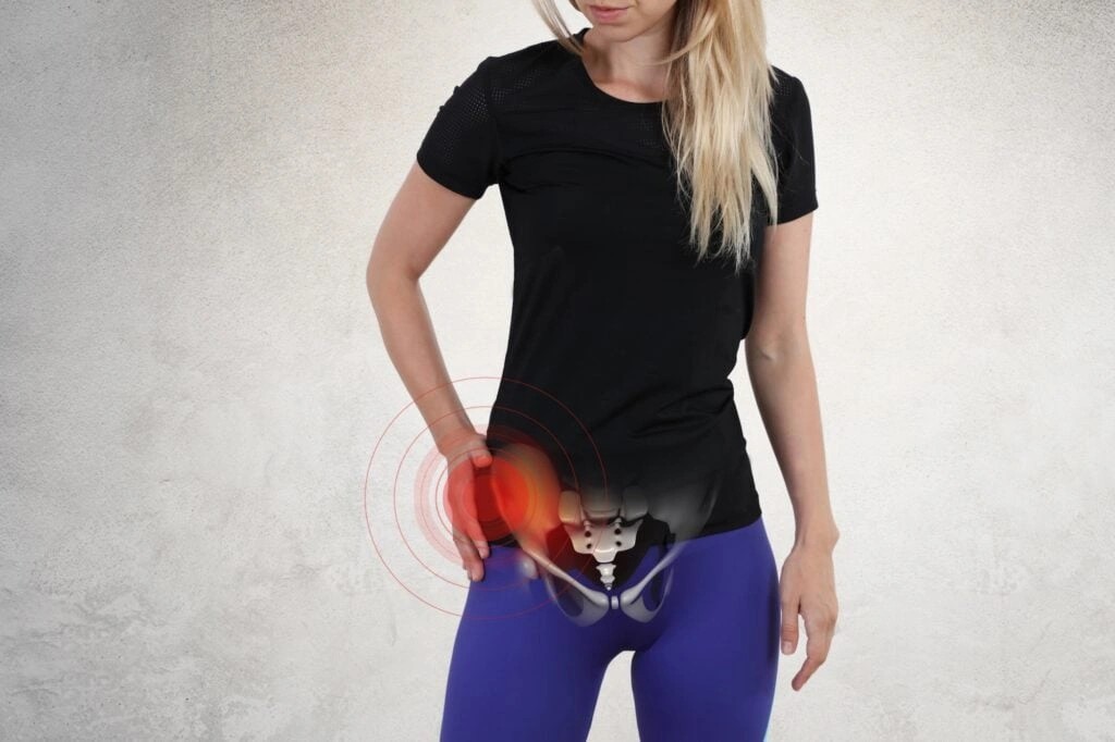
Hip patients often describe their pain using the characteristic “C sign”—placing their index finger near the anterior superior iliac spine (ASIS) and their thumb over the posterior trochanteric region to indicate the wraparound nature of their discomfort.
The consequences of misdiagnosis can be serious. In the aforementioned study, twelve patients had undergone surgical knee interventions with minimal to no relief before their hip condition was properly identified. Subsequently, fourteen patients experienced complete resolution of their knee pain after receiving appropriate hip treatment.
Conditions That Cause Hip Pain to Radiate
Several distinct hip conditions can cause pain to travel down your leg, with each having characteristic symptoms and radiation patterns. Understanding these conditions helps pinpoint the true source of discomfort.
Osteoarthritis and joint degeneration
Hip osteoarthritis develops when protective cartilage wears away while bone around the joint changes shape. This degenerative process typically causes pain that begins in the groin area but often radiates to the thigh, buttocks, or knee. Indeed, studies show that hip pain can radiate below the knee in approximately 47% of hip osteoarthritis cases.
The most telling symptom is groin pain, which occurs in 84.3% of patients with hip dysfunction. Many people experience worse pain in the morning or after sitting for extended periods. Other symptoms include stiffness, decreased range of motion, and a grinding sensation during movement. Curiously, the severity of radiographic hip deterioration doesn’t necessarily correlate with pain intensity or distribution.
Labral tears and impingement
The labrum is cartilage that surrounds your hip socket, providing stability and cushioning. When torn, it typically causes pain in the front of the hip or groin that may travel down to the knee. Femoroacetabular impingement (FAI) occurs when hip bones rub or pinch against each other, often leading to labral tears.
Common symptoms include pain during bending movements, stiffness, and a clicking sensation when moving your hip. FAI symptoms frequently worsen with activities like riding a bike, tying shoes, or sitting for extended periods. Without treatment, this condition can damage cartilage and eventually lead to arthritis.
Hip bursitis and snapping hip syndrome
Trochanteric bursitis involves inflammation of the fluid-filled sac that cushions the upper, outer part of your femur. Pain typically occurs on the outer hip, buttock, or side of your upper thigh. This pain often worsens when lying on the affected side or after sitting.
Snapping hip syndrome causes a snap or pop sensation during hip movement. The external type affects the outside hip area, whereas internal snapping hip causes discomfort near the groin. Although usually harmless, snapping hip can eventually lead to bursitis.
Avascular necrosis and deep hip pain
Avascular necrosis (AVN) results from interrupted blood supply to bone tissue, causing bone death. This serious condition primarily affects the hips, knees, and shoulders. The pain often radiates to the groin, thigh, or buttock, making it difficult to pinpoint the exact source.
Without treatment, AVN worsens as bone tissue dies, potentially leading to bone collapse and severe arthritis. Risk factors include corticosteroid use, excessive alcohol consumption, joint injuries, and certain medical conditions like sickle cell anemia. Early diagnosis is crucial since the progression can be quite rapid.
How Doctors Diagnose the True Source of Pain
Pinpointing the true source of radiating hip pain requires specialized medical expertise and diagnostic techniques. Doctors employ a systematic approach combining detailed examinations, specialized tests, and advanced imaging to differentiate between hip pathology and other conditions.
Physical exam and movement tests
Doctors typically begin with a thorough patient interview, as research indicates patient history plays a vital role in differential diagnosis of hip pain and sometimes proves superior to objective tests. The physician will ask about pain location, nature, patterns throughout the day, and activities that trigger discomfort.
Several specialized movement tests help identify hip joint issues:
- FABER test (Flexion, ABduction, External Rotation): With the patient supine, the hip is flexed, abducted, and externally rotated. Positive findings indicate potential hip pathology or sacroiliac joint dysfunction
- FADIR test: The hip is flexed, adducted, and internally rotated to provoke pain suggestive of labral tears or impingement
- Log roll test: Passive internal/external rotation of the leg while supine can reveal hip joint pathology
- Thomas test: Identifies hip flexion contractures by examining if the opposite hip lifts off the examination table
Gait analysis primarily helps identify conditions like antalgic gait (painful shortened stance) or Trendelenburg gait (indicating abductor weakness).
Imaging for hip vs knee pain
Generally, standing anteroposterior hip and pelvic radiography serves as the initial imaging study for chronic hip pain. Nevertheless, fractures aren’t always visible on initial X-rays – MRI shows higher sensitivity for detecting subtle fractures.
MRI proves especially valuable for diagnosing soft tissue problems around the hip joint. Meanwhile, ultrasound effectively evaluates joint effusion, synovial thickening, bone/cartilage contour issues, plus conditions like trochanteric bursitis and hamstring tendinopathy.
When to consider referred pain
Referred pain should be suspected whenever:
- Pain patterns seem disproportionate to clinical findings
- Knee pain fails to improve with knee-focused treatments
- Hip injections provide relief from knee symptoms
- Patients report posterior hip pain alone (57.1%) or both anterior and posterior pain (21.4%)
Importantly, studies confirm that disk space narrowing at L1/L2 or L2/L3 spine levels significantly correlates with hip pain.
Case example: misdiagnosed hip arthritis
One revealing case study highlights this diagnostic challenge. A patient presented with localized hip/groin pain, positive FABER/FADDIR tests, morning stiffness, and abductor weakness. Initially, spine-related causes were suspected.
Upon X-ray examination, severe hip osteoarthritis with complete joint space loss was discovered. This exemplifies how objective findings like pain during squats, referred groin pain, abduction weakness, and restricted hip movements can achieve a positive likelihood ratio of 15.4 for correct hip diagnosis.
Managing and Treating Radiating Hip Pain
Effective management of radiating hip pain requires a multi-faceted approach tailored to your specific condition and symptoms. Treatment options range from simple exercises to surgical interventions depending on severity.
Exercises for hip joint pain relief
Targeted exercises strengthen muscles supporting your hip joint, improving stability and function. Physical therapists often recommend leg raises, bridging, and hip extensions to build strength around the joint. The butterfly stretch and double hip rotations help improve flexibility and range of motion. Start with gentle movements—hip flexion exercises where you march in place can improve mobility without excessive strain. Importantly, cease any exercise that increases pain rather than relieves it.
Non-surgical treatments for hip arthritis
Low-impact activities like swimming or cycling maintain fitness without stressing painful joints. Over-the-counter medications such as NSAIDs (ibuprofen, naproxen) effectively reduce inflammation and pain. For persistent discomfort, corticosteroid injections provide short-term improvements in pain, function, and range of motion. Hyaluronic acid injections offer additional joint lubrication. Physical therapy remains valuable as it presents minimal risk of adverse events compared to medications.
When surgery is needed
Consider surgical options primarily when hip pain interferes with daily activities despite trying non-surgical treatments. Hip replacement surgery (total hip arthroplasty) becomes appropriate if pain: doesn’t respond to medications, worsens with walking even when using support, interferes with sleep, or makes climbing stairs difficult. Remarkably, more than 90% of people who undergo hip replacement experience significant pain reduction.
Preventing future flare-ups
Maintain a healthy weight to reduce unnecessary stress on your hip joints. Modify activities that trigger pain—avoid low chairs which bend the hip more acutely. Use walking aids correctly if recommended by healthcare professionals. Apply the RICE method (Rest, Ice, Compression, Elevation) for acute flare-ups. Finally, strengthen thigh and leg muscles through regular exercise as everything is connected.
Conclusion
Understanding why hip pain travels down your leg empowers you to seek appropriate care rather than treating symptoms at the wrong location. Hip joint problems frequently masquerade as knee pain, thigh discomfort, or even foot issues due to the complex network of nerves connecting these areas. This referral pattern explains why treating the apparent pain site often fails to provide lasting relief.
Accurate diagnosis stands as the cornerstone of effective treatment. Your doctor should conduct thorough physical examinations and movement tests before considering advanced imaging. Consequently, this comprehensive approach helps differentiate between true knee problems and hip-originated pain that merely presents in the knee area.
Hip osteoarthritis, labral tears, bursitis, and avascular necrosis represent the primary culprits behind radiating hip pain. Each condition creates distinctive pain patterns that may extend well beyond the hip joint itself. Therefore, describing your exact symptoms—including pain location, triggers, and timing—helps your healthcare provider make the correct diagnosis.
The good news? Most hip conditions respond well to appropriate treatment. Conservative approaches such as targeted exercises, medication, and lifestyle modifications provide relief for many patients. Nonetheless, surgical interventions like hip replacement offer excellent outcomes when necessary, with over 90% of patients experiencing significant pain reduction afterward.
Your proactive participation remains essential for long-term hip health. Maintaining healthy weight, modifying problematic activities, and strengthening supporting muscles all contribute to preventing future flare-ups. Additionally, early intervention prevents minor problems from developing into more serious conditions that might eventually require surgery.
Though radiating hip pain can significantly impact your quality of life, proper diagnosis and treatment can restore your mobility and eliminate discomfort. Armed with this knowledge about pain referral patterns, you can advocate for thorough evaluation if you suspect your knee pain might actually originate from your hip.
FAQs
Q1. What conditions can cause hip pain to radiate down the leg?
Hip osteoarthritis, labral tears, femoroacetabular impingement, bursitis, and avascular necrosis are common conditions that can cause hip pain to travel down the thigh, knee, or even lower leg areas. This is due to the complex network of nerves connecting the hip to other parts of the leg.
Q2. How can doctors accurately diagnose the source of radiating hip pain?
Doctors use a systematic approach involving detailed patient history, physical exams with specialized movement tests like FABER and FADIR, and advanced imaging techniques like X-rays, MRI, and ultrasound to pinpoint whether the pain originates from the hip joint or other areas.
Q3. What are some effective non-surgical treatments for hip joint pain?
Non-surgical options include low-impact exercises to strengthen supporting muscles, over-the-counter anti-inflammatory medications, corticosteroid or hyaluronic acid injections for temporary relief, physical therapy, and activity modification to reduce joint stress.
Q4. When is hip replacement surgery recommended for radiating hip pain?
Hip replacement surgery may be considered if the hip pain significantly interferes with daily activities despite trying non-surgical treatments, worsens with walking, disturbs sleep, or makes tasks like climbing stairs difficult. Over 90% of hip replacement patients experience significant pain reduction.
Q5. How can I prevent future flare-ups of radiating hip pain?
Maintaining a healthy weight, modifying activities that trigger pain, using walking aids correctly if recommended, applying the RICE method for acute flare-ups, and regularly exercising to strengthen the muscles supporting the hip joint can help prevent future episodes of radiating hip pain.