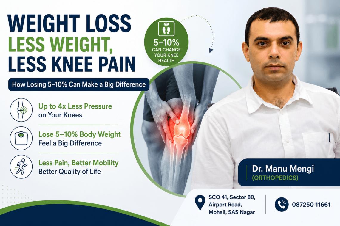
Why Autoimmune Diseases Affect Women More Than Men

What Are Autoimmune Diseases

Why do autoimmune diseases affect women more than men? The answer lies in a complex interplay between hormones and immunity. About 75% of people with autoimmune illnesses are women, and over 80% of autoimmune disorder patients are female. Women face up to a fourfold increase in risk for autoimmune disease compared to men. This striking gender gap isn’t coincidental. Estrogen and immune system interactions create stronger immune responses in women, but this advantage comes with a steep price. In this article, I’ll explore the hormone immunity connection, examine how estrogen, progesterone, and testosterone influence autoimmune risk, and uncover why women’s biology creates unique vulnerabilities to conditions like lupus and rheumatoid arthritis.

Why Women Are More Affected by Autoimmune Disorders

Statistics on autoimmune diseases in women

The numbers paint a stark picture of gender disparity in autoimmune disorders. Up to 50 million people in the U.S. live with an autoimmune disease. Four of every five people diagnosed with an autoimmune disease are female. Autoimmune diseases present with a clear gender bias, occurring at a rate of 2 to 1 among women. Nearly 80% of autoimmune disease cases occur in women, particularly during their reproductive years.

The gap becomes even more pronounced when examining specific conditions. Women face up to a fourfold increase in risk for autoimmune disease compared to men. This disparity isn’t a statistical anomaly. It reflects fundamental biological differences between male and female immune systems that researchers are still working to fully understand.

Common Autoimmune Diseases in Women

Some autoimmune disorders show dramatically skewed female-to-male ratios:

  • Sjögren’s syndrome: 9:1 or as high as 19:1
  • Lupus: 9:1[11]
  • Hashimoto’s disease: 9:1
  • Rheumatoid arthritis: 3:1
  • Multiple sclerosis: 2:1

Other conditions significantly more prevalent among women include autoimmune thyroid diseases like Graves’ disease, scleroderma, and celiac disease. Rheumatoid arthritis is not only more common in women but also more severe.

Understanding the immune system differences between men and women

Women have two X chromosomes, while men have one X and one Y. The X chromosome carries a high number of genes responsible for immune regulation. In women, gene duplication and sometimes gene overexpression from the second X chromosome may trigger autoimmune reactions.

Women are biologically designed to protect future generations, meaning their immune systems are naturally stronger and more reactive. This helps them better fight infections compared to men. However, an immune system that is too strong is more likely to malfunction and attack the body’s own tissues. Males and females differ in their immunological responses to foreign and self-antigens and show distinctions in innate and adaptive immune responses.

By the same token, women’s immune systems offer superior protection against pathogens but create a higher risk of misfiring against the body itself.

The Hormone Connection Explained

Estrogen and immune system activation

Estrogen acts on all cellular subsets of the immune system through receptor-dependent and independent mechanisms. The hormone enhances immune surveillance by modulating neutrophils, macrophages, dendritic cells, T cells, and B cells. Estrogen receptors are widely dispersed in many immunomodulatory cells and regulate immune cell function.

The hormone’s effect on T cells varies by subset. Estrogen modulates IFNγ-secreting Th1 cells by enhancing IFNγ expression. High estrogen levels, such as during pregnancy, skew the immune response from Th1 to Th2. This shift has protective effects in some autoimmune conditions like multiple sclerosis and rheumatoid arthritis, since estrogen promotes Treg expansion and activation.

For B cells, estrogen increases plasma cell and autoantibody-producing cell numbers. This enhancement of humoral immunity partly explains why women mount stronger antibody responses but also face higher autoimmune risk. The outcome varies depending on estrogen levels, cell type, activation state, and local environment.

Progesterone’s protective role

Progesterone acts in an immunosuppressive way, favoring a Th2-like cytokine profile. The hormone inhibits inflammatory innate immune responses by suppressing macrophage and dendritic cell activation. When bound to its receptor, progesterone interferes with the NF-κB pathway, a key driver of inflammation.

Progesterone decreases production of proinflammatory cytokines like TNF-α, IFN-γ, and IL-12 while increasing anti-inflammatory IL-10. The hormone skews naive T cells away from Th1 responses toward Th2 responses, with increased IL-4, IL-5, and IL-10 production. During pregnancy, high progesterone levels shift the immune response toward a CD4+ Treg phenotype.

Testosterone’s protective effect on immunity

Men with relatively high testosterone levels benefit less from influenza vaccination than men with lower testosterone and women. Women had a generally stronger antibody response to the vaccine than men, but men with low testosterone showed responses equivalent to women.

Testosterone interacts with a gene cluster called Module 52 in a way that dampens immune response. In high-testosterone men, elevated Module 52 gene activity correlated with reduced post-vaccination antibody levels. Several Module 52 genes accelerate differentiation of cells that suppress rather than foster immune response.

Cortisol and stress response in autoimmune conditions

Cortisol plays a central role in immune regulation through the HPA axis. In acute stress, cortisol enhances immune surveillance. However, chronic stress leads to sustained cortisol elevation, suppressing immune function by inhibiting T-cell activity, reducing cytokine production, and impairing antibody responses.

Prolonged cortisol exposure leads to glucocorticoid resistance, where immune cells become less responsive to cortisol’s anti-inflammatory effects[38]. This results in persistent inflammation despite cortisol presence. Chronic stress can also cause cortisol depletion over time, and these low levels fail to control inflammation in autoimmune diseases, exacerbating symptoms.

Hormonal Fluctuations Throughout a Woman’s Life

Puberty and autoimmune disease onset

Puberty marks a critical turning point where disease susceptibility changes dramatically. Before puberty, multiple sclerosis affects males and females equally. After puberty, MS becomes two to three times more common in females. Allergic asthma shows the opposite pattern: prevalence decreases in males while increasing in females after puberty.

Girls with premature puberty face additional risks. A quarter of individuals with premature puberty also presented with autoimmune thyroiditis. Puberty may accelerate onset of Type 1 diabetes in genetically susceptible females, mediated by estrogen’s effect on the Interleukin-6 promoter.

Pregnancy and autoimmune diseases: improvement or flare?

Pregnancy’s impact on autoimmune conditions varies widely. Rheumatoid arthritis and multiple sclerosis may temporarily improve during pregnancy, while lupus often worsens. Women with lupus and antiphospholipid syndrome are five times more likely to experience preeclampsia, preterm birth, and stillbirth.

Postpartum flares represent the most common time for disease activation. Approximately one of five babies born to women with myasthenia gravis is born with the disorder, though muscle weakness in the baby is usually temporary.

Early Warning Signs You Should Not Ignore

Estrogen decline during menopause can trigger new autoimmune diseases or worsen existing ones. Early menopause may induce rheumatoid arthritis onset and worsen RA symptoms, possibly due to the loss of estrogen’s immunomodulatory effects. Women with multiple sclerosis notice increased fatigue and potential acceleration of disease progression as estrogen declines.

Why autoimmune symptoms flare during the menstrual cycle

Women with rheumatoid arthritis and systemic lupus erythematosus experience disease flares that correlate with their menstrual cycles, occurring just prior to or at the start of menses. During the follicular phase, increasing estrogen levels cause an immune response with increasing antibodies and inflammation. The late luteal phase through menstruation brings a worsening of autoimmune symptoms with increased inflammatory responses.

Beyond Hormones: The X Chromosome Factor

What role does the X chromosome play in autoimmune disorders

Women carry two X chromosomes while men have one X and one Y. Each female cell must shut down one X chromosome to prevent lethal protein overproduction through a process called X-chromosome inactivation. A molecule called Xist achieves this shutdown by coating sections of one X chromosome.

In reality, this protective mechanism creates vulnerability. Xist consists of long noncoding RNA that binds with proteins and DNA fragments, forming complexes that resemble autoimmune targets. These RNA-protein-DNA complexes can trigger strong immune responses. Research in male mice engineered to produce Xist showed elevated autoantibody levels and extensive tissue damage.

Genetic factors in female autoimmune risk

Several immune-related genes on the X chromosome escape inactivation, contributing to heightened immune reactivity in females. The VGLL3 gene on the X chromosome links directly to inflammation. When X-chromosome inactivation maintenance is disrupted, genes like TLR7 become expressed at higher levels from the inactive X chromosome.

Furthermore, blood samples from autoimmune patients revealed autoantibodies targeting many Xist-associated complexes. Some autoantibodies were specific to particular disorders, indicating potential for early disease identification.

How biology creates double the vulnerability

Susceptibility stems from the gene dosage effect of having two X chromosomes. Turner’s syndrome patients with X chromosome monosomy show high prevalence of juvenile arthritis, thyroiditis, inflammatory bowel diseases, and diabetes.

Conclusion

Women’s biology creates a double-edged sword. Stronger immune systems protect against infections but increase autoimmune vulnerability. Hormonal fluctuations throughout life, combined with the X chromosome factor, explain why women account for nearly 80% of autoimmune cases. This knowledge empowers you to recognize patterns in your own health. Without a doubt, understanding how estrogen, progesterone, and your menstrual cycle affect immunity helps you anticipate flares and work with your healthcare provider to manage symptoms more effectively.

Key Takeaways

Understanding why autoimmune diseases disproportionately affect women reveals crucial insights about biology, hormones, and health management that every woman should know.

• Women account for nearly 80% of autoimmune disease cases, with some conditions like lupus and Sjögren’s syndrome showing 9:1 female-to-male ratios due to biological differences.

• Estrogen enhances immune responses, making women better at fighting infections but more prone to autoimmune attacks on their own tissues.

• Hormonal fluctuations during menstrual cycles, pregnancy, and menopause directly trigger autoimmune symptom flares and disease progression changes.

• The X chromosome carries immune-regulating genes, and having two X chromosomes creates genetic vulnerability through overactive immune responses.

• Recognizing these hormonal patterns empowers women to anticipate symptom changes and collaborate more effectively with healthcare providers for better disease management.

This biological understanding transforms how women can approach their health, moving from reactive symptom management to proactive awareness of their body’s unique immune patterns throughout different life stages.

FAQs

Q1. Why are women more likely to develop autoimmune diseases than men?

Women face up to a fourfold increase in autoimmune disease risk due to several biological factors. Their stronger immune systems, influenced by estrogen and the presence of two X chromosomes, make them better at fighting infections but also more prone to immune system misfiring. Hormonal fluctuations throughout life, combined with immune-regulating genes on the X chromosome, create unique vulnerabilities that explain why nearly 80% of autoimmune cases occur in women.

Q2. How do hormones contribute to autoimmune disease development in women?

Estrogen enhances immune responses by activating various immune cells, which helps fight infections but increases the risk of the immune system attacking the body’s own tissues. Progesterone acts as an immunosuppressant and provides some protective effects, while testosterone in men offers protective benefits against autoimmune conditions. These hormonal differences, particularly the immune-activating effects of estrogen during reproductive years, significantly contribute to women’s higher autoimmune disease rates.

Q3. Do autoimmune symptoms change during different stages of a woman’s life?

Yes, hormonal fluctuations throughout a woman’s life significantly impact autoimmune symptoms. Many autoimmune diseases emerge after puberty when estrogen levels rise. During pregnancy, some conditions like rheumatoid arthritis may improve while others like lupus often worsen. Menopause can trigger new autoimmune diseases or worsen existing ones due to declining estrogen levels. Even monthly menstrual cycles can cause symptom flares, typically occurring just before or at the start of menstruation.

Q4. What role does the X chromosome play in autoimmune disease risk?

Women have two X chromosomes compared to men’s one, and the X chromosome carries numerous immune-regulating genes. During X-chromosome inactivation, a molecule called Xist creates complexes that can trigger autoimmune responses. Additionally, some immune-related genes on the X chromosome escape inactivation, leading to heightened immune reactivity in women. This genetic double dose creates increased vulnerability to autoimmune conditions.

Q5. Which autoimmune diseases show the highest female predominance?

Sjögren’s syndrome shows the most dramatic gender disparity with a 9:1 to 19:1 female-to-male ratio. Lupus and Hashimoto’s disease both affect women at a 9:1 ratio, while rheumatoid arthritis occurs three times more often in women than men. Multiple sclerosis affects women at a 2:1 ratio. These conditions not only occur more frequently in women but often present with more severe symptoms as well.

Q6. Why do autoimmune diseases affect women more than men?

Women have a more active immune system due to hormonal influences (especially estrogen) and genetic factors like two X chromosomes. While this stronger immunity helps fight infections, it also increases the risk of the immune system mistakenly attacking the body’s own tissues.

Q7. How does estrogen influence the immune system?

Estrogen enhances immune activity by stimulating immune cells and antibody production. This heightened response can sometimes overshoot, increasing susceptibility to autoimmune diseases such as lupus and rheumatoid arthritis.

Q8.Does testosterone protect against autoimmune diseases?

Yes, testosterone generally has an immunosuppressive effect. It dampens immune responses, which may explain why men have a lower risk of developing many autoimmune conditions compared to women.

Q9. Why is lupus more common in women?

Lupus is strongly influenced by estrogen, which amplifies immune responses and antibody formation. Additionally, genetic factors linked to the X chromosome contribute to the higher prevalence of lupus in women.

Q10. Can pregnancy affect autoimmune diseases?

Pregnancy can either improve or worsen autoimmune conditions. Some diseases like rheumatoid arthritis may improve due to immune suppression, while others like lupus may flare due to hormonal and immune changes.

Q11. Do autoimmune diseases worsen during menstruation?

Yes, some women experience symptom flares around menstruation due to hormonal fluctuations, particularly changes in estrogen and progesterone levels that influence immune activity.

Q12. Does menopause impact autoimmune diseases?

Menopause leads to a drop in estrogen levels, which can alter immune function. Some autoimmune diseases may improve, while others may worsen or newly appear after menopause.

Q13. What role does the X chromosome play in autoimmunity?

The X chromosome contains many immune-related genes. Women have two X chromosomes, and incomplete inactivation of one X may lead to overexpression of these genes, increasing autoimmune risk.

Q14. Are autoimmune diseases hereditary in women?

Autoimmune diseases are not strictly hereditary but have a genetic predisposition. Women with a family history are at higher risk, especially when combined with hormonal and environmental triggers.

Q15. Can lifestyle factors influence autoimmune diseases in women?

Yes, factors like stress, diet, infections, smoking, and poor sleep can trigger or worsen autoimmune conditions by affecting immune regulation and inflammation levels.

Consult Dr. Manu Mengi, the best orthopedic doctor in Mohali, for all your orthopedic problems.

Is Your Joint Pain Autoimmune or Mechanical? Early Warning Signs to Watch For – Dr. Manu Mengi

Arthritis affects nearly 60 million American adults and 300,000 children, yet many of us struggle with a critical question: is my joint pain autoimmune or mechanical, and how can I tell the difference early? Understanding whether your discomfort stems from autoimmune conditions, such as rheumatoid arthritis affecting over 1.3 million Americans, or mechanical wear and tear is essential for proper treatment. In this guide, we’ll walk you through the key differences between autoimmune vs mechanical joint pain, early warning signs to watch for, and when you should seek medical attention.

Understanding the Two Main Types of Joint Pain

Pain classification forms the foundation of proper diagnosis. Doctors broadly categorize joint pain into two distinct types: mechanical pain and inflammatory (autoimmune) pain. This distinction shapes everything from your treatment plan to your long-term prognosis.

What Is Mechanical Joint Pain?

Mechanical pain stems from wear and tear, overload, strain, or structural problems in bones, joints, muscles, ligaments, or disks. The term “mechanical” indicates the pain relates directly to movement and physical stress on tissues.

Common conditions that produce mechanical joint pain include:

  • Osteoarthritis
  • Disk problems
  • Muscle strain
  • Ligament injury
  • Plantar fasciitis
  • Degenerative knee pain
  • Posture-related back pain

The defining characteristic of mechanical joint pain is its relationship to activity. Pain intensifies when you move or use the affected joint, then improves with rest. Morning stiffness exists but typically resolves within 20 to 30 minutes. The pain usually localizes to one or two specific joints, often those bearing the most weight or previously injured. You won’t typically see major swelling or warmth unless you’ve experienced an acute injury. Doctors call this pattern “activity pain” because symptoms worsen by day’s end after accumulated use.

The continuous wear on cartilage that cushions bones leads to deterioration, causing bones to rub against each other. This friction results in pain and further joint damage. Over time, the body attempts to restore stability by remodeling bone, which can create undesirable bony growths called osteophytes.

What Is Autoimmune Joint Pain?

In contrast, autoimmune joint pain occurs when your immune system mistakenly targets your own body’s tissues, including joints, muscles, skin, and organs. This misguided immune response triggers inflammation, leading to pain, swelling, stiffness, and potentially long-term damage without prompt treatment.

Autoimmune-related joint pain differs markedly from mechanical pain. The inflammation often affects joints symmetrically, meaning both sides of your body equally. You’ll experience stiffness, particularly in the morning, that lasts considerably longer than the brief stiffness from mechanical issues. The pain persists and progresses without treatment.

Unlike mechanical pain that worsens with activity, inflammatory joint pain often improves with movement. Pain at rest is common with autoimmune conditions but rare with mechanical problems. Night pain appears more frequently in inflammatory conditions. During physical examination, inflammatory joints feel boggy and warm, whereas mechanical pain areas feel tender but not inflamed.

Why the Difference Matters

Treatment approaches diverge dramatically between these two pain types. Mechanical pain treatment focuses on physiotherapy, load correction, posture improvement, weight management, and when needed, interventional procedures or regenerative options. Autoimmune conditions require immune-modulating treatments to control the underlying inflammatory process.

Treating inflammatory pain like mechanical pain, or vice versa, often leads to poor results and prolonged suffering. Given that over 80 types of autoimmune diseases exist affecting various body parts, accurate identification becomes critical. Without proper diagnosis and treatment, autoimmune conditions can cause irreversible joint damage and raise your risk for complications like heart disease.

Key Differences: How Autoimmune and Mechanical Pain Feel Different

Recognizing specific patterns helps us distinguish between autoimmune and mechanical joint pain. The way your joints feel throughout the day, respond to movement, and appear visually provides critical diagnostic clues.

Morning Stiffness: Duration Tells the Story

The duration of morning stiffness serves as one of the most telling indicators. Inflammatory joint pain causes morning stiffness lasting over 30 to 45 minutes. In many cases with rheumatoid arthritis, this stiffness persists for hours. The prolonged stiffness can interfere with basic self-care tasks like dressing, buttoning clothes, combing hair, or making a fist.

In contrast, mechanical joint pain produces stiffness lasting under 20 to 30 minutes. This brief morning tightness improves quickly as you move around. While osteoarthritis stiffness appears most noticeable upon awakening or after inactivity, it resolves faster than inflammatory stiffness.

Autoimmune conditions also create a phenomenon called “gelling,” where joints stiffen after periods of rest, such as difficulty getting up from sitting on the floor. This pattern repeats throughout the day, not just in the morning.

Pain Pattern: Rest vs Movement

The relationship between pain and activity differs dramatically between the two types. Inflammatory pain improves with gentle movement, while inactivity causes gelling. You’ll notice pain even at rest is common with autoimmune conditions, whereas rest pain rarely occurs with mechanical problems.

Mechanical pain follows the opposite pattern. Pain increases with activity and improves with rest. Loading the joint during walking, lifting, or other activities triggers discomfort that peaks by day’s end after accumulated use. Night pain appears more common with inflammatory conditions compared to mechanical issues.

Swelling Characteristics

The quality and persistence of swelling reveal important differences. Inflammatory arthritis produces joints that feel puffy, warm, and tender, with swelling that’s soft and may appear red. These clear inflammation signs persist for several days to weeks, waxing and waning over months. The swelling associates with warmth and redness.

Mechanical pain presents differently. If swelling exists, it’s usually mild and hard (bony) rather than soft and inflamed. The enlargement comes from bony growths rather than inflammatory fluid. You won’t typically see major swelling or warmth except following acute injury. When mechanical pain causes swelling, it worsens with and after activity.

Symmetry and Joint Distribution

Rheumatoid arthritis demonstrates a distinctive symmetrical pattern. If one hand or foot becomes affected, the other side likely will too. Symptoms happen on both sides of the body simultaneously. For example, someone with symmetrical knee arthritis experiences symptoms in both knees at the same time.

Mechanical pain typically affects large joints or those with injury history, often with one side worse than the other. Noninflammatory arthritis like osteoarthritis produces asymmetrical symptoms, frequently occurring in the spine and weight-bearing joints such as the knee or hip. Inflammatory conditions hit multiple joints in this mirror pattern, commonly affecting hands, wrists, ankles, and feet.

Early Warning Signs of Autoimmune Joint Disease

Catching autoimmune joint disease in its earliest stages can prevent irreversible damage and preserve your mobility. Several distinct warning signs emerge before full-blown disease develops, and recognizing them prompts timely medical intervention.

Prolonged Morning Stiffness (Over 1 Hour)

Morning stiffness lasting more than one hour signals potential rheumatoid arthritis. This prolonged stiffness is characteristic of RA and associates with markers of systemic and local inflammation in RA patients. In contrast to brief mechanical stiffness, inflammatory arthritis includes morning stiffness that lasts for an hour.

The duration of your morning stiffness serves as a useful gage of inflammatory activity in your body. In some cases, this stiffness persists for several hours. You might first notice stiffness in one or more joints, usually accompanied by pain on movement and tenderness. This extended stiffness distinguishes inflammatory conditions from mechanical wear and tear, where symptoms resolve within 20 to 30 minutes.

Joint Pain That Improves With Movement

A hallmark of inflammatory arthritis involves joint pain and stiffness after periods of rest or inactivity, particularly in the morning. Paradoxically, this pain improves slowly with movement. If your stiffness lasts longer than 30 minutes and improves with activity, it may indicate inflammation inside the joints.

Pain that worsens after rest rather than activity points toward autoimmune origins. Unlike mechanical pain that intensifies with use, inflammatory joint pain often diminishes as you move throughout the day. This pattern helps distinguish autoimmune conditions from injury-related pain.

Systemic Symptoms Beyond the Joints

Inflammatory arthritis is usually systemic, meaning symptoms related to inflammation may occur in other parts of the body. Besides joint involvement, you might experience:

  • Skin rashes
  • Eye inflammation
  • Hair loss
  • Dry mouth
  • Fever

Nonspecific systemic symptoms, primarily fatigue, malaise, and depression, may commonly precede other symptoms of the disease by weeks to months. Inflammatory arthritis does not just affect joints but also causes constant tiredness, weakness, and low energy. This happens because the immune system is continuously active.

Autoimmune diseases cause inflammation, which includes a feeling of warmth or heat, discoloration or redness on your skin, swelling, and pain. Many autoimmune diseases cause symptoms that come and go. These episodes of more noticeable or more severe symptoms are called flares or attacks. During flares, you may experience an increase in systemic symptoms more than discrete joint swelling or tenderness.

Multiple Joints Affected Symmetrically

Unlike injury-related pain, inflammatory arthritis usually affects multiple joints at the same time. For example, both wrists, both knees, or both hands. This pattern is a key warning sign.

The number of joints involved is highly variable, but the process is eventually polyarticular, involving five or more joints. Symmetrical joint swelling is characteristic of rheumatoid arthritis that has been persistent for a period of time. Your symptoms often happen on both sides of the body in a symmetrical pattern. As an illustration, if you have RA in the right hand, you may also have it in the left hand.

Common Autoimmune Conditions That Cause Joint Pain

Several distinct autoimmune conditions share joint pain as a primary symptom, yet each presents unique characteristics that help us identify them.

Rheumatoid Arthritis

Rheumatoid arthritis stands as one of the most well-known autoimmune diseases, with a prevalence of 1 to 2% worldwide. Prevalence increases with age, approaching 5% in women over age 55. Both incidence and prevalence are two to three times greater in women than in men.

RA primarily targets joints, causing painful swelling and progressive damage. The disease most frequently affects the proximal interphalangeal and metacarpophalangeal joints of the hands, the wrists, and small joints of the feet including the metatarsophalangeal joints. The shoulders, elbows, knees, and ankles are also affected in many patients. RA often starts in the small joints of the hands and feet and can eventually affect larger joints.

In addition to joint involvement, RA may affect other systems of the body such as skin, eyes, lungs, heart, and blood vessels. The subcutaneous nodule is the most characteristic extra-articular lesion of the disease, occurring in 20 to 30% of cases. Approximately 10 to 15% of patients with rheumatoid arthritis develop Sjogren’s syndrome.

Lupus (Systemic Lupus Erythematosus)

Lupus is a systemic disease that can inflame joints as well as the skin, kidneys, heart, and lungs. Arthritis in systemic lupus erythematosus is one of the most common disease manifestations. Joint pain and swelling are among the earliest signs of lupus, along with fatigue, rash, and sensitivity to sunlight.

Nearly all joints can be affected by SLE, but hand and knee involvement are the most typical. The typical presentation of lupus arthritis is a symmetric polyarthritis that preferentially involves the small joints over the large joints although any joint may be affected. While the majority of lupus patients with arthritis have a non-deforming course, lupus arthritis is less likely to cause permanent joint damage than rheumatoid arthritis.

Psoriatic Arthritis

Psoriatic arthritis affects some people with psoriasis, a skin disease. It’s estimated that around 30% of people with psoriasis will develop psoriatic arthritis. Most people who develop psoriatic arthritis already have psoriasis.

This condition causes joint pain, stiffness, and swelling, often along with patches of scaly skin. Common symptoms include pain, stiffness, and swelling in joints on one or both sides of the body, pain and swelling in other parts of the body such as hands, feet, and low back, and itchy or sore patches of thick, red, scaling on the skin. Dactylitis is the swelling of a whole finger or toe, giving them a sausage-like appearance.

Ankylosing Spondylitis

Ankylosing spondylitis primarily affects the spine and sacroiliac joints, leading to chronic back pain and reduced flexibility. The most common symptom is lower back and hip pain and stiffness. The pain typically worsens during periods of rest or inactivity, which may cause some people to experience more pain during the middle of the night or after prolonged sitting. Moving and exercise can help improve pain.

In severe cases, inflammation may cause the vertebrae to fuse, leading to a rigid and inflexible spine. More than 90% of white people who have AS also have a mutated HLA-B27 gene. In addition to spinal symptoms, other symptoms may develop depending on which other areas of the body are affected, including eye disease (uveitis), skin disease (psoriasis), or gut disease (inflammatory bowel disease).

When to See a Doctor and What to Expect

Knowing when your joint pain requires professional evaluation can prevent serious complications. Schedule an appointment if joint symptoms persist for three days or more, or if you experience several episodes within a month.

Red Flags That Need Medical Attention

Certain symptoms demand immediate medical attention. When a joint suddenly becomes red, swollen, and tender, it signals more than a minor issue. Fever paired with joint pain, particularly without other cold or flu symptoms, could indicate infection or an autoimmune response. Unexplained weight loss accompanying joint discomfort may point to rheumatoid arthritis or other systemic conditions. Pain that disrupts your sleep often indicates underlying inflammatory conditions that activate during rest. Watch for skin changes like rashes, redness, or nail pitting, which can signal psoriatic arthritis. If a joint suddenly locks up or won’t move, seek care immediately.

Diagnostic Tests Your Doctor May Order

Your primary care doctor will perform a physical examination, checking joints for swelling, redness, warmth, and assessing reflexes and muscle strength. Rheumatoid arthritis can be difficult to diagnose in early stages since symptoms mimic other conditions. If your doctor suspects autoimmune disease, they’ll refer you to a rheumatologist who specializes in making complex arthritis diagnoses.

Blood Tests and Imaging Studies

Blood tests measure inflammation markers like erythrocyte sedimentation rate (ESR) and C-reactive protein (CRP). Your doctor may also check for rheumatoid factor and anti-cyclic citrullinated peptide (anti-CCP) antibodies. X-rays track disease progression over time, while MRI and ultrasound scans help diagnose early disease and assess severity.

Conclusion

Joint pain doesn’t have to remain a mystery. We’ve shown you the critical differences between autoimmune and mechanical joint pain, particularly morning stiffness duration, pain patterns with movement, and symmetrical involvement. These distinctions matter because they determine your treatment path and long-term outcomes.

Essentially, early recognition saves you from irreversible damage. If your morning stiffness lasts over an hour, your pain improves with movement, or you notice symmetrical joint involvement, don’t wait. See your doctor promptly for proper evaluation.

Your body sends clear signals. Learn to recognize them, act quickly, and you’ll have the best chance of preserving your joint health and maintaining your quality of life for years to come.

Key Takeaways

Understanding the difference between autoimmune and mechanical joint pain can prevent irreversible damage and guide you toward the right treatment approach.

• Morning stiffness lasting over 1 hour signals autoimmune disease, while mechanical pain stiffness resolves within 20-30 minutes

• Autoimmune joint pain improves with movement and occurs at rest, while mechanical pain worsens with activity and improves with rest

• Symmetrical joint involvement affecting both sides of the body simultaneously indicates autoimmune conditions like rheumatoid arthritis

• Seek immediate medical attention if joint pain persists over 3 days, includes fever, or disrupts sleep patterns

• Early diagnosis and treatment of autoimmune joint disease prevents permanent joint damage and preserves long-term mobility

The key is recognizing these warning signs early. Your body provides clear signals about whether inflammation or wear-and-tear is causing your discomfort. Acting on these signs promptly can mean the difference between manageable symptoms and irreversible joint damage.

FAQs

Q1. What are the typical symptoms of autoimmune joint pain?

Autoimmune joint pain typically presents with swelling, stiffness, and tenderness affecting multiple joints simultaneously. You’ll often experience prolonged morning stiffness lasting over an hour, pain that occurs even at rest, and symptoms appearing symmetrically on both sides of your body. Many people also notice systemic symptoms like extreme fatigue, low-grade fever, and general malaise accompanying their joint discomfort.

Q2. How long does morning stiffness last with different types of joint pain?

Morning stiffness duration is a key distinguishing factor between joint pain types. With autoimmune conditions like rheumatoid arthritis, stiffness typically lasts over 30-45 minutes and can persist for several hours, often interfering with daily tasks. In contrast, mechanical joint pain from conditions like osteoarthritis causes stiffness that resolves within 20-30 minutes as you begin moving around.

Q3. Does joint pain that improves with movement indicate an autoimmune condition?

Yes, joint pain that improves with gentle movement is a hallmark sign of inflammatory autoimmune conditions. Unlike mechanical joint pain that worsens with activity and improves with rest, autoimmune joint pain often feels worse after periods of inactivity and gradually lessens as you move throughout the day. This paradoxical pattern helps distinguish autoimmune conditions from injury-related or wear-and-tear joint problems.

Q4. What does symmetrical joint involvement mean in autoimmune diseases?

Symmetrical joint involvement means that symptoms affect the same joints on both sides of your body simultaneously. For example, if you have pain and swelling in your right wrist, you’ll likely experience similar symptoms in your left wrist. This mirror pattern is characteristic of autoimmune conditions like rheumatoid arthritis and typically affects multiple joints including hands, wrists, ankles, and feet.

Q5. When should I see a doctor about my joint pain?

You should seek medical attention if joint pain persists for three days or more, or if you experience multiple episodes within a month. Immediate evaluation is necessary if you notice sudden redness and swelling in a joint, fever accompanying joint pain, unexplained weight loss, pain that disrupts your sleep, skin changes like rashes, or if a joint suddenly locks up and won’t move.

Q6. How can I tell if my joint pain is autoimmune or mechanical?

Autoimmune pain is usually inflammatory—it comes with swelling, warmth, prolonged morning stiffness, and may affect multiple joints symmetrically. Mechanical pain is more related to use and wear, worsens with activity, and improves with rest.

Q7. What is the biggest early clue of autoimmune joint pain?

Morning stiffness lasting more than 30–60 minutes is a key early sign. Mechanical pain typically loosens up within a few minutes of movement.

Q8. Does autoimmune joint pain occur at rest?

Yes. Autoimmune pain often persists even at rest and may even worsen at night. Mechanical pain usually reduces when the joint is not being used.

Q9. Can mechanical joint pain cause swelling?

Yes, but it is usually mild and localized. In autoimmune conditions, swelling tends to be more persistent, soft (boggy), and associated with warmth.

Q10. Which joints are commonly affected in autoimmune arthritis?

Autoimmune arthritis often starts in small joints like fingers, wrists, and toes, usually on both sides of the body. Mechanical pain more often affects weight-bearing joints like knees, hips, or spine.

Q11. Is symmetrical joint pain always autoimmune?

Symmetry raises suspicion for autoimmune causes, but it is not absolute. Clinical pattern, duration, and associated symptoms matter more than symmetry alone.

Q12. Do I need blood tests to confirm autoimmune joint pain?

Blood tests like rheumatoid factor or anti-CCP can help, but diagnosis is clinical first. Some patients may have autoimmune disease even with normal blood tests early on.

Q13. Why does my pain improve after I start moving?

This “start-up stiffness” can be seen in both, but if relief is significant and stiffness is prolonged, it leans toward inflammatory (autoimmune). Mechanical pain improves briefly but worsens with continued use.

Q14. When should I worry about my joint pain?

If pain is persistent, involves multiple joints, has swelling, or morning stiffness >30 minutes, it’s worth getting evaluated early to avoid long-term joint damage.

Q15. Can autoimmune joint pain be treated if detected early?

Yes—and this is crucial. Early diagnosis allows timely use of disease-modifying treatments, which can slow or even halt progression and preserve joint function.

consult with manu manegi for all the types of pain like joint pain ankle pain he is Best orthopedic doctor in Tricity

Rheumatoid Factor Positive But No Symptoms: What Your Blood Test Really Means – Dr. Manu Mengi

Why do your blood tests show rheumatoid factor positive when you feel perfectly fine? This question worries many people who receive unexpected lab results. Actually, rheumatoid factor has been found in up to 4% of young, healthy individuals, and it’s particularly common in older adults. A positive RF test doesn’t automatically mean rheumatoid arthritis. We’ll explain what rheumatoid factor positive meaning really is, why asymptomatic rheumatoid factor occurs, how it differs from anti-CCP testing, and when you should be concerned about your results.

What Does a Positive Rheumatoid Factor Test Mean?

What Does a Positive Rheumatoid Factor Test Mean?

Understanding Rheumatoid Factor and Its Normal Function

Rheumatoid factor is an antibody your immune system produces, but it behaves differently from the antibodies that protect you from illness. Most antibodies act as defenders, targeting germs, viruses, and other harmful invaders in your bloodstream. Rheumatoid factor, on the other hand, attacks healthy cells in your body.

Specifically, RF targets the Fc portion of immunoglobulin G (IgG), one of the most common antibodies your body makes to fight infections. Your immune system creates rheumatoid factor either by mistake or when it becomes overactive while fighting an infection. In reality, this protein doesn’t have a specific protective target like other antibodies, which is why it sometimes damages healthy tissue throughout your body.

The presence of RF isn’t always problematic. Many people have small amounts in their blood and remain perfectly healthy. RF can be part of your body’s normal response to various triggers, including bacterial toxins like lipopolysaccharides or viruses such as Epstein-Barr virus. In these cases, the RFs produced are low-affinity, transient antibodies that form immune complexes, which are then cleared by inflammatory cells. Their role could actually be protective in this context.

How RF Tests Work and What They Measure

An RF test measures the amount of rheumatoid factor in your bloodstream through a simple blood sample. A healthcare professional draws blood from a vein in your arm using a small needle, then sends the sample to a laboratory for analysis.

Labs typically measure rheumatoid factor in units per milliliter of blood (U/mL). Most assays detect IgM rheumatoid factors, although RF can occur in all immunoglobulin classes, including IgG and IgA. The test results come back either as a numerical value showing how many units of RF exist in each milliliter of your blood, or as a simple positive or negative designation.

A negative result means you have little or no rheumatoid factor in your blood. Correspondingly, a positive result indicates higher levels were detected. The test itself takes only a few minutes, though results may not return for several days.

Rheumatoid Factor Normal Range and Interpretation

A normal, healthy amount of rheumatoid factor is less than 20 units per milliliter (<20 U/mL)[7]. Some laboratories use slightly different thresholds, with results below 14 IU/mL considered normal, while others use higher cutoffs. Values may be slightly higher in older patients.

RF above 20 IU/mL is not considered enough to diagnose RA on its own, as other factors can elevate these levels. In fact, about 20% of people with confirmed RA will not have an abnormal RF test, while 5% of people who do not have RA will show elevated RF. This demonstrates why a positive result serves as a clue rather than a definitive diagnosis.

The sensitivity of RF testing in RA patients ranges from 60% to 90%, with a specificity of 85%. However, RF results are positive in less than 40% of patients with early RA. Higher RF levels generally indicate a greater likelihood of having a condition linked to rheumatoid factors, but people with rheumatoid arthritis can have little or no detectable RF. Similarly, people without rheumatoid arthritis can have substantial amounts of RF in their blood.

Your provider considers your RF levels alongside other test results, your symptoms, and your medical history to determine what’s causing any changes in your blood work.

Why Your RF Can Be Positive Without Any Symptoms

Rheumatoid Factor in Healthy Individuals

Perfectly healthy people can have elevated rheumatoid factor without any disease. In fact, RF appears in up to 4% of young, healthy individuals. Many people carry small amounts of RF in their blood and remain completely healthy throughout their lives.

Some people have high RF levels and never experience any symptoms. High amounts aren’t always a sign of a health condition. Your body might produce rheumatoid factor for no apparent reason or without a specific cause. This happens because RF can form as a normal immune response to various triggers, not just autoimmune conditions.

Most asymptomatic persons with a positive RF do not progress to RA. This bears repeating because it addresses the primary concern when you receive unexpected results. The presence of RF in your blood doesn’t predict that you’ll develop rheumatoid arthritis or any other condition. Your immune system sometimes produces these antibodies temporarily or maintains low levels indefinitely without causing tissue damage.

Age-Related Increase in Rheumatoid Factor

RF becomes increasingly common as you get older. The prevalence of RF can be as high as 14.1% in apparently healthy people aged 67-95, with a mean age of 81. This dramatic increase with age explains why many seniors receive positive results during routine blood work.

RF is also 3.5 times more common in healthy elderly subjects aged over 65 than in their younger counterparts. Your immune system changes as you age, producing more autoantibodies even when you’re healthy. These age-related changes don’t necessarily indicate disease development.

All these factors may alter the natural history of arthritis in elderly patients who have RF either in good health or in a non-arthritic presentation. Where you experience symptoms and their severity depends on where your immune system damages healthy tissue in your body.

The Timeline: When Symptoms May or May Not Develop

Some patients actually develop RF preceding the symptomatic disease. The earlier onset of RF in such patients has been associated with more severe disease. However, this represents only a subset of people with positive RF results.

Alternatively, there is a subset of patients in whom the appearance of RF follows symptoms. About 80% of all patients with rheumatoid arthritis will eventually be seropositive for rheumatoid factor, while only 40% are positive at clinical onset. This timeline variation demonstrates that RF positivity can occur at different stages or not at all.

People without rheumatoid arthritis can have substantial amounts of RF in their blood. Correspondingly, people with rheumatoid arthritis can have little or no rheumatoid factor. Your RF status today doesn’t determine your health status tomorrow.

Other Conditions That Cause Elevated Rheumatoid Factor

Other Conditions That Cause Elevated Rheumatoid Factor

Autoimmune Diseases Beyond Rheumatoid Arthritis

Rheumatoid arthritis isn’t the only autoimmune condition that triggers RF production. Several other autoimmune disorders cause elevated rheumatoid factor levels, sometimes even higher than those seen in RA patients.

Patients with Sjögren’s syndrome commonly show positive RF results. In effect, patients with primary Sjögren’s often have a positive RF test without having RA. Other rheumatic diseases that elevate RF include systemic lupus erythematosus (SLE), scleroderma, mixed connective tissue disease, and mixed cryoglobulinemia. Juvenile idiopathic arthritis also produces elevated RF in affected children.

These conditions share a common feature: uncontrolled immune system activity that becomes misdirected toward our own tissues rather than protecting us from infectious organisms. We believe some people inherit the tendency to develop these conditions, and that the diseases develop when something in the environment, perhaps an infection, stimulates the immune system to react.

Chronic Infections and RF Positivity

Chronic infections frequently cause RF elevation even when you don’t have any autoimmune disease. Hepatitis C stands out as a particularly common trigger. Hepatitis C could have very high levels of RFs, as high as 76%. Elevated rheumatoid factor levels and systemic immune activation are highly prevalent during chronic hepatitis C virus infection. This connection is so strong that it has been suggested that HCV status should be checked in all patients with increased RF levels.

The explanation lies in the nature of cryoglobulins produced during hepatitis C infection. They are cold precipitating IgM antibodies against IgG, which matches the basic definition of rheumatoid factor. By the same token, the positive rheumatoid factor could be from hepatitis C, particularly when you have arthritis and vasculitis.

Tuberculosis and subacute infective endocarditis also elevate RF. Infective endocarditis is associated with high inflammatory markers including rheumatoid factor. Actually, the latex test was positive in 45% of patients with infective endocarditis. More severe infection cases, as evidenced by duration of fever after initiation of antibiotic therapy, were more likely to develop rheumatoid factor.

Sarcoidosis and certain malignancies like primary sclerosing cholangitis also show RF elevation. Certain types of cancer, including leukemia, can cause high RF levels.

Temporary vs Persistent RF Elevation

The persistence of your elevated RF depends largely on what’s causing it. It’s not certain how a subset of chronic infections and inflammatory diseases lead to increased RF production. One explanation is chronic and persistent stimulation of the immune system by these disorders leading to a state of chronic inflammation.

Successful treatment of the underlying disorder frequently leads to the disappearance of these autoantibodies. Your RF levels can return to normal once the infection clears or the inflammatory condition resolves.

However, RF increases can be relatively stable in participants who have still not developed RA, especially in those with a combined increase of IgM and IgA RF at 74%. Unlike RA where no erosive joint changes occur, rheumatoid factor may be present in up to 50-85% of patients with certain chronic infections without causing joint damage.

Anti-CCP vs Rheumatoid Factor: Understanding the Difference

Anti-CCP vs Rheumatoid Factor: Understanding the Difference

Why Anti-CCP Is More Specific for RA

Anti-CCP stands for anti-cyclic citrullinated peptide, an autoantibody that appears early in RA development. The immune system produces anti-CCP when it mistakenly attacks certain peptides in joint linings, causing inflammation that leads to pain, swelling, and eventual joint damage. This antibody targets something fundamentally different from what RF targets, which explains why anti-CCP performs better as a diagnostic marker.

The specificity of anti-CCP reaches 95-98%, meaning it’s rarely positive in other conditions. Some studies report specificity at 96% with a positive likelihood ratio of about 14. In contrast, RF shows considerably lower specificity, ranging from 18% in some studies to 80-90% in early inflammatory arthritis. Given that RF appears in healthy individuals and numerous other conditions, anti-CCP delivers far more reliable results when diagnosing RA.

Anti-CCP antibodies may appear years before symptoms develop. Elevated levels indicate a more aggressive and destructive form of disease, serving as a marker for disease progression. In reality, anti-CCP remains negative in normal individuals, whereas RF is present in 5% of healthy people. This distinction matters when you’re trying to understand unexpected positive results without symptoms.

What It Means When RF Is Positive But Anti-CCP Is Negative

This specific combination suggests you’re unlikely to have RA, though clinical evaluation remains necessary. Studies show that 24.2% of anti-CCP-negative patients tested positive for RF. The interpretation changes based on your clinical presentation and other test results.

When anti-CCP is positive but RF is negative, you may be in the early stages of RA or could develop it in the future. This pattern appears in approximately 7.3% of cases. Correspondingly, when both tests come back negative, RA becomes highly unlikely, though seronegative RA still requires clinical evaluation to rule out.

Combining Both Tests for Better Accuracy

Doctors order both tests together, along with C-reactive protein and erythrocyte sedimentation rate, for complete diagnosis. Double positivity for RF and anti-CCP notably increases the positive likelihood ratio of RA. When both tests show positive results, you probably have rheumatoid arthritis, occurring in about 11.3% of cases.

Testing for both RF and anti-CCP is ideal for diagnosis. If testing for only one marker, anti-CCP is most recommended. The combined approach provides more accurate diagnosis than using RF testing results alone. Studies have shown that anti-CCP is positive even in RA patients who are RF negative.

When to Worry About Your Positive Rheumatoid Factor and What to Do Next

Warning Signs That Require Medical Attention

Joint symptoms warrant immediate medical attention. Pain, swelling, stiffness, and tenderness in more than one joint signal potential rheumatoid arthritis development. Morning stiffness or pain after sitting for long periods indicates inflammatory arthritis. RA typically affects joints on both sides of your body, so if one hand or knee hurts, watch the other.

Increased joint pain and swelling, new joint deformities, or feeling more tired and stiff for longer periods suggest your condition may be worsening. Fatigue, weakness, and low-grade fever accompanying joint problems require evaluation. Hard bumps under your skin near joints, called rheumatoid nodules, are red flags.

Patients with inflammatory joint disease should be referred to a rheumatology subspecialist, especially if symptoms last more than six weeks.

When to Repeat or Further Investigate Your Test Results

Inappropriate repeat testing of RF is common, and the results only rarely change. Patients who have clearly tested positive for RF or anti-CCP should not be retested routinely. RF and anti-CCP have no role in assessing disease activity.

Your clinician may check your RF level again if you were initially negative, but ongoing monitoring is based on symptoms, inflammation markers, and imaging. However, most asymptomatic persons with a positive RF do not progress to RA.

Do You Need Treatment If RF Is Positive But You Have No Joint Pain?

Treatment depends on the underlying cause. Without symptoms, you don’t need treatment for RA. Early diagnosis and effective treatment are important only when symptoms develop.

Working With Your Doctor to Monitor Your Condition

Track any new or worsening symptoms and share details with your clinician. This helps them adjust your treatment plan if needed. Regular monitoring focuses on symptom development rather than repeatedly checking RF levels.

Conclusion

We’ve demystified what rheumatoid factor positive really means when you have no symptoms. By and large, your positive RF test doesn’t predict rheumatoid arthritis development, particularly if your anti-CCP remains negative. Many healthy people carry RF in their blood without ever experiencing problems. Age-related increases, chronic infections, and various autoimmune conditions can all elevate your RF levels.

Focus on your symptoms rather than the numbers. You should worry only on the condition that you develop joint pain, swelling, or morning stiffness affecting multiple joints. Work with your doctor to monitor your health, but remember that most asymptomatic people with positive RF never progress to disease. Your blood test is just one piece of your overall health picture.

Key Takeaways

Understanding your positive rheumatoid factor test without symptoms can ease unnecessary worry and guide appropriate next steps.

• Positive RF doesn’t equal rheumatoid arthritis – Up to 4% of healthy young people and 14% of elderly individuals have elevated RF without any disease

• Anti-CCP testing is more reliable – Anti-CCP has 95-98% specificity for RA compared to RF’s lower accuracy, making it the better diagnostic marker

• Multiple conditions cause RF elevation – Chronic infections like hepatitis C, autoimmune diseases, and even normal aging can trigger positive results

• Symptoms matter more than numbers – Focus on joint pain, swelling, and morning stiffness rather than RF levels; most asymptomatic people never develop RA

• Monitor symptoms, not RF levels – Routine retesting of positive RF is unnecessary; work with your doctor to watch for actual disease signs instead

The key insight is that RF positivity without symptoms rarely indicates future disease development, especially when combined with negative anti-CCP results and absence of joint inflammation.

FAQs

Q1. Is it possible to have elevated rheumatoid factor without experiencing any symptoms?

Yes, it’s entirely possible to have high RF levels without any symptoms. Many healthy individuals have elevated rheumatoid factor in their blood and never develop any health problems. Up to 4% of young, healthy people and as many as 14% of elderly individuals have positive RF tests without any disease. The presence of RF alone doesn’t predict that you’ll develop rheumatoid arthritis or other conditions.

Q2. What does it mean if my rheumatoid factor is positive but my anti-CCP test is negative?

When RF is positive but anti-CCP is negative, it generally suggests you’re unlikely to have rheumatoid arthritis, though clinical evaluation is still important. Anti-CCP is much more specific for RA (95-98% specificity) compared to RF, which can be elevated in many other conditions including chronic infections, other autoimmune diseases, and normal aging. This combination is relatively common and doesn’t necessarily indicate disease.

Q3. How common is rheumatoid factor positivity in people with confirmed rheumatoid arthritis?

Approximately 60-80% of people with RA will have positive RF during the course of their disease. However, only about 40% of patients with early RA test positive for RF. This means that RF can be absent even when someone has rheumatoid arthritis, and conversely, it can be present in people who don’t have the disease, which is why doctors use it alongside other tests and clinical symptoms for diagnosis.

Q4. What symptoms should prompt me to see a doctor if I have a positive rheumatoid factor?

You should seek medical attention if you develop joint pain, swelling, stiffness, or tenderness affecting multiple joints, especially on both sides of your body. Morning stiffness lasting more than 30 minutes, fatigue, low-grade fever, or hard bumps under your skin near joints are warning signs. If joint symptoms persist for more than six weeks, a referral to a rheumatologist is recommended.

Q5. Besides rheumatoid arthritis, what other conditions can cause elevated rheumatoid factor?

Many conditions can elevate RF levels, including chronic infections like hepatitis C (which shows positive RF in up to 76% of cases), tuberculosis, and infective endocarditis. Other autoimmune diseases such as Sjögren’s syndrome, lupus, and scleroderma also cause RF elevation. Even certain cancers and normal aging can increase RF levels without indicating rheumatoid arthritis.

Q6. What does a positive rheumatoid factor (RF) mean?

A positive RF means that a specific antibody is detected in your blood. While it is commonly associated with Rheumatoid Arthritis, it is not exclusive to it. Many people can have a positive RF without having any disease.

Q7. Can I have a positive RF and still be completely healthy?

Yes. A significant number of people, especially older adults, may have a positive RF without any symptoms or illness. In such cases, it may not have clinical significance.

Q8. Does a positive RF always mean I will develop rheumatoid arthritis?

No. A positive RF does not guarantee that you will develop rheumatoid arthritis. It is just one piece of the puzzle and must be interpreted along with symptoms and other tests.

Q9. What other conditions can cause a positive RF?

RF can also be elevated in other conditions such as:

  • Chronic infections (like hepatitis or tuberculosis)
  • Other autoimmune diseases like Systemic Lupus Erythematosus
  • Certain lung or liver diseases

Q10. What is the difference between RF and anti-CCP?

RF is less specific, meaning it can be positive in multiple conditions. Anti-CCP (anti-cyclic citrullinated peptide) is more specific for rheumatoid arthritis and helps confirm the diagnosis when symptoms are present.

Q11. Should I start treatment if my RF is positive but I have no symptoms?

No treatment is needed based on RF alone. Treatment decisions are made only if you have symptoms like joint pain, swelling, or stiffness.

Q12. Can rheumatoid factor levels go back to normal?

Yes, RF levels can fluctuate and may even return to normal over time, especially if they were elevated due to a temporary cause like an infection.

Q13. When should I be concerned about a positive RF?

You should consult a doctor if you develop:

  • Persistent joint pain or swelling
  • Morning stiffness lasting more than 30 minutes
  • Fatigue or unexplained weakness

Q14. Do I need further tests if my RF is positive?If you have no symptoms, often no immediate action is required. However, your doctor may advise:

  • Anti-CCP test
  • ESR/CRP (inflammation markers)
  • Clinical follow-up over time

Q15. How often should I repeat the RF test?

Routine repetition is usually not necessary if you are asymptomatic. Follow-up testing is guided by clinical changes rather than lab numbers alone.

Consult Dr. Manu Mengi for all types of joint pain and orthopedic problems. A Top orthopedic doctor in Chandigarh, he provides care for bone and joint.

Seronegative Arthritis: Can You Have Rheumatoid Arthritis Even If Blood Tests Are Normal? – Dr. Manu Mengi

Can you have rheumatoid arthritis even if blood tests are normal? The answer might surprise you. In fact, about 15% to 20% of people with rheumatoid arthritis have negative rheumatoid factor (RF) test results. This condition is called seronegative rheumatoid arthritis, accounting for roughly 20% to 30% of all RA cases. Because standard blood tests don’t detect the usual antibodies in these patients, diagnosing seronegative RA requires a different approach. We’ll walk you through what seronegative rheumatoid arthritis is, how doctors diagnose it without positive blood tests, the key differences between seropositive and seronegative RA, and what treatment options are available for managing this form of inflammatory arthritis.

What Is Seronegative Rheumatoid Arthritis?

When you receive a diagnosis of rheumatoid arthritis but your blood work shows no signs of the typical antibodies, you have what doctors call seronegative rheumatoid arthritis. This form of RA presents the same joint pain and inflammation as the more common seropositive type, yet the standard markers simply don’t appear in blood tests.

Understanding rheumatoid factor and anti-CCP antibodies

Your immune system produces antibodies to protect you from infections and harmful invaders. Rheumatoid factor (RF) is a protein your immune system makes by mistake or when it becomes overactive. Unlike regular antibodies that fight germs, RF attacks healthy tissue in your body, particularly immunoglobulin G (IgG), one of your most common protective antibodies.

Anti-cyclic citrullinated peptide (anti-CCP) antibodies work similarly. These autoantibodies invade certain peptides in the lining of joints, triggering inflammation that leads to pain, swelling, and eventual joint damage. Around 60% to 80% of people diagnosed with RA test positive for anti-CCP. What makes anti-CCP particularly valuable for diagnosis is its specificity. Unlike RF, which appears in various conditions including hepatitis C, tuberculosis, and even in healthy older people[2], anti-CCP is very specific to rheumatoid arthritis.

Why blood tests come back negative

Several factors explain why your blood tests might not detect these antibodies. The levels of RF and anti-CCP vary considerably from person to person. If your levels are very low, standard blood tests simply won’t pick them up. In effect, you’re producing the antibodies, but in amounts too small for detection.

The timing of testing matters as well. Anti-CCP may be present in only 23% of people with early stage RA, increasing to 50% at diagnosis and 53% to 70% two years after diagnosis. This progression means early testing might miss antibodies that will appear later.

How common is seronegative RA

Seronegative RA accounts for approximately 20% to 30% of all rheumatoid arthritis cases. Between 60% and 80% of people diagnosed with RA test positive for rheumatoid factor, meaning the remaining 20% to 40% fall into the seronegative category.

While seronegative RA typically remains seronegative, changes can occur. Future blood tests sometimes reveal that rheumatoid factor has developed over time. However, it remains rare for someone who is seronegative to become seropositive.

How Is Seronegative Rheumatoid Arthritis Diagnosed?

Diagnosing seronegative rheumatoid arthritis requires more detective work than the seropositive form. Without positive antibody markers, doctors rely on clinical signs, inflammation markers, and imaging evidence to piece together an accurate diagnosis.

Symptoms doctors look for in seronegative RA

Your symptoms provide the first clues. Doctors assess whether you experience tenderness, swelling, and redness in your joints, particularly in the hands, knees, ankles, hips, and elbows. Morning stiffness lasting longer than 30 minutes signals inflammatory arthritis rather than ordinary joint wear.

The pattern matters just as much as the symptoms themselves. Persistent inflammation affecting joints on both sides of your body points toward RA rather than other conditions. Fatigue often accompanies these joint symptoms. Doctors also check for red or dry eyes, since RA can affect more than just your joints.

Physical examination findings

During your physical exam, doctors examine your joints for signs of synovitis, the hallmark of inflammatory arthritis. They look for swollen joints, particularly in your hands and feet, and assess how many joints show active inflammation. In reality, the number and pattern of affected joints heavily influence the diagnosis.

Blood tests that show inflammation

While RF and anti-CCP tests come back negative, other blood markers reveal inflammation. Erythrocyte sedimentation rate (ESR) and C-reactive protein (CRP) levels indicate active inflammation in your body. ESR values correlate with radiological damage in seronegative RA. Normal acute phase reactants are infrequent in untreated RA and should prompt consideration of alternative diagnoses.

Imaging tests: X-rays, ultrasound, and MRI

X-rays detect erosion and damage to bone and cartilage. Seronegative RA shows more carpal fusion on hand and feet radiographs compared to seropositive RA. Ultrasound proves particularly useful, revealing a higher proportion of tenosynovitis in seronegative patients. MRI confirms diagnosis when physical examination cannot determine the presence of synovitis.

Diagnostic criteria used by rheumatologists

Rheumatologists use the ACR/EULAR classification criteria, which score various factors including joint involvement, serology results, acute-phase reactants, and symptom duration. Seronegative RA remains a clinical diagnosis that may take longer to confirm. Doctors must rule out conditions like psoriatic arthritis, ankylosing spondylitis, and osteoarthritis that produce similar symptoms.

Seropositive vs. Seronegative RA: Key Differences

The distinction between seropositive and seronegative RA extends beyond blood work. These two forms differ in severity, complications, and how they respond to specific medications.

Blood test results

Seropositive RA shows the presence of rheumatoid factor, anti-CCP antibodies, or both in blood tests. Between 60% and 80% of people with RA fall into this category. Seronegative RA means you don’t have these antibodies in your blood at all, or you don’t have much of them. RA seronegativity is defined as the absence of both RF and anti-CCP, while seropositivity requires at least one of the two antibodies.

Disease severity and progression

Seronegative RA has been considered a less severe disease subset than seropositive RA, with less radiographic damage. However, seropositive patients face significantly higher all-cause mortality with an odds ratio of 1.24. Kaplan-Meier analysis showed lower survival probabilities in the seropositive group at 76.51% versus 81.68%.

People with seropositive RA experience more extra-articular complications. They commonly develop firm lumps on or near affected joints called rheumatoid nodules, while seronegative patients generally don’t get these nodules. Seropositive patients show higher rates of vasculitis, inflamed blood vessels, rheumatoid lung issues, and cardiovascular disease. In contrast, interstitial lung disease occurs with an odds ratio of 2.42 in seropositive patients.

Risk of joint damage

During follow-up periods, 37.1% of 116 patients with seronegative RA showed radiographic damage. Seropositive patients had a higher risk of RA-related joint damage with an odds ratio of 1.24.

Response to treatment

Most medications work the same whether you’re seropositive or seronegative. However, seronegative patients may not respond as well to rituximab as seropositive patients. DMARD use was more frequent in seropositive patients. Because seronegative RA takes longer to diagnose, treatment delays might reduce your chances of achieving remission.

Treatment and Long-Term Outlook for Seronegative RA

Treatment for seronegative RA mirrors the approach used for seropositive disease. Your rheumatologist will prescribe medications from the same categories regardless of your antibody status.

Medications used to treat seronegative arthritis

NSAIDs like ibuprofen or naproxen reduce inflammation and pain during flare-ups. Corticosteroids manage severe symptoms but aren’t suitable for long-term use on account of adverse effects. Methotrexate serves as the first-line DMARD for most patients. Your doctor may add biologic DMARDs if methotrexate alone doesn’t control your symptoms. However, evidence suggests seronegative patients may not respond as well to rituximab compared to seropositive patients.

Importance of early treatment

Seronegative RA requires intensive treatment similar to seropositive RA. Starting methotrexate within three months of diagnosis more than doubles your chances of achieving a good or moderate therapeutic response. Basically, early intervention prevents joint damage and improves long-term outcomes.

Can seronegative RA become seropositive over time

About 13% of individuals with seronegative RA experience a change in diagnosis within 10 years. In similar fashion, 25% achieve drug-free remission during this period. While blood tests sometimes show rheumatoid factor development over time, this remains uncommon.

When to consider getting a second opinion

Seek another rheumatologist’s perspective if your diagnosis remains unclear, your current treatment isn’t working, or you’re facing major treatment decisions. Second opinions are common in rheumatology and provide peace of mind.

Conclusion

Seronegative rheumatoid arthritis proves you don’t need positive blood tests to have RA. While diagnosing this condition requires more detective work from your rheumatologist, the treatment approach remains equally effective. Undoubtedly, early intervention with DMARDs gives you the best chance at managing symptoms and preventing joint damage. If you’re experiencing persistent joint pain and swelling despite negative antibody tests, don’t dismiss the possibility of seronegative RA. Work closely with your rheumatologist to get the right diagnosis and treatment plan.

Key Takeaways

Understanding seronegative rheumatoid arthritis can help you recognize that normal blood tests don’t rule out RA, and proper diagnosis and treatment remain crucial for joint health.

• You can have RA with normal blood tests – 20-30% of RA cases are seronegative, meaning standard antibody tests (RF and anti-CCP) come back negative despite active disease.

• Diagnosis relies on clinical evidence – Doctors use joint swelling patterns, inflammation markers (ESR/CRP), imaging tests, and physical symptoms rather than antibody results.

• Seronegative RA is generally less severe – Patients typically experience fewer complications, less joint damage, and better survival rates compared to seropositive cases.

• Early treatment is critical for both types – Starting methotrexate within three months doubles your chances of good therapeutic response, regardless of antibody status.

• Most treatments work equally well – Standard RA medications like methotrexate and biologics are effective for seronegative patients, though rituximab may be less responsive.

The key message: Don’t let negative blood tests delay proper evaluation if you have persistent joint symptoms. Seronegative RA requires the same aggressive treatment approach as seropositive disease for optimal outcomes.

FAQs

Q1. Can I have rheumatoid arthritis even if my blood tests come back normal?

Yes, you can have rheumatoid arthritis with normal blood test results. This is called seronegative RA, and it accounts for about 20-30% of all rheumatoid arthritis cases. While standard antibody tests (RF and anti-CCP) may be negative, you can still have active disease with joint inflammation and pain.

Q2. What does “seronegative” actually mean?

“Seronegative” simply means that commonly used blood markers like RF and anti-CCP are not detected in the blood, even though the disease process may still be present.

Q3. What are the early symptoms of seronegative rheumatoid arthritis?

Early signs often include:

  • Morning stiffness
  • Pain and swelling in small joints (hands, wrists, feet)
  • Fatigue

Difficulty making a fist or gripping objects

Q4. How is rheumatoid arthritis diagnosed without positive blood tests ?

Diagnosis is clinical. It is based on:

  • Persistent joint pain and swelling
  • Morning stiffness (typically >30–60 minutes)
  • Symmetrical joint involvement

Imaging (X-ray, ultrasound, MRI)
Blood tests support the diagnosis, but they are not mandatory.

Q5. How do doctors diagnose seronegative rheumatoid arthritis without positive antibody tests?

Doctors diagnose seronegative RA by examining your symptoms, conducting physical examinations of your joints, checking for inflammation markers like ESR and CRP, and using imaging tests such as ultrasound, X-rays, or MRI. They look for patterns of joint swelling, morning stiffness lasting over 30 minutes, and inflammation affecting both sides of your body.

Q6. Is seronegative RA less severe than seropositive RA?

Generally, seronegative RA is considered less severe than seropositive RA. People with seronegative disease typically experience less joint damage, fewer extra-articular complications like rheumatoid nodules, and better survival rates. However, both types require early and aggressive treatment to prevent progression.

Q7. Will the same medications work for seronegative rheumatoid arthritis?

Most RA medications work equally well for both seronegative and seropositive patients. Methotrexate is typically the first-line treatment, and biologic DMARDs can be added if needed. The main exception is rituximab, which may not be as effective for seronegative patients compared to those who are seropositive.

Q8. Can seronegative RA change to seropositive over time?

While it’s possible for seronegative RA to become seropositive, this is uncommon. About 13% of people with seronegative RA may experience a change in diagnosis within 10 years, and blood tests may occasionally show rheumatoid factor development later. However, most people who are seronegative remain seronegative throughout their disease course.

Q9. Can my blood tests become positive later ?

Yes. Some patients initially test negative but may become seropositive over time as the disease evolves.

Q10. Which tests help when blood reports are normal?

Imaging plays a key role:

  • Ultrasound can detect early synovitis

MRI can show inflammation before X-rays
These help confirm inflammation when blood tests are inconclusive.

Q11. What conditions can mimic seronegative rheumatoid arthritis?

Several conditions can look similar, including:

  • Osteoarthritis
  • Psoriatic arthritis
  • Ankylosing spondylitis
  • Viral arthritis
  • Lupus

Proper evaluation is essential to avoid misdiagnosis.

Q12. Should I delay treatment if my reports are normal?

No. If clinical suspicion is high, early treatment is crucial. Waiting for blood tests to turn positive can delay care and increase the risk of joint damage.

Q13. Is treatment different for seronegative rheumatoid arthritis?

The treatment principles are the same as seropositive RA:

Lifestyle modifications

DMARDs (like methotrexate)

Physiotherapy

The goal remains early control of inflammation and prevention of joint damage.

Why Does My Body Feel Stiff After Sleeping? Your Mattress, Posture, or Something Else? – Dr. Manu Mengi

Why does my body feel stiff after sleeping? This frustrating question affects countless people each morning. In fact, research shows that 75% of Brits have been woken by pain or discomfort. Morning body stiffness causes range from your mattress and sleeping posture to underlying medical conditions. A mattress causing back pain and stiffness, poor sleeping posture symptoms, or even early signs of arthritis could be responsible. We’ll explore the common stiffness after waking up reasons, including how to identify whether your discomfort is mechanical or inflammatory, and most importantly, how to reduce morning stiffness naturally.

What Causes Morning Body Stiffness After Sleep?

Why Your Body Feels Stiff When You Wake Up

Blaming age for morning stiffness misses the real story. Old age alone does not cause morning joint stiffness. Several physiological processes occur during sleep that create that tight, achy feeling when you first get out of bed.

During sleep, your body remains static for hours. This lack of movement means reduced blood flow to muscles and less lubrication in joints. Your joints produce synovial fluid, which acts as a lubricant. The joint lining produces less of this fluid during periods of inactivity. Without regular movement to keep joints limber, you wake up feeling creaky.

Your body undergoes other changes too. The spongy cushion of cartilage in your joints begins to dry out and stiffen as joints age. Weak muscles and stiff tendons also tend to tighten during sleep. Sleeping position plays a role as well. Too much bending or twisting in your sleep can strain muscles and restrict blood flow, leading to stiffness and discomfort when you wake up.

Dehydration compounds the problem. If you’re not drinking enough water throughout the day, your body struggles to lubricate your joints effectively, leading to stiffness and discomfort in the morning. Water maintains the balance of electrolytes crucial for muscle contraction and relaxation. Going to bed slightly dehydrated, then spending all night without water, can result in muscle cramps or soreness.

Temporary Stiffness vs. Chronic Morning Stiffness

The type of stiffness you experience reveals what’s happening inside your body. Morning stiffness falls into two categories: mechanical and inflammatory.

Mechanical stiffness relates to normal changes from wear and tear in joints, muscles, or tendons. This type feels more like tightness than deep pain. It occurs after physical activity, poor sleep, or long periods of inactivity. Joints with known osteoarthritis or prior injury commonly experience this type.

Inflammatory stiffness, in effect, stems from immune system activity inside the joints. Conditions like rheumatoid arthritis trigger this type. Your immune system attacks the synovium (the tissue lining your joints) and causes inflammation. Osteoarthritis and rheumatoid arthritis both can trigger morning stiffness, but they behave differently.

The stiffness goes away as you move and warm up the joints and muscles. This improvement with movement signals mechanical stiffness. Inflammatory stiffness, by comparison, takes longer to ease and may be accompanied by joint swelling, warmth, or redness.

How Long Should Morning Stiffness Last?

Duration separates normal stiffness from potential health concerns. For osteoarthritis, the average episode lasts only about 10 to 15 minutes. The stiffness often stops after a few minutes and eases once you start moving.

Stiffness from rheumatoid arthritis may last more than an hour. Some sources indicate inflammatory arthritis stiffness can take an hour or longer to go away. Morning stiffness that lasts over 30 minutes warrants attention. Longer periods of morning stiffness may mean your inflammation is higher or that your disease is more active.

If you wake up feeling seriously stiff and sore for more than 3 days in a row, make an appointment with your doctor. Your doctor may check your joints, run bloodwork, or test your joint fluid to check for inflammation.

Can Your Mattress Cause Body Stiffness After Sleeping?

Your mattress might be the last thing you suspect when investigating morning stiffness. Before blaming everything else, consider that the surface you spend 6-8 hours on each night plays a significant role in how you feel when you wake up.

Signs Your Mattress Is Causing Stiffness

Waking up with new aches and pains each day signals that your mattress no longer provides adequate support. An old mattress causes pain in your neck, shoulders, lower back, and hips. Your body offers clear warning signs when your mattress becomes the problem.

Look for physical evidence first. Body impressions visible on your mattress surface mean the supportive structure has failed. Beds are supposed to return to their natural state, but when your mattress loses its shape, it no longer supports your spine’s natural curvature. This adds stress to other parts of your body during sleep.

Pain that disappears once you’re up and moving points directly to your mattress. While lying on the mattress, inadequate support creates pressure on joints, muscles, or spine, which results in pain. Once you get up and move around, the pressure is relieved, and the pain subsides.

Constant tossing and turning reveals your inability to find a comfortable sleeping position. This continuous movement contributes to back pain and signals that your mattress isn’t providing proper support. If you feel like you’re sinking into your mattress and your spine cannot maintain a neutral position, that’s likely the reason behind your back issues.

Feeling the springs or support bars through the mattress surface clearly indicates the mattress no longer provides proper support. Visible sagging, especially in the middle, proves particularly harmful for spine health.

Too Soft vs. Too Hard: How Mattress Firmness Affects Your Body

Mattress firmness operates on a spectrum, and both extremes create problems. A too-soft mattress allows your body to sink excessively. This doesn’t offer ideal spinal alignment and causes back and neck pain. Back sleepers and stomach sleepers should avoid extremely soft mattresses.

When a mattress feels too firm, it puts excessive pressure on certain body areas like hips, shoulders, and lower back. A firm mattress creates pressure points, which cause stiffness and soreness when you wake up. The lower back cannot reach the mattress when lying down on overly firm surfaces, leading to abnormal spine positioning.

Medium-firm mattresses strike a balance between support and comfort. They offer enough firmness to keep the spine aligned while still providing cushioning. Research shows that sleeping on a medium-firm surface for four weeks can reduce back pain by roughly 48% while boosting sleep quality by 55%.

Spine Alignment Issues from Poor Mattress Support

Poor mattress support fundamentally compromises your body’s ability to achieve neutral spinal alignment. A too-soft surface causes the heavy midsection (hips and torso) to sink disproportionately, resulting in an unnatural hammocking effect.

Excessive sinking pulls the lumbar spine out of alignment, creating an intensified arch. This sustained, unnatural posture stretches muscles, ligaments, and nerves surrounding the spine, leading to morning stiffness, chronic lower back pain, and sciatica-like symptoms.

Your muscles must work overtime when mattresses fail to provide necessary support. Instead of relaxing for repair, deep postural muscles must engage throughout the night to stabilize the spine. As a result, this creates inflammation, fatigue, and the sensation of being tired even after a full night in bed.

When to Replace Your Mattress

Different mattresses have different lifespans. A waterbed lasts anywhere from 12 to 15 years. Foam beds (latex or gel) usually last around 10 to 15 years. Memory foam beds can last eight to 10 years if properly rotated. Innerspring mattresses typically last eight years or so.

Most sleep experts recommend replacing your mattress every 7-8 years. The mattress likely wears out during this time. By the same token, your body weight, sleeping habits, and bone density change with time. This calls for replacing the mattress with a new one that has optimum firmness and support.

Start looking for a new mattress as soon as you’re no longer comfortable. Listen to what your body tells you. If you sleep significantly better on a different bed for one night, that’s a good indication to start looking for a new mattress.

How Sleeping Position Affects Morning Stiffness

Poor Sleeping Posture Symptoms

Sleep posture affects your health just as much as standing and sitting posture. If you wake up in pain that you don’t feel during the day, your sleep position is likely playing a role. Poor sleeping postures create undue pressure on the neck, shoulders, lower back, hips, and knees.

Research found 10% of Americans wake up with pain each day. Considering we sleep for approximately eight hours a night, certain sleeping positions may cause strain on our shoulders and body, resulting in stiffness and soreness over time. Neck pain, stiffness, or soreness when you wake up in the morning signals that your sleep posture needs adjustment.

People with spinal pain spend more of the night in provocative sleep postures and have lower sleep quality. If something in the body is crooked, twisted, pinned under another body part, or held at a strange angle, it can stay that way for far longer than it would when you’re awake.

Stomach Sleeping and Spine Stiffness

Stomach sleeping places large amounts of stress on your neck and lower back joints and soft tissues. The sleeping position of the stomach imposes too much pressure on the lower back and neck. Most of your weight concentrates in the middle of your body, which makes it difficult to maintain a neutral spine position.

This position causes the spine to take an unnatural curve, increasing pain in the joints. Stomach sleepers experience morning back pain more frequently since your stomach sinks into the bed, putting pressure and stress on the joints and muscles in your back. Unless you’ve figured out how to breathe through your pillow, you need to turn your head to the side, which puts your head and spine out of alignment, twisting your neck.

Sleeping on your stomach with your head twisted to one side is a recipe for neck strain. You might not notice the damage this causes after one episode, but over time neck problems can develop.

Side Sleeping: Hip and Shoulder Pressure Points

Side sleeping is often the culprit for morning shoulder pain. Shoulder pain from sleeping is caused by prolonged pressure on the shoulder joint, most often from sleeping on your side. When you sleep on your side, your shoulder bears a lot of your upper body weight.

Some people may even develop shoulder impingement or tendinitis from their bone pressing against the sensitive tendons of the shoulder. If you sleep with your arm underneath your pillow, this can also pinch your rotator cuff tendon. Side sleeping can worsen rotator cuff injury symptoms, including dull pain or aches in the shoulder and reduced range of motion from stiffness.

Pillowing in between the knees puts the hips into position and eliminates the pressure on the spine. A supportive pillow of the head and neck is also important to prevent neck stiffness.

Back Sleeping: Best Position for Reducing Stiffness

The back position is usually recommended as the best posture for the spine. Sleeping on your back keeps the spine in a neutral position and prevents excess stress on the shoulders. It balances body weight and eliminates pressure points.

Lying flat is the nearest approximation to standing in a relaxed posture with your head, neck, and spine aligned. This position evenly distributes your weight across your body. A small pillow placed under the knees can support the natural lumbar curve and minimize lumbar strain.

Does Pillow Height Affect Neck Stiffness?

Pillow height matters significantly. If your pillow is too high or too low, too firm or too soft, your head will bend away from the neutral spine position. A pillow that’s too high pushes your neck upward, and your muscles end up having to work harder to hold that position, even while you’re asleep.

Research shows that inappropriate pillow height increases muscle activity during sleep and disrupts the alignment your cervical spine needs to stay neutral. A pillow that’s too flat lets your neck bend downward, flattening the natural curve. Too much height pushes the chin toward the chest, a common cause of morning stiffness. If you prefer sleeping on your back, it is recommended to use a thinner pillow to maintain the alignment of your neck with your spine.

Medical Conditions That Cause Morning Stiffness

Morning Stiffness vs. Arthritis: What’s the Difference?

Not all morning stiffness points to arthritis. Rheumatoid arthritis affects joints symmetrically, meaning both sides of the body at the same time and to the same extent. The small joints in hands and feet are often the first to be affected. Joint pain associated with rheumatoid arthritis is usually a throbbing and aching pain, often worse in the mornings and after a period of inactivity.

Morning stiffness that is a symptom of osteoarthritis usually wears off within 30 minutes of getting up, but morning stiffness in rheumatoid arthritis often lasts longer than this. Rheumatoid arthritis-related joint stiffness often lasts an hour or more in the morning.

Inflammatory Stiffness vs. Mechanical Stiffness

The immune system drives inflammation in inflammatory arthritis. Inflammation builds within your joints as you rest overnight, reaching peak levels in the morning. Swelling overnight in an immobile joint causes surrounding muscles and tissues to tighten up.

Mechanical stiffness stems from osteoarthritis. The joint isn’t moving much at night, so it’s not getting the lubrication it needs to slide smoothly. Many patients describe a rusty hinge sensation when they first start moving.

Early Signs of Arthritis in Morning Stiffness

Morning stiffness precedes the development of rheumatoid arthritis. Joints affected by rheumatoid arthritis become inflamed, which can cause them to swell and become hot and tender to touch. Symptoms vary from person to person and may come and go, or change over time.

When Should You Worry About Morning Stiffness?

Stiffness lasting more than 30 to 45 minutes warrants evaluation. Seek medical attention if you notice swelling, warmth, or redness around joints. Pain that disrupts sleep, numbness or tingling in limbs, or gradual worsening over weeks requires professional assessment. Muscle stiffness occurring with fever, muscle weakness, neck stiffness, or swelling can point to serious infections like meningitis.

How to Reduce Morning Body Stiffness Naturally

Why Stiffness Improves After You Start Moving

Movement stimulates circulation, warms tissues, and redistributes lubrication. During sleep, synovial fluid settles in your joints. When you begin moving, this natural lubricant flows again, reducing that initial resistance. Blood flow increases to muscles and connective tissue, delivering oxygen and nutrients while removing waste products. The first few steps feel hardest because your body is transitioning from stillness to activity.

Simple Stretches to Reduce Morning Stiffness

Gentle stretching before bed keeps muscles flexible. Focus on your neck, shoulders, hips, and lower back. A warm shower before or after stretching helps you feel more limber. Hold each stretch for about 30 seconds and repeat 2 to 3 times on both sides. Heat treatments boost blood circulation in your joints, easing stiffness.

Choosing the Right Mattress and Pillow for Your Body

Research shows that a medium-firm mattress works best for most people. Your body weight affects how firm a mattress feels. Lightweight sleepers need softer surfaces, while heavier sleepers require firmer support. Pillow height must match your sleeping position. Back sleepers need a thinner pillow, while side sleepers require a taller, firmer one to keep the head level with the spine.

Sleep Position Adjustments That Help

Back sleeping with a pillow under your knees supports the natural lumbar curve. Side sleepers should place a firm pillow between the knees to maintain natural alignment of the hips, pelvis, and spine. Alternating sides helps avoid muscle imbalance.

When to See a Doctor About Morning Stiffness

Consider booking a consultation if morning stiffness lasts more than three weeks. Pain increasing in intensity, restricted daily activities, or symptoms that provide only temporary relief with over-the-counter medication warrant professional evaluation. Muscle stiffness with fever, muscle weakness, or swelling can point to serious infections.

Conclusion

Morning stiffness affects most of us, but you don’t have to accept it as inevitable. On the whole, mechanical stiffness from your mattress or sleeping position responds quickly to simple adjustments. Evaluating your mattress firmness, adjusting your pillow height, and switching to back sleeping can deliver noticeable improvements within days.

Inflammatory stiffness, by comparison, requires medical attention. If your stiffness lasts more than 30 minutes or worsens over time, schedule an appointment with your doctor. The right diagnosis makes all the difference between temporary discomfort and managing a chronic condition effectively. Take action now, and you’ll likely wake up feeling better sooner than you think.

Key Takeaways

Understanding the root cause of morning stiffness helps you choose the right solution, whether it’s adjusting your sleep setup or seeking medical care.

• Morning stiffness lasting under 30 minutes is usually mechanical and improves with movement, while stiffness over an hour may indicate inflammatory conditions like arthritis.

• Your mattress significantly impacts morning stiffness – medium-firm surfaces provide optimal spine alignment, while too-soft or too-hard mattresses create pressure points and poor posture.

• Back sleeping with a pillow under your knees offers the best spine alignment, while stomach sleeping creates the most stress on your neck and lower back.

• Simple changes like gentle stretching, proper hydration, and heat therapy can naturally reduce mechanical stiffness within days of implementation.

• Seek medical evaluation if morning stiffness lasts over 3 weeks, worsens over time, or includes swelling, warmth, or fever – these may signal underlying health conditions.

The key distinction between temporary discomfort and chronic conditions lies in duration and accompanying symptoms. Most morning stiffness resolves with better sleep hygiene and ergonomics, but persistent symptoms deserve professional attention to rule out inflammatory arthritis or other medical issues.

FAQs

Q1. Is it normal to wake up feeling stiff in the morning?

Yes, it’s normal to experience some stiffness after sleeping. During the night, your body remains inactive for hours, which reduces blood flow to muscles and decreases joint lubrication. Additionally, your body’s natural anti-inflammatory responses are suppressed during sleep and haven’t fully activated yet when you first wake up. This stiffness typically improves once you start moving around.

Q2. How long should morning stiffness typically last?

Normal morning stiffness usually lasts about 10 to 15 minutes and improves as you move and warm up your joints and muscles. If your stiffness persists for more than 30 minutes to an hour, or if you experience stiffness for more than three consecutive days, it may indicate an underlying condition that requires medical attention.

Q3. Can my mattress be causing my morning stiffness?

Yes, your mattress can definitely cause morning stiffness. Signs include waking up with new aches in your neck, shoulders, lower back, or hips that disappear once you’re up and moving. Visible body impressions, sagging, or feeling springs through the surface indicate your mattress no longer provides proper support. A medium-firm mattress typically works best for maintaining proper spine alignment.

Q4. Which sleeping position is best for reducing morning stiffness?

Back sleeping is generally the best position for reducing stiffness because it keeps your spine in a neutral position and evenly distributes body weight. Placing a small pillow under your knees can support the natural lumbar curve. Stomach sleeping creates the most stress on your neck and lower back, while side sleeping can cause shoulder and hip pressure points.

Q5. When should I see a doctor about morning stiffness?

You should consult a doctor if your morning stiffness lasts more than three weeks, persists for over 30-45 minutes daily, or worsens over time. Also seek medical attention if you notice joint swelling, warmth, redness, pain that disrupts sleep, numbness, tingling, or if stiffness is accompanied by fever or muscle weakness, as these may indicate serious conditions.

Q6. Why does my body feel stiff after sleeping?

Morning stiffness usually happens because your body stays still for hours. During sleep, joints receive less lubrication and muscles remain inactive, leading to temporary tightness. It’s usually harmless if it improves within a few minutes of movement.

Q7. Is morning stiffness normal or a sign of a problem?

Mild stiffness that settles within 5–15 minutes is generally normal. But if stiffness lasts more than 30–60 minutes, it may indicate inflammation, such as early arthritis.

Q8. Can my mattress really cause morning stiffness?

Yes. A mattress that is too soft can misalign your spine, while one that is too hard can create pressure points. Both can lead to muscle tension and stiffness on waking.

Q9. Does sleeping posture affect stiffness?

Absolutely. Poor posture like sleeping without proper neck support or with a twisted spine can strain muscles overnight, leading to stiffness in the neck, back, or hips.

Q10. Why does stiffness improve after I start moving?

Movement increases blood flow and joint lubrication (synovial fluid), helping muscles and joints “warm up.” This is typical of mechanical stiffness, not inflammatory disease.

Q11. What is the difference between normal stiffness and arthritis stiffness?

  • Normal stiffness: Short-lasting, improves quickly with movement
  • Arthritis-related stiffness: Lasts longer (often >30 minutes), may be associated with swelling, pain, or fatigue

Q12. Can dehydration or lifestyle habits cause stiffness?

Yes. Poor hydration, lack of exercise, prolonged sitting, and even stress can contribute to muscle tightness and reduced flexibility, making morning stiffness worse.

Q13. Why do I feel stiff even after a good night’s sleep?

Even if sleep duration is adequate, factors like poor mattress support, wrong pillow height, or underlying inflammation can still cause stiffness.

Q14. When should I worry about morning stiffness?

You should seek medical advice if:

  • Stiffness lasts more than 30–60 minutes daily
  • There is joint swelling or redness
  • Symptoms persist for weeks or worsen over time

Q15. How can I reduce stiffness after waking up?

Simple steps help significantly:

  • Gentle stretching in bed
  • Staying hydrated
  • Using a supportive mattress and pillow
  • Regular exercise to keep joints mobile

Visit us and consulted with our specialist Dr Manu Mengi he the Top orthopedic doctor in Chandigarh