Early Physiotherapy Intervention in Toe-Walking in Kids is crucial when you notice your child consistently walking on their tiptoes. If this walking pattern sounds familiar, you’re not alone—the prevalence of idiopathic toe walking varies considerably, affecting between 5% to 12% of healthy children, with some reports suggesting rates as high as 7-24%.
While many children experiment with toe walking during development, persistent toe walking beyond age 2 may indicate developmental concerns. Additionally, research shows that many children who toe walk also experience speech delays and other developmental challenges. Children who primarily walk on their toes often struggle with balance and coordination, making everyday activities like running, jumping, and navigating uneven surfaces more difficult. However, there’s good news—a cohort study found that by age 10, 79% of children who toe-walked had spontaneously developed a typical gait.
In this article, we’ll explore what causes toe walking, when you should consult a physiotherapist, how early intervention works, and why addressing this walking pattern promptly matters for your child’s long-term development.
What is idiopathic toe walking (ITW)?
Idiopathic toe walking (ITW) refers to a walking pattern where a child consistently walks on their toes or forefoot without any heel strike, despite having no underlying neurological, orthopedic, or psychiatric conditions. This particular gait pattern occurs in otherwise healthy, ambulant children and is diagnosed only after other potential causes have been ruled out – making it a diagnosis of exclusion.
How ITW differs from other types of toe walking
Unlike pathological toe walking, which stems from specific medical conditions, idiopathic toe walking has no identifiable medical cause. Toe walking associated with conditions like cerebral palsy, muscular dystrophy, or autism spectrum disorders involves distinct neurological or orthopedic components.
In contrast, children with ITW typically have normal developmental milestones and can often walk flat-footed when prompted. What distinguishes ITW from other types is its symmetrical, bilateral nature. Furthermore, children with ITW generally display normal strength, coordination, and muscle tone.
Research shows that approximately 2-5% of all children continue walking on their toes with no apparent neuropsychiatric, neurodevelopmental, or orthopedic cause. Some studies suggest even higher prevalence rates, ranging from 7% to 24% of the childhood population.
Common age of onset and diagnosis criteria
For a toe-walking pattern to be classified as ITW, several criteria must be met:
- The child begins toe walking at the onset of independent walking or shortly after
- The pattern appears bilaterally, not just on one side
- No signs of neurological, orthopedic, or psychiatric conditions are present
Most clinicians consider toe walking to be normal in children under 3 years of age as part of immature gait development. Consequently, a diagnosis of ITW is typically considered only when toe walking persists beyond age 2-3.
Notably, family history plays a significant role – studies indicate a positive family predisposition in approximately 40% of ITW cases. In fact, one study found that children with positive family predisposition were significantly more affected than those without family history.
Why early identification matters
Early identification of toe walking is crucial because prolonged toe walking may lead to structural changes over time. These changes can include shortening of the Achilles tendon and underdevelopment of the calcaneus (heel bone).
Specifically, children who persist in toe walking face a three times higher risk of developing severely restricted range of motion in their ankle joints compared to children who don’t toe walk. This reduced ankle dorsiflexion can predispose children to future ankle injuries and ongoing mobility issues.
Despite these concerns, there’s a clinical tradition of “watchful waiting” based on the assumption that ITW often resolves spontaneously. Unfortunately, this approach may delay intervention during critical developmental periods.
Since toe walking can be associated with various underlying issues, focused early intervention addressing the specific cause typically yields better outcomes. Early detection enables physiotherapists to implement targeted treatments before structural adaptations become permanent, preserving normal gait development and preventing long-term complications.
What causes toe walking in children?
Many parents wonder what’s causing their child to walk on tiptoes. Understanding the underlying causes is essential for appropriate Early Physiotherapy Intervention in Toe-Walking in Kids.
Neurological causes of toe-walking
Neurological conditions represent a significant portion of toe-walking cases. According to research, approximately 62% of children referred for toe walking have an underlying neurological etiology. The most common neurological causes include:
Cerebral palsy accounts for 37% of neurological toe walking cases, making it the most frequent cause. Children with cerebral palsy often toe walk due to increased muscle tone and spasticity affecting their lower limbs.
Other neurological conditions include muscular dystrophy, which weakens muscles over time, and peripheral neuropathy, responsible for about 16.7% of neurologically-based toe walking. Hereditary spastic paraparesis (15.7%) and spinal cord abnormalities (2.8%) are less common but important causes to consider.
Sensory issues leading to toe-walking
Sensory processing challenges play a substantial role in persistent toe walking. The relationship between toe walking and sensory integration has been extensively studied, particularly regarding three key sensory systems:
The vestibular system, which provides feedback about motion and position, may contribute significantly to toe walking patterns. A dysfunctional vestibular system, particularly common in autism, can lead children to toe walk.
Moreover, children with sensory processing differences might toe walk as a way to seek additional proprioceptive input through joint receptors. Some children demonstrate tactile defensiveness—an oversensitivity to touch sensations—which may exacerbate toe walking.
Nevertheless, recent research presents conflicting evidence on this relationship, with some studies finding no difference in sensory profiles between toe walkers and non-toe walkers.
Tight calf muscles and Achilles tendon tightness
Physical factors often contribute to persistent toe walking. A short Achilles tendon, which connects calf muscles to the heel bone, can physically prevent the heel from touching the ground.
For many toe walkers, this muscle-tendon combination may have been shorter at birth or shortened over time. This condition, known as equinus deformity, makes it physically impossible or painful for children to achieve a flat-footed gait.
Interestingly, most children with idiopathic toe walking can temporarily walk with heels down when reminded, suggesting that in some cases, the physical limitation may develop secondary to the habitual toe walking pattern.
Developmental delay and autism-related toe walking
The connection between toe walking and developmental conditions is particularly noteworthy. Autism spectrum disorder (ASD) has a strong association with toe walking, with studies showing that approximately 9% of children with autism toe walk, compared to less than 0.5% of children without autism.
This higher prevalence may relate to differences in sensory processing or motor planning in children with ASD. Some researchers suggest toe walking in autism represents a persistent primitive walking pattern rather than a sensory response.
Furthermore, toe walking correlates with broader developmental challenges. Studies have found significant language delays in more than 75% of persistent toe walkers, with additional delays in fine motor, visuomotor, and gross motor abilities.
The connection between language development and toe walking appears consistent across studies, with toe walkers demonstrating consistently lower language quotients, although the clinical significance of this association requires further research.
When should you see a physiotherapist?
Recognizing when toe-walking requires professional attention enables parents to seek Early Physiotherapy Intervention in Toe-Walking in Kids at the optimal time. As your child develops their walking pattern, knowing when to consult experts can prevent future complications.
Early signs of toe-walking problem
Watching your child walk on tiptoes occasionally isn’t necessarily concerning, especially during the early walking phase. Yet persistent toe-walking might indicate underlying issues requiring attention. Look for these warning signs:
- Your child walks or stands consistently on their toes (either one foot or both)
- They cannot put their feet flat on the ground even when asked
- There’s a noticeable difference between walking barefoot versus with shoes
- Your child frequently trips or falls due to their toe-walking pattern
- You observe balance concerns like difficulty standing still or navigating stairs
- There are changes in foot shape or structure, including abnormal calluses
- Your child experiences leg or foot pain related to their walking pattern
The presence of these signs doesn’t automatically indicate a serious problem, but they do warrant professional evaluation—especially if the toe-walking persists.
Age milestones and red flags
Timing is crucial when determining whether toe-walking requires intervention. Initially, toe-walking can be normal in children learning to walk. Throughout development, be mindful of these critical age milestones:
First, understand that toe-walking is considered normal in children under age 2 as they explore different walking patterns. After this age, heel-to-toe walking should become more consistent.
Key red flags that indicate the need for immediate assessment include:
- Toe-walking that persists beyond 2 years of age
- Continuation for more than 6 months after establishing independent walking[48]
- Toe-walking that appears suddenly after a period of normal walking[48]
- Presence of tight leg muscles or stiffness in the Achilles tendon
- Lack of coordination alongside toe-walking
Importantly, these red flags should “never be underestimated and must be considered a sign of a pathological condition until proven otherwise”[48].
How to prepare for a physiotherapy assessment
Once you’ve decided to seek professional help, preparing for the physiotherapy assessment ensures you get maximum benefit from the consultation. Prior to your appointment:
First, document your observations of your child’s walking patterns—note when toe-walking occurs, how frequently, and any associated behaviors. Consider recording a video of your child walking naturally, as this provides valuable information for the physiotherapist.
Second, gather your child’s medical history, including birth details (especially if premature or breech), developmental milestones, and family medical history.
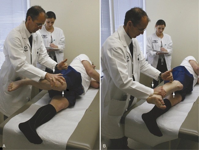
The physiotherapist will likely conduct a thorough examination including:
- Assessment of reflexes, flexibility, and range of motion
- Evaluation of muscle strength and tension
- Analysis of standing and walking patterns
- Tests for balance and coordination
They may ask questions about whether your child has other medical conditions, if there’s a family history of muscular dystrophy or autism spectrum disorder, and whether your child can walk on their heels when instructed.
Following this assessment, the physiotherapist will determine whether physical therapy alone can address the toe-walking or if additional specialist referrals are needed.
How physiotherapy helps correct toe walking
Physiotherapy offers various effective approaches to help children transition from toe-walking to a normal heel-toe gait pattern. Once diagnosed, Early Physiotherapy Intervention in Toe-Walking in Kids can make a substantial difference in correcting this walking pattern before it leads to long-term complications.
Gait training for toe-walking
Gait training focuses on retraining the brain and muscles to adopt proper walking mechanics. Physical therapists utilize techniques that encourage weight-bearing on the heels during various positions and activities. For young children, this often involves playful exercises like “duck walking” – where they walk forward bearing weight only on their heels with toes off the ground. This exercise strengthens the anterior leg muscles while simultaneously stretching the posterior calf muscles.
Another effective approach involves balance beam exercises, where children walk slowly along a line or beam taking large steps with deliberate heel-toe patterns. Subsequently, this reinforces proper weight transfer from heel to toe during normal walking.
Stretching and strengthening exercises
Consistent stretching helps improve flexibility in tight calf muscles and Achilles tendons. Some effective exercises include:
- Squats: With feet flat and shoulder-width apart, children perform deep squats while keeping toes and knees pointed straight ahead. Typically performed in sets of 10 repetitions.
- Seated toe taps: Children sit on a child-sized seat with hips and knees at 90-degrees and feet flat on the ground, then repeatedly tap their toes on the ground to improve foot control.
Equally important are strengthening exercises that target weak muscle groups, particularly those in the legs and core, which help establish proper biomechanical patterns during walking.
Foot alignment correction in toddlers
Proper foot alignment forms the foundation for correcting toe-walking. As children grow, their feet and legs constantly change, sometimes leading to alignment issues that contribute to abnormal gait patterns.
Supportive shoes with firm heel counters can provide stability and encourage proper foot positioning. Henceforth, physiotherapists might recommend orthopedic rigid high-top shoes, sometimes with heel lifts, to ensure sensory input through the heel and achieve a flat-foot position.
Role of orthotic devices in toe-walking
Orthotic devices play a crucial role in toe-walking treatment. For milder cases, a rigid carbon fiber footplate attached to a foot orthosis may be sufficient. These devices limit plantar flexion and toe extension by stiffening the sole, making toe-walking physically difficult.
For moderate cases, supramalleolar orthoses (SMOs) with no cut-out for the Achilles tendon serve as a “kinesthetic reminder” during gait. In persistent cases, articulated ankle-foot orthoses (AFOs) provide more restrictive support.
Research shows that both AFOs and carbon fiber footplates significantly improve kinematics compared to baseline. Interestingly, foot orthosis treatments, though less restrictive, demonstrated better sustained improvement even when removed.
What are the long-term effects of untreated toe walking?
Untreated toe walking leaves lasting effects that can impact a child’s quality of life. Left unaddressed, this walking pattern may create significant physical challenges beyond childhood.
Impact on balance and coordination
Persistent toe walking interferes with normal balance development. Children who toe walk typically experience decreased balance and coordination. These difficulties make everyday activities challenging, from navigating uneven surfaces to climbing stairs. Over time, these limitations can hinder participation in sports and physical activities.
Risk of falls and injuries
Children who consistently toe walk face a heightened risk of falling. This increased instability creates a concerning safety issue. Research shows toe walkers have less stability during gait, making them more prone to slipping. Furthermore, limited ankle dorsiflexion predisposes children to ankle injuries.
Muscle imbalances and posture issues
Continual toe walking creates profound musculoskeletal changes. The calf muscles and Achilles tendons gradually tighten, potentially leading to abnormal posture. These muscle imbalances affect overall posture and can create compensatory movements throughout the body.
Future complications of toe-walking
As children grow, untreated toe walking may cause progressive complications. The condition can eventually make it difficult or impossible to walk flat-footed. Some children develop foot pronation and excessive tibial torsion. In severe cases, surgery might be required to correct the tightened heel cord. Beyond physical effects, children may face social challenges and self-esteem issues as they stand out among peers.
Conclusion
Toe-walking represents a common childhood development pattern that deserves proper attention and intervention when it persists beyond age two. Throughout this article, we’ve explored how Early Physiotherapy Intervention in Toe-Walking in Kids can make a significant difference in your child’s development. Parents must remember that while occasional toe-walking might appear harmless, persistent patterns without treatment could lead to serious long-term consequences affecting balance, coordination, and overall mobility.
Early detection certainly makes all the difference. Children who receive timely physiotherapy often transition to normal heel-toe walking with minimal complications, whereas untreated cases might require more intensive interventions later. Physiotherapists offer valuable expertise through specialized gait training, targeted stretching exercises, and personalized orthotic solutions that address your child’s specific needs.
We encourage you to trust your parental instincts. If you notice your child consistently walking on their toes beyond age two, scheduling an assessment with a qualified physiotherapist should become a priority. Their comprehensive evaluation will determine whether the toe-walking stems from sensory issues, muscle tightness, or potentially underlying developmental concerns.
Life offers many challenges for our children – walking shouldn’t be one of them. With proper intervention, most toe-walking children develop normal gait patterns, allowing them to run, jump, and play without limitations. Your vigilance combined with professional guidance provides the foundation for your child’s healthy development and future mobility.
Key Takeaways
Understanding toe-walking and when to seek help can prevent long-term complications and ensure your child develops healthy movement patterns.
• Toe-walking beyond age 2 requires professional evaluation, as 5-12% of children experience persistent patterns that won’t resolve naturally
• Early physiotherapy intervention prevents muscle tightness and structural changes that make correction more difficult later
• Untreated toe-walking increases fall risk and can lead to permanent ankle restrictions requiring surgical correction
• Physiotherapy uses gait training, stretching exercises, and orthotic devices to successfully retrain normal heel-toe walking patterns
• Children with toe-walking often have associated developmental delays, making comprehensive assessment crucial for optimal outcomes
Early intervention is key—most children who receive timely physiotherapy develop normal walking patterns, while delayed treatment may require more intensive interventions and potentially surgery.
FAQs
Q1. At what age should parents be concerned about toe walking? Parents should be concerned if toe walking persists beyond age 2-3. While it’s normal for toddlers to experiment with walking on their toes, consistent toe walking after this age may indicate a need for professional evaluation.
Q2. How can physiotherapy help correct toe walking? Physiotherapy can help correct toe walking through various methods, including gait training exercises, stretching and strengthening activities, and the use of orthotic devices. These techniques aim to improve muscle flexibility, strengthen weak muscle groups, and encourage proper heel-to-toe walking patterns.
Q3. What are the potential long-term effects of untreated toe walking? Untreated toe walking can lead to decreased balance and coordination, increased risk of falls and injuries, muscle imbalances, posture issues, and potential complications in foot and ankle development. It may also affect a child’s participation in sports and physical activities.
Q4. Are there any exercises parents can do at home to help with toe walking? Yes, there are several exercises parents can try at home. These include “duck walking” (walking on heels with toes up), squats, seated toe taps, and balance beam exercises. However, it’s important to consult with a physiotherapist for a personalized exercise plan.
Q5. Is toe walking always a sign of a serious problem? Not necessarily. While toe walking can be associated with certain neurological or developmental conditions, it can also occur in otherwise healthy children. However, persistent toe walking beyond age 2-3 should be evaluated by a professional to rule out underlying issues and prevent potential long-term complications.

Dr. Manu Mengi is a best orthopedic doctor in Mohali, specializing in joint pain, arthritis, and sports injuries. With qualifications in orthopedics and advanced training in joint replacement, he provides effective care for bone and joint conditions, helping patients improve mobility and manage pain with the right treatment approach.