Ozone Therapy – The Natural Solution for Chronic Joint Pain Management

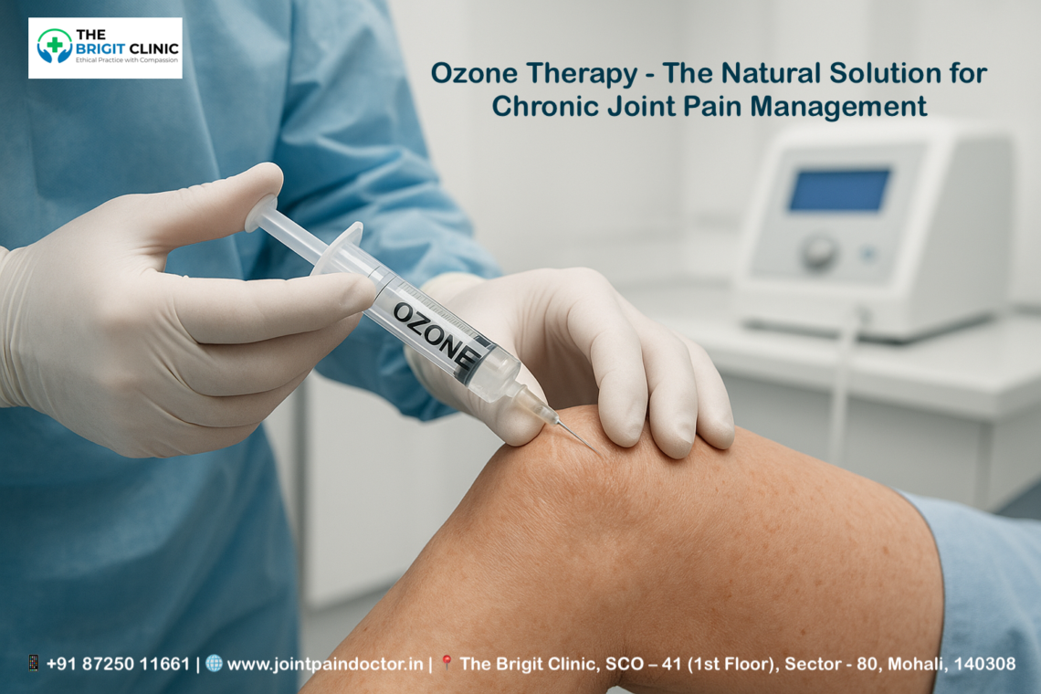
Are you struggling with persistent joint pain? Ozone Infusion Therapy has been studied and utilised for more than a century, offering a potential alternative approach to managing chronic joint discomfort. Despite the U.S. Food and Drug Administration (FDA) issuing a warning against using ozone therapy in 2019, this controversial alternative medicine practice continues to attract attention for its possible health benefits.

Although currently not FDA-approved as a medical treatment, ozone therapy demonstrates considerable therapeutic applications in the management of musculoskeletal disorders, including osteoarthritis and chronic pain syndromes. Research has shown limited but promising evidence that this treatment might help with various conditions, such as arthritis and diabetes. For example, its anti-inflammatory properties and potential to improve joint mobility make it an intriguing option for those seeking relief from joint pain. However, it’s important to note that studies have raised concerns regarding its potential toxicity, emphasising the need for stringent administration protocols to ensure safety.

In this comprehensive guide, you’ll discover what ozone therapy is, how it works for joint pain, its potential benefits, and what scientific evidence says about its effectiveness. You’ll also learn about important safety considerations to help you make an informed decision about whether this alternative treatment might be right for your joint health needs.

Considering advanced pain management solutions? Consult with the Best Orthopaedics Doctors in Mohali at The Brigit Clinic to discuss if ozone therapy is a suitable option for your joint health. Call +91 87250 11661 to book an appointment.

What is Ozone Therapy and How Does It Work?

Ozone gas consists of three oxygen atoms (O3) arranged in an unstable molecular structure, making it significantly different from the oxygen we breathe (O2). Discovered in the 1840s, ozone’s potential as more than just another atmospheric gas has evolved considerably over time. Its unique properties and therapeutic applications have made ozone therapy an intriguing option for those seeking alternative approaches to managing joint conditions.

Understanding the O3 molecule

The O3 molecule has a bent structure with an O–O–O angle of 116.78° and O–O distances of 127.2 pm. This unstable arrangement creates what scientists call “mesomeric states,” which contribute to ozone’s highly reactive nature. Unlike stable oxygen, ozone is pale blue as a gas, turns dark blue when condensed into liquid form at extremely low temperatures, and becomes violet-black when solidified.

Ozone’s instability is both a challenge and an advantage in medical applications. Due to its unstable molecular configuration, ozone readily dissolves in water and reacts quickly with biological components. This high reactivity makes it difficult to maintain concentrated forms, as ozone rapidly undergoes transient reactions with itself or water. Furthermore, ozone is approximately 10 times more soluble in water than regular oxygen, enabling it to interact effectively with body tissues and fluids.

How ozone interacts with the body

When introduced to the body, ozone functions similarly to a prodrug—it reacts with molecules to create more active substances that trigger a cascade of biological responses. Upon contact with the aqueous component of plasma, ozone dissolves and reacts with polyunsaturated fatty acids (PUFAs) and water to form hydrogen peroxide (H2O2) and lipid ozonation products (LOPs).

This moderate oxidative stress initiates several important mechanisms:

  1. Activation of antioxidant systems – Ozone stimulates the transcription factor Nrf2, which activates antioxidant response elements (ARE). This leads to increased production of antioxidant enzymes such as superoxide dismutase (SOD), glutathione peroxidase (GPx), and catalase (CAT).
  2. Enhanced oxygen metabolism – Ozone increases the transmembrane flow of oxygen and makes the mitochondrial respiratory chain more efficient. In red blood cells, it boosts glycolysis, raising ATP and 2,3-diphosphoglycerate levels, which allows haemoglobin to release oxygen more readily to tissues.
  3. Anti-inflammatory effects – Through modulation of inflammatory mediators, ozone reduces prostaglandin synthesis and decreases joint oxidative stress. It also activates key signalling pathways that support tissue regeneration and repair.

The therapeutic principle resembles hormesis, where low-dose exposure to a stressor triggers beneficial adaptive responses. Much like exercise, moderate ozone exposure can be beneficial, whereas excessive exposure can be harmful. This controlled oxidative stress preconditions cells to become more resilient against future oxidative damage, a phenomenon known as “oxidative preconditioning”.

Why ozone is considered for joint pain

Joint pain, particularly in conditions like osteoarthritis, is characterised by inflammation, oxidative stress, and tissue degeneration. Ozone therapy addresses these fundamental aspects through multiple mechanisms, making it potentially valuable for managing joint conditions.

Firstly, ozone’s ability to reduce inflammation helps mitigate the inflammatory response that drives joint pain and degeneration. Additionally, by improving tissue oxygenation and cellular metabolism, ozone therapy may support the repair processes within damaged joint tissues.

The anti-inflammatory effects are especially relevant in knee osteoarthritis, where ozone can inhibit inflammatory mediators like IL-1β and Nerve Growth Factor (NGF), which contribute significantly to pain sensations. Moreover, ozone’s capacity to stimulate antioxidant systems helps neutralise the harmful free radicals that accumulate in inflamed joints.

Clinical applications further demonstrate ozone’s versatility for joint conditions through various administration methods. Ozone can be delivered via intra-articular injections directly into affected joints, or through systemic approaches like major autohemotherapy (MAH), where blood is drawn, exposed to ozone, and reinfused. These delivery methods ensure that ozone’s therapeutic benefits reach the target areas effectively, potentially offering relief from chronic joint pain with minimal side effects when administered according to precise protocols.

Understanding the science behind treatments is key. Our Top Orthopaedic Doctors in Mohali SAS Nagar at The Brigit Clinic are experts in advanced therapies and can provide a clear, personalised explanation for your condition. Visit us in Sector 80, Mohali (Get Directions), to learn more.

How Ozone Therapy is Administered for Joint Pain

Medical professionals employ various methods to deliver ozone therapy for joint pain, each tailored to specific conditions and patient needs. The route of administration significantly impacts how ozone interacts with joint tissues and the resulting therapeutic effects.

Ozone injection therapy explained

Joint pain treatment typically involves direct ozone administration through several approaches. Practitioners primarily use intra-articular injections, where a mixture of oxygen-ozone (95-96% oxygen and 4-5% ozone) is delivered directly into the affected joint cavity. For knee osteoarthritis, a common protocol involves injecting 20 ml of ozone-oxygen mixture at a concentration of 20 μg/ml.

Periarticular injections target tissues surrounding the joint, while paravertebral injections help with spine-related pain conditions. These targeted approaches enable ozone to directly influence the biochemical environment of the joint, addressing inflammation at its source.

In cases like frozen shoulder or hip bursitis, ozone infiltration can significantly reduce both pain and inflammation through peritrochanteric routes. Before the procedure, local anaesthetics such as 1% lidocaine are often applied to minimise discomfort.

Topical and systemic applications

Beyond injections, practitioners utilise several external and whole-body approaches. Topical applications involve exposing affected areas to ozone gas under protective coverings (sometimes called “ozone sauna”) or applying ozonised solutions directly to the skin. These methods are particularly valuable for addressing superficial joint issues.

Systemic ozone delivery can occur through insufflation techniques, where ozone gas is introduced into body cavities. This approach allows ozone to be absorbed through mucous membranes, creating whole-body effects that complement localised treatments.

Autohemotherapy and IV methods

Autohemotherapy stands out as one of the most extensively studied systemic ozone administration methods. During major autohemotherapy (MAH), approximately 200 ml of blood is withdrawn from the patient, mixed with 40 ml of medical ozone at a concentration of 30 μg/ml, and then reinfused within 15 minutes. This procedure is typically performed three times weekly for two weeks.

Minor autohemotherapy involves smaller blood volumes (60-80 ml) mixed with ozone and then injected intramuscularly into the buttock. IV ozone therapy follows similar principles to other infusions, with ozone-enriched blood returning to circulation via drip methods.

A modern advancement called hyperbaric ozone/oxygen treatment modifies traditional autohemotherapy to deliver larger ozone quantities in fewer sessions. This approach can significantly reduce the number of treatments needed while maximising therapeutic benefits.

Safety protocols during administration

Proper safety measures are essential during ozone therapy. Fundamentally, ozone should never be inhaled, as this can cause severe lung irritation and fluid buildup. Medical-grade equipment with precise concentration controls and sterile application techniques is mandatory.

Qualified healthcare professionals must administer treatments in controlled clinical environments. The oxygen-ozone mixture should be produced through specialised medical generators that ensure precise concentrations. Practitioners should follow established protocols, with ozone concentrations for systemic therapy typically ranging between 10-40 μg/ml.

Most importantly, each treatment plan should be individually tailored. As no two patients are alike, dosage and administration protocols require personalisation based on specific conditions and responses.

The correct administration technique is critical for safety and effectiveness. Ensure you are in expert hands. Schedule a consultation with an Orthopedic Doctor in Mohali from our team to discuss the best administration method for you.

Ozone Therapy Benefits for Chronic Joint Conditions

Research demonstrates that Ozone Infusion Therapy offers multiple benefits for patients suffering from chronic joint conditions. Studies reveal its effectiveness across various joint disorders, making it an increasingly popular alternative treatment option.

Ozone therapy for arthritis and osteoarthritis

Clinical studies show that intra-articular ozone injections can effectively alleviate pain and inhibit cartilage degeneration in osteoarthritis (OA). OA affects over 346 million people globally and is characterised by inflammation in cartilage and synovium. In a study of patients with knee osteoarthritis, approximately 50% improvement in health status was observed after just five weekly sessions of ozone therapy. Notably, treatments using concentrations between 20-30 μg/mL have demonstrated significant pain reduction and functional improvement over six-month periods.

Reducing inflammation and oxidative stress

The therapeutic effects of ozone stem from its ability to modulate inflammation through multiple pathways. Ozone decreases the production of pro-inflammatory cytokines—particularly IL-6, IL-1β, and TNF-α—which are primarily responsible for cartilage degradation. In fact, studies have demonstrated that ozone therapy reduces these inflammatory markers at 1, 2, and 6 months after treatment, with effects more stable than steroid injections.

Consequently, ozone inhibits the NF-κB pathway while enhancing the Nrf2 pathway, which generates antioxidant response elements, including superoxide dismutase and glutathione peroxidase. This dual action creates what scientists call “oxidative preconditioning,” preparing cells to better resist future oxidative damage.

Improving joint flexibility and mobility

Patients receiving ozone treatment experience significant improvements in joint function and mobility. Clinical trials report notable increases in Lysholm scores (measuring knee function) alongside decreases in WOMAC and VAS pain scores following treatment. Essentially, these improvements occur because ozone enhances oxygen metabolism and blood circulation to affected joints.

Supporting cartilage and ligament healing

Ozone therapy actively supports tissue repair mechanisms. Research shows it reduces the release of matrix metalloproteinases (MMPs) that degrade cartilage while simultaneously stimulating chondrocytes to facilitate the synthesis of proteoglycans, glycosaminoglycans, and collagen. This balanced approach helps repair damaged joint structures and potentially slows disease progression.

Enhancing immune response in chronic disease

Beyond local effects, ozone therapy provides systemic immune benefits. It activates the immune system by functioning like an antioxidant or anti-inflammatory agent. Studies reveal that ozone can regulate immune functions, particularly enhancing macrophage activity crucial for immune reactions. Furthermore, ozone’s ability to improve liver mitochondrial function and modulate antioxidant responses may contribute to its systemic therapeutic effects.

These multifaceted benefits make Ozone Therapy worthy of consideration for those seeking alternative approaches to managing chronic joint conditions.

Ready to explore the benefits of ozone therapy for your arthritis or joint pain? Talk to the Best Orthopedic Surgeon in Mohali at The Brigit Clinic to see if you are a candidate. Find relief and improve your mobility today!

Scientific Evidence and Effectiveness in Orthopaedic Use

Scientific research increasingly supports the effectiveness of Ozone Infusion Therapy across various orthopaedic applications. Multiple clinical trials highlight its potential as both a primary and complementary treatment option.

Clinical trials on ozone therapy for knee osteoarthritis

Recent systematic reviews examining ozone therapy for knee osteoarthritis (KOA) revealed statistically significant effects in pain reduction compared to placebo groups. Intra-articular ozone at concentrations of 20-30 μg/mL demonstrated substantial improvements in both pain management and functional mobility over six-month periods. Remarkably, one double-blind randomised trial found that both 20 μg/mL and 40 μg/mL doses effectively reduced pain without significant differences between concentrations, suggesting a threshold effect where lower doses sufficiently activate therapeutic pathways.

Ozone therapy for spinal arthritis and back pain

For back pain treatment, ozone therapy has shown success rates of 70-80% in patients with disk herniation. Intramuscular paravertebral ozone injections produced Visual Analogue Scale (VAS) reductions in 79% of patients while simultaneously improving Oswestry Disability Index scores in nearly all cases. Interestingly, these treatments can be performed without computed tomography guidance or an anesthesiologist’s support, making them accessible options for many practitioners.

Post-surgical recovery and pain management

Ozone therapy shows promise in post-surgical recovery scenarios. Indeed, in one preliminary study involving patients with methicillin-resistant Staphylococcus aureus (MRSA) infections following surgery, ozone therapy substantially reduced bacterial counts by at least 2 logs CFU/mL (p = 0.00093097). Subsequently, inflammation markers decreased dramatically—C-reactive protein dropped by 98.95% (p = 0.0007) and erythrocyte sedimentation rate by 75.78% (p = 0.0002).

Comparisons with traditional treatments

When compared with conventional therapies, ozone shows comparable or superior results in several studies. In comparison with hyaluronic acid for KOA treatment, both treatments significantly improved pain, stiffness, and function at 1, 3, and 6-month follow-ups with no significant differences between groups. Nevertheless, ozone demonstrated superiority over corticosteroid injections in longer-term outcomes—at 12 weeks, ozone produced significantly better pain reduction (mean difference = 3.9, p < 0.01). Correspondingly, clinical studies found ozone marginally favoured over periradicular steroid infiltrations for medium and long-term pain management in back pain patients.

Evidence-based practice is our priority. Our Orthopedic Doctors in Mohali stay updated with the latest research to offer you the most effective treatments. Contact us to discuss the scientific evidence supporting your care plan.

Risks, Side Effects, and Regulatory Considerations

Though ozone therapy shows promise for joint pain, understanding its risks is vital for patient safety. Several adverse effects have been documented, ranging from mild to severe.

Potential side effects and contraindications

Air embolism represents a serious risk during IV ozone therapy, potentially causing stroke or heart attack. Patients commonly experience discomfort, cramping, and occasional flu-like symptoms (Herxheimer reaction) as the body responds to treatment. Altogether, contraindications include glucose-6-phosphate dehydrogenase deficiency, pregnancy, hyperthyroidism, thrombocytopenia, cardiovascular instability, and recent myocardial infarction. Patients taking ACE inhibitors should exercise caution, as sudden hypotension may occur.

Why is inhalation dangerous?

Primarily, ozone should never be inhaled. Even brief exposure to small amounts can cause severe lung damage. Initially, patients might experience burning eyes, coughing, nausea, or headaches, alongside throat irritation and chest tightness. Over time, inhaled ozone may lead to chronic conditions like asthma or COPD.

FDA stance and lack of standardisation

The FDA prohibits ozone use “in any medical condition for which there is no proof of safety and effectiveness”. Rather than approving it, the FDA has prosecuted individuals selling ozone therapy products as medical cures. Currently, no standardised protocols exist—concentrations, dosages, and administration methods vary widely across practitioners.

How to find a qualified provider

Before treatment, verify that the provider uses medical-grade equipment with precise concentration controls. Qualified practitioners should follow established protocols with ozone concentrations typically between 10-40 μg/ml for systemic therapy. Additionally, they must be trained in emergency procedures and have equipment like an Ambu, medical oxygen, and emergency medications.

Your safety is non-negotiable. It is essential to choose a qualified and experienced provider. Trust the Top Orthopedics Doctor in Mohali at The Brigit Clinic, where stringent safety protocols are always followed. Call us to address any concerns.

Conclusion

Ozone therapy represents a fascinating alternative approach for those suffering from chronic joint pain. Throughout this article, we’ve explored how this unique three-atom oxygen molecule works within the body to potentially reduce inflammation, combat oxidative stress, and support tissue healing. Although not FDA-approved, research suggests ozone therapy may offer significant benefits for conditions like osteoarthritis through various delivery methods such as intra-articular injections, topical applications, and autohemotherapy.

Scientific evidence increasingly supports ozone therapy’s effectiveness for joint conditions. Clinical trials demonstrate promising results for knee osteoarthritis patients, with many experiencing substantial pain reduction and improved mobility. Similarly, back pain sufferers have shown positive outcomes comparable to traditional treatments in several studies. Nevertheless, these benefits must be weighed against potential risks.

Safety considerations remain paramount when exploring ozone therapy. Under no circumstances should you inhale ozone gas, as this can cause severe respiratory damage. Additionally, treatment should only be sought from qualified practitioners using medical-grade equipment. While the FDA has not approved ozone for medical treatment, many patients still pursue this alternative therapy under proper medical supervision.

Before trying ozone therapy, carefully consider your specific health condition, consult with healthcare providers familiar with both conventional and alternative approaches, and thoroughly research qualified practitioners. Ultimately, ozone therapy offers an intriguing option that might complement traditional joint pain management strategies for some individuals. The decision rests with you and your healthcare team to determine if this alternative treatment aligns with your joint health needs and overall wellness goals.

Key Takeaways

Ozone therapy offers a promising alternative approach to chronic joint pain management through multiple therapeutic mechanisms, though it requires careful consideration of safety protocols and regulatory status.

• Ozone therapy reduces joint inflammation by decreasing pro-inflammatory cytokines (IL-6, IL-1β, TNF-α) and activating antioxidant pathways for tissue repair.

• Clinical studies show 50% improvement in knee osteoarthritis patients after five weekly sessions, with effects lasting up to six months.

• Multiple delivery methods exist, including intra-articular injections (20-30 μg/mL), autohemotherapy, and topical applications for targeted treatment.

• Never inhale ozone gas as it causes severe lung damage; only seek treatment from qualified practitioners using medical-grade equipment.

• FDA has not approved ozone therapy for medical use, emphasising the importance of consulting healthcare providers before pursuing treatment.

While research demonstrates ozone therapy’s potential for managing chronic joint conditions like arthritis and osteoarthritis, patients must weigh promising clinical outcomes against safety risks and regulatory considerations. The therapy’s ability to support cartilage healing and improve joint mobility makes it an intriguing complement to traditional treatments when administered properly by qualified professionals.

FAQs

Q1. What is ozone therapy, and how does it work for joint pain?

A1. Ozone therapy is a treatment that uses medical-grade ozone gas to reduce inflammation and promote healing in joints. It works by stimulating the body’s antioxidant systems, improving oxygen metabolism, and modulating inflammatory responses in affected areas.

Q2. How is ozone therapy administered for joint conditions?

A2. Ozone therapy can be administered through various methods, including intra-articular injections directly into the affected joint, autohemotherapy (where blood is mixed with ozone and reinfused), and topical applications. The specific method depends on the condition being treated and the practitioner’s approach.

Q3. What are the potential benefits of ozone therapy for chronic joint pain?

A3. Potential benefits include reduced inflammation, improved joint mobility, decreased pain, enhanced tissue repair, and better overall joint function. Some patients report significant improvements in conditions like osteoarthritis and chronic back pain after undergoing ozone therapy.

Q4. Are there any risks or side effects associated with ozone therapy?

A4. While generally considered safe when administered properly, ozone therapy can have side effects such as temporary discomfort, flu-like symptoms, and, in rare cases, more serious complications like air embolism. It’s crucial never to inhale ozone gas as it can cause severe lung damage.

About Dr. Manu Mengi – Best Orthopaedic Doctors in Mohali

Dr. Manu Mengi is a distinguished name in orthopaedics, renowned as one of the Best Orthopaedic Doctors in Mohali. Practicing at The Brigit Clinic in Sector 80, Mohali, Dr. Mengi is committed to providing compassionate and comprehensive care for a wide range of joint, bone, and musculoskeletal conditions. With a patient-centric approach, he combines advanced treatments like ozone therapy with proven surgical and non-surgical interventions to develop personalised recovery plans. If you are seeking expert guidance for chronic joint pain from a trusted Orthopaedic Doctor in Mohali, schedule a consultation with Dr. Manu Mengi to take a decisive step towards a pain-free, active life.

#OzoneTherapy #JointPainRelief #ChronicPainManagement #MohaliOrtho #BestOrthopedicSurgeonInMohali #DrManuMengi #TheBrigitClinic #OrthopedicCare #NaturalPainRelief #OzoneTreatment #ArthritisRelief #Osteoarthritis #HealthTech #AlternativeMedicine #MohaliDoctors

When Shoulder Ages – The Complete Guide to Shoulder Arthritis

Shoulder arthritis affects up to 32.8% of adults over the age of sixty, making it one of the most common causes of shoulder pain and disability in older adults. When your shoulder joint’s smooth cartilage wears down, the resulting condition can severely limit your comfort and function. Most often occurring in people over age 50, shoulder arthritis can also develop in younger individuals following an injury or trauma, such as a shoulder fracture or dislocation.

Pain is the most common symptom you’ll experience with shoulder arthritis, often accompanied by stiffness, weakness, and a noticeable grinding sensation during shoulder movement. However, you don’t need to simply accept these limitations as inevitable. From non-surgical approaches like gentle stretching and anti-inflammatory medications to surgical options for severe cases, effective treatments are available. Throughout this guide, we’ll explore everything you need to know about shoulder arthritis, including its various types, how it differs from other shoulder conditions, and the best strategies for managing your symptoms and maintaining your quality of life.

Understanding Shoulder Arthritis

The degradation of cartilage within your shoulder joint defines shoulder arthritis. Your shoulder contains two primary joints, but shoulder arthritis typically refers to the larger ball-and-socket glenohumeral joint that connects your upper arm bone (humerus) to your shoulder blade (scapula). This protective cartilage covers both the ball (humeral head) and socket (glenoid), allowing smooth movement.

What is shoulder arthritis?

Shoulder arthritis occurs when the smooth cartilage lining your joint surfaces begins to break down, eventually leading to bone-on-bone contact. This cartilage breakdown creates a rough, uneven surface that causes pain, stiffness, and reduced function. As the condition progresses, bone spurs may form, further limiting motion. Patients often report that shoulder arthritis impacts their quality of life comparably to serious conditions like congestive heart failure or diabetes.

Types: Osteoarthritis, Rheumatoid, Post-traumatic

Several distinct types of shoulder arthritis exist, each with different origins:

  • Osteoarthritis – The most common form, primarily affecting people over 50, this “wear and tear” arthritis gradually degrades cartilage through normal ageing processes. It affects up to 32.8% of patients over sixty and appears more common in women than men.
  • Rheumatoid Arthritis – An autoimmune condition where your body attacks the synovial lining of joints, causing inflammation that damages cartilage. Unlike osteoarthritis, rheumatoid arthritis often affects both shoulders simultaneously.
  • Post-Traumatic Arthritis – Develops after shoulder injuries like fractures or dislocations, which damage the cartilage surface.

Additional types include rotator cuff tear arthropathy (occurring after large rotator cuff tears) and avascular necrosis (when blood supply to the humeral head is disrupted).

Shoulder arthritis vs frozen shoulder

Despite being commonly confused, shoulder arthritis and frozen shoulder (adhesive capsulitis) are entirely different conditions. The primary distinction lies in their underlying mechanisms:

Frozen shoulder specifically involves joint stiffness and chronic pain due to thickening and tightening of the joint capsule. In contrast, shoulder arthritis stems from cartilage deterioration. Furthermore, while arthritis causes pain but still allows some joint movement, frozen shoulder severely restricts mobility regardless of pain levels.

Understanding these differences is crucial since their treatments differ significantly, despite some overlapping symptoms.

Is your shoulder stiffness more than just ageing? Don't guess about your joint health. Get a precise diagnosis from an experienced Orthopedic Doctor in Mohali. Book Your Consultation Today or call us at +91 87250 11661.

Recognising the Signs and Symptoms

Recognising symptoms early helps manage shoulder arthritis effectively. Symptoms vary between individuals, with severity not always matching the degree of cartilage damage.

Early signs of shoulder arthritis

Initially, you might notice a dull ache in your shoulder that worsens after activities like lifting or reaching. Pain typically begins intermittently, appearing only during certain movements before becoming more constant as the condition progresses. Another early indicator is mild stiffness, particularly after periods of inactivity such as sleeping. Some individuals report occasional clicking sounds when moving their arms, even without pain.

Common symptoms: pain, stiffness, grinding

Pain remains the most common symptom of shoulder arthritis. As your condition advances, discomfort may occur at rest or during the night, making sleep difficult. The pain typically feels deep within the joint and can radiate down your arm toward your elbow or wrist.

Stiffness and reduced range of motion gradually increase, making everyday tasks like bathing or dressing challenging. Many patients experience crepitus—grinding, clicking, or popping sensations—when moving their shoulders. This occurs because cartilage loss creates uneven joint surfaces where bones rub together.

Shoulder arthritis vs rotator cuff tear

Although these conditions can coexist, they affect different structures. Shoulder arthritis primarily involves cartilage deterioration, whereas rotator cuff tears damage the tendons that position and power your joint. With rotator cuff tears, you’ll typically experience more pronounced weakness, especially during overhead activities. Moreover, rotator cuff tear arthropathy represents an advanced condition combining both problems, causing severe weakness, pain, and grinding.

Shoulder arthritis vs bursitis and tendinitis

Distinguishing between these conditions requires careful assessment. Bursitis involves inflammation of fluid-filled sacs (bursae) that cushion your joints. Comparatively, tendinitis affects the tendons connecting muscles to bones. Both can cause pain similar to arthritis, but differ in several ways:

  • Location: Bursitis pain typically occurs at the top and outside of your shoulder, whereas arthritis pain originates within the joint itself.
  • Duration: Bursitis generally resolves within weeks with proper care, while arthritis progressively worsens over time.
  • Onset: Tendinitis often develops suddenly after repetitive activities, unlike the gradual onset of arthritis.
Pain and grinding in your shoulder shouldn't be ignored. Early intervention is key to preserving your mobility. Schedule an appointment with a Best Ortho Doctor in Mohali to discuss your symptoms and create a personalised management plan. Contact Our Ortho Clinic in Mohali Now.

Diagnosis and Imaging Techniques

Accurate diagnosis forms the cornerstone of effective shoulder arthritis treatment. Your doctor will employ a series of progressive diagnostic steps to pinpoint the exact nature and extent of your condition.

Physical examination and history

The diagnostic journey typically begins with a detailed conversation about your symptoms and medical history. Subsequently, your physician will conduct a thorough physical examination, checking for muscle weakness, tenderness to touch, and limited range of motion. They’ll assess both passive (assisted) and active (self-directed) shoulder movements. Notably, the doctor will listen for crepitus—a grinding sensation inside the joint during movement—which often indicates cartilage damage. The examination may include specific manoeuvres to evaluate your shoulder from multiple angles, including having you place your hands behind your head and back to assess rotation.

X-ray findings in shoulder arthritis

X-rays remain the first-line imaging technique for diagnosing shoulder arthritis. Key findings visible on X-rays include:

  • Joint space narrowing (indicating cartilage loss)
  • Bone spurs (osteophytes) along joint margins
  • Subchondral sclerosis (increased bone density)
  • Bone cysts beneath the joint surface

The axillary view provides the best angle to evaluate joint space narrowing, accordingly helping rule out dislocations. Your doctor may use the Kellgren-Lawrence score or Samilson and Prieto classification to grade the severity of your condition.

Shoulder arthritis MRI findings

Although not always necessary, MRI offers an exceptionally detailed evaluation of both soft tissues and bone. Typical MRI findings include cartilage thinning, bone marrow oedema, synovial thickening, and joint effusion. Furthermore, MRI excels at detecting rotator cuff tears that often accompany shoulder arthritis. This comprehensive assessment proves invaluable for surgical planning and detecting underlying causes of your condition.

When to consider lab tests

Laboratory tests become essential primarily when inflammatory arthritis is suspected. In certain cases, your doctor may analyse blood, urine, or joint fluid samples. Joint fluid analysis involves numbing the area before inserting a needle to withdraw fluid. These tests help differentiate between various forms of arthritis, particularly identifying inflammatory conditions like rheumatoid arthritis or ruling out infection.

Ready for a clear path to relief? Our clinic is equipped with advanced imaging technology to accurately diagnose your shoulder pain. Let the Best Orthopedician in Mohali guide your treatment. Visit our Google My Business Page to see reviews and call us to get started.

Treatment Options and Management

Treatment approaches for shoulder arthritis range from conservative options to surgical interventions, depending on pain severity and functional limitations. A comprehensive management plan typically begins with the least invasive methods first.

Non-surgical management of shoulder arthritis

First and foremost, optimising your overall health can improve the quality of life with shoulder arthritis. Regular aerobic exercise, maintaining a healthy weight, and avoiding activities that worsen pain form the foundation of management. Gentle stretching exercises help maintain range of motion, thus preventing further stiffness. Applying ice reduces inflammation, while heat can loosen stiff joints. Physical therapy often plays a crucial role in preserving mobility without aggravating symptoms.

Shoulder arthritis injection – Steroid, PRP, Hyaluronic Acid

Corticosteroid injections can provide significant short-term relief by reducing inflammation. Nevertheless, most doctors limit these to three injections yearly to prevent cartilage damage. Hyaluronic acid injections mimic your joint’s natural lubricant, potentially offering pain relief lasting up to six months. Meanwhile, platelet-rich plasma (PRP) therapy uses your own blood components to potentially promote healing, though research shows mixed results.

Pain relief for shoulder arthritis

Over-the-counter medications like acetaminophen offer basic pain control with fewer side effects than other options. Nonsteroidal anti-inflammatory drugs (NSAIDs) such as ibuprofen reduce both pain and inflammation. Certainly, medication should complement rather than replace other treatment approaches. For nighttime pain, icing the shoulder before bed may improve sleep quality.

Shoulder arthritis supplements

Certain supplements show promise for arthritis management. Glucosamine and chondroitin have produced mixed research results. In fact, turmeric/curcumin demonstrates anti-inflammatory properties, with one study showing effectiveness comparable to ibuprofen. Additionally, fish oil contains omega-3 fatty acids that help block inflammatory compounds. Remember to discuss any supplements with your physician, as they may interact with medications.

Shoulder arthritis surgical options

When conservative measures fail, surgical options include arthroscopic debridement for early arthritis, which removes loose tissue fragments. Total shoulder replacement replaces both the ball and socket with artificial components, effectively eliminating arthritis pain. For patients with combined rotator cuff tears and arthritis, reverse total shoulder replacement may provide better outcomes.

Shoulder arthritis recovery time and prognosis

Recovery from shoulder replacement typically requires wearing a sling for 4-6 weeks. Most people resume light activities after 2-3 weeks. Physical therapy continues for approximately 12 weeks, focusing initially on flexibility, then progressing to strength. Fortunately, 95% of patients experience pain-free function one year after surgery. Many prosthetic shoulders last at least 15 years, often for the remainder of a patient’s life.

From non-surgical therapies to advanced joint replacement, explore all your options for Orthopedic Treatment in Mohali. Don't let pain dictate your life. Take the first step towards a pain-free shoulder. Click here to consult with the Best Orthopedic Doctor in Mohali.

Conclusion

Living with shoulder arthritis certainly presents challenges, but knowledge empowers you to take control of your condition. Throughout this guide, we’ve explored how shoulder arthritis develops when protective cartilage deteriorates, leading to pain, stiffness, and reduced mobility. Remember that several types exist—osteoarthritis, rheumatoid arthritis, and post-traumatic arthritis—each requiring specific approaches.

Early recognition of symptoms allows for prompt intervention. Pain, stiffness, and grinding sensations warrant medical attention, especially when they persist or worsen. Your doctor will likely use physical examinations, X-rays, and sometimes MRIs to accurately diagnose your condition.

Treatment options span a wide spectrum based on your specific needs. Conservative approaches like gentle stretching, heat and ice application, and physical therapy often provide significant relief. Medications and injections serve as effective tools for managing pain and inflammation. When these methods prove insufficient, surgical interventions such as debridement or joint replacement can dramatically improve your quality of life.

Many patients worry about their future with shoulder arthritis. However, modern treatment approaches yield excellent outcomes. After surgery, most people return to normal activities within weeks, and the long-term prognosis remains positive with proper care. Whether you’re newly diagnosed or have battled shoulder pain for years, effective management strategies exist.

Shoulder arthritis doesn’t need to define your life. Armed with proper information and working alongside healthcare providers, you can minimise pain, maintain mobility, and continue enjoying meaningful activities despite this condition. Your journey with shoulder arthritis may require adjustments, but it need not end the activities that bring you joy.

Key Takeaways

Understanding shoulder arthritis empowers you to recognise symptoms early and pursue effective treatment options that can significantly improve your quality of life.

• Early recognition matters: Watch for persistent shoulder pain, stiffness after inactivity, and grinding sensations during movement—these often signal developing arthritis.

• Multiple treatment paths exist: From gentle stretching and anti-inflammatory medications to injections and surgery, effective options are available for every severity level.

• Conservative care works first: Physical therapy, ice/heat application, and maintaining a healthy weight can provide significant relief without invasive procedures.

• Surgery offers excellent outcomes: 95% of patients experience pain-free function one year after shoulder replacement, with most prosthetics lasting 15+ years.

• Don’t accept limitations: With proper diagnosis and treatment, shoulder arthritis doesn’t have to prevent you from enjoying meaningful activities and maintaining an active lifestyle.

The key is working with healthcare providers to develop a personalised management plan that matches your specific type of arthritis and lifestyle needs. Whether through conservative care or surgical intervention, effective relief is achievable.

FAQs

Q1. At what age does shoulder arthritis typically develop?

A1. Shoulder arthritis most commonly affects individuals over 50 years old. However, it can also occur in younger people, particularly following shoulder injuries or trauma. The condition tends to progress gradually over time.

Q2. What are the early warning signs of shoulder arthritis?

A2. Early signs include a dull ache in the shoulder that worsens with activity, mild stiffness (especially after periods of inactivity), and occasional clicking sounds during arm movement. As the condition progresses, pain may become more constant and affect sleep.

Q3. How is shoulder arthritis diagnosed?

A3. Diagnosis typically involves a physical examination, review of medical history, and imaging tests. X-rays are the primary diagnostic tool, showing joint space narrowing and bone spurs. In some cases, MRI scans may be used for a more detailed evaluation of soft tissues and bone.

Q4. What non-surgical treatments are available for shoulder arthritis?

A4. Non-surgical options include gentle stretching exercises, physical therapy, ice/heat application, and pain relief medications. Corticosteroid injections can provide short-term relief. Some patients find benefit from supplements like glucosamine or turmeric, though results vary.

Q5. What is the recovery process like after shoulder replacement surgery?

A5. After shoulder replacement, patients typically wear a sling for 4-6 weeks. Light activities can often resume after 2-3 weeks. Physical therapy continues for about 12 weeks, focusing first on flexibility, then strength. Most patients experience pain-free function within a year, with prosthetic shoulders often lasting 15 years or more.

Your journey to a pain-free life starts with a single step. Trust your care to a specialist dedicated to restoring your mobility. For expert Orthopedic Treatment in Mohali, reach out to us.
📞 Call Now: +91 87250 11661 | ⭐ See Patient Reviews: Our Google My Business Profile


Gout Treatment in Mohali | Dr. Manu Mengi Explains

Have you ever woken up in the middle of the night with your big toe feeling like it’s on fire? This intense pain is often a hallmark of gout, a painful form of arthritis that affects about 1-2% of adults. Finding effective Gout Treatment in Mohali is the first step toward lasting relief. Historically known as “the disease of kings,” gout has actually become more common in recent decades.

Gout occurs when urate crystals accumulate in your joints, causing inflammation and intense pain during an attack. This condition most commonly affects your big toe joint, though it can target other joints as well. High uric acid levels in your body are the primary culprit, often resulting from eating or drinking foods high in purines. Additionally, certain risk factors increase your likelihood of developing gout, including being overweight, having high blood pressure, diabetes, kidney disease, or heart failure. Men are three times more likely to develop this condition than women, though anyone can be affected.

Fortunately, with early diagnosis, appropriate treatment, and lifestyle changes, gout is one of the most controllable forms of arthritis. In this comprehensive guide, Dr. Manu Mengi explains everything you need to know about managing and preventing gout flares.

> > > CTA: Is your big toe swollen and red? Don't ignore the signs. Consult with Dr. Manu Mengi, a leading Red Painful Toe Specialist in Mohali, for an accurate diagnosis. Book your appointment today: +91 87250 11661

Understanding Gout: What It Is and How It Starts

Gout represents a complex form of inflammatory arthritis that occurs when sharp, needle-shaped crystals form in and around your joints. These crystals cause intense inflammation, resulting in sudden attacks of severe pain, swelling, and tenderness.

What is gout and how it affect joints

Gout is characterised by the formation of monosodium urate (MSU) crystals in joints and surrounding tissues. These crystals trigger highly inflammatory responses in the affected areas. The inflammation doesn’t just cause pain—it can eventually lead to joint damage if left untreated. Furthermore, gout progresses through several stages, beginning with asymptomatic hyperuricemia, followed by acute gout flares, intervals between attacks, and eventually, in some cases, chronic gout with tophi (crystal deposits under the skin).

The role of uric acid and purines

At the heart of gout is uric acid, the final product of purine metabolism. Purines are chemicals naturally found in your body and certain foods. Normally, uric acid dissolves in your blood and passes through your kidneys into urine. However, problems arise in two scenarios: either your body produces too much uric acid, or your kidneys don’t excrete enough.

Consequently, uric acid can build up in your bloodstream—a condition called hyperuricemia. When levels exceed 6.8 mg/dL (the saturation threshold), uric acid can form those painful MSU crystals. Essentially, these crystals are what cause the inflammation and intense pain of a gout attack.

Why the big toe is often the first target

The first metatarsophalangeal joint (your big toe joint) is ground zero for approximately 50% of initial gout attacks, with about 90% of patients experiencing at least one attack in this location. This preference for the big toe isn’t random—there are scientific reasons behind it.

Temperature plays a significant role in crystal formation. A reduction of even 2°C is sufficient to lower the solubility point of urate from 6.8 to 6.0 mg/dL. The big toe, being farther from your body’s core and having less tissue coverage, naturally maintains a slightly lower temperature. Additionally, this joint experiences repetitive trauma from walking, creating an ideal environment for crystal formation.

Unlike other forms of arthritis, gout is largely controllable with proper management of uric acid levels. Understanding this connection between uric acid and gout symptoms is the first step toward effective treatment.

> > > CTA: Struggling with high uric acid? Get personalised advice from the Best Gout Doctor in Mohali. Dr. Mengi creates tailored management plans to prevent crystal formation and stop the pain. Call now: +91 87250 11661

Recognising the Signs: Symptoms and Triggers

The sudden, excruciating pain of a gout attack often strikes without warning, leaving you searching for relief in the middle of the night. Recognising the symptoms early can help you seek treatment promptly and potentially reduce the severity of flares.

Gout symptoms in the foot and other joints

While the big toe is gout’s favourite target, the condition doesn’t limit itself to just one location. Gout can also affect your ankles, knees, elbows, wrists, and fingers. The pain is typically most severe within the first 4-12 hours after it begins. As gout progresses, you might experience a limited range of motion in the affected joints.

Swollen big toe joint and severe tenderness

The joint at the base of your big toe often bears the brunt of a gout attack. During a flare, this joint becomes so tender that even the light touch of a bedsheet can cause excruciating pain. You’ll notice significant swelling along with this tenderness, making it difficult to wear shoes or walk normally.

Redness & heat in joints during a flare

Beyond pain and swelling, visual cues help identify a gout attack. The affected joint typically becomes noticeably red or discoloured. Moreover, you’ll feel warmth or even a sensation like the joint is “on fire”. This inflammation is your body’s response to the sharp urate crystals irritating the joint.

Gout attack signs and how long they last

Gout flares often begin suddenly at night, with pain intense enough to wake you from sleep. A typical gout attack lasts between one and two weeks. Between flares, you usually won’t experience any symptoms. Nevertheless, without proper treatment, attacks may become more frequent and last longer over time.

Alcohol binge and other common triggers

Research shows alcohol consumption is the most common trigger for gout attacks, responsible for 14% of flares in one study. Specifically, drinking more than one alcoholic beverage in a day raises your risk of experiencing a gout attack. Beer appears to pose a greater risk than spirits, due to its high purine content.

Other common triggers include:

  • Consuming red meat or seafood
  • Dehydration
  • Physical injury or excessive activity
  • Extremely warm or cold weather

For those with gout, experts recommend avoiding alcoholic beverages entirely, despite the potential benefits of moderate consumption for other health conditions.

> > > CTA: Woken up by a sudden, fiery joint pain? You don't have to suffer through a gout flare alone. Contact Dr. Manu Mengi's Clinic in Mohali for urgent consultation and effective pain relief strategies.

What Causes Gout: Risk Factors and Underlying Conditions

Understanding what causes gout begins with identifying its root mechanism – disrupted uric acid balance in your body. Let’s explore the factors that put you at risk.

High uric acid levels and hyperuricemia

Hyperuricemia occurs when uric acid levels exceed 6.8 mg/dL in your blood. This condition happens when your body either produces too much uric acid or your kidneys don’t excrete enough. Although one-quarter of people with hyperuricemia develop gout, most remain symptom-free.

Risk factors for gout – diet, alcohol, obesity

Your dietary choices significantly impact gout risk. Foods high in purines, such as red meat, organ meats, and certain seafood, can raise uric acid levels. Beer consumption poses a greater risk than other alcoholic beverages because of its high guanosine content. Weight gain is another major factor – obesity increases your risk of gout by 3.26 times compared to a normal weight.

Genetics & gout: family history matters

Gout runs in families, with heritability estimated at approximately 60%. About 20% of people with gout have a relative with the condition. Research shows that an additive genetic score of high-risk alleles can increase gout risk up to 41-fold.

Medications that trigger gout

Certain medications raise uric acid levels in your blood. Diuretics increase the relative risk of gout by almost 80%. Other culprits include low-dose aspirin, cyclosporine, and some transplant medications.

Hormonal changes in gout (e.g., menopause)

For women, menopause significantly increases gout risk. The hormone estrogen helps eliminate uric acid through urine. After menopause, estrogen production declines, causing uric acid levels to rise.

Gout & kidney disease: a two-way link

A bidirectional relationship exists between gout and kidney disease. Kidney disease impairs uric acid excretion, raising gout risk. Conversely, high uric acid can damage the kidneys by forming stones and causing scarring.

> > > CTA: Confused about how your diet or medications affect your gout? Get a personalised Uric Acid Diet Consultation in Mohali with Dr. Mengi to understand your triggers and build a prevention plan.

Diagnosis and Treatment Options Explained

Getting an accurate diagnosis is crucial for effective gout management. Modern diagnostic techniques have improved physicians’ ability to detect and monitor this painful condition.

Ultrasound for gout detection

Ultrasound has emerged as a valuable tool for diagnosing gout, offering high-resolution imaging without radiation. The characteristic “double contour sign” (an irregular hyperechoic line over cartilage) has 43.7% sensitivity but 99% specificity for gout. Ultrasound can also identify tophaceous deposits in soft tissues and joints that might not be evident during clinical examination.

X-ray changes in chronic gout

X-rays typically show normal findings early in the disease. In chronic gout, hallmark features include marginal erosions with overhanging edges and sclerotic margins. Soft-tissue nodules (tophi) may appear as dense masses with higher attenuation than surrounding tissues.

Corticosteroids in gout treatment

Corticosteroids effectively treat acute gout flares, especially for patients who cannot tolerate NSAIDs. Options include oral prednisone (30-40mg daily for 5 days), intravenous administration, or direct joint injections. For single-joint gout, intra-articular injections offer fewer systemic side effects.

Gout treatment without medications

Non-pharmaceutical approaches include applying ice to affected joints, resting the inflamed area, and drinking plenty of water. Maintaining a healthy weight through regular exercise helps prevent flares, with low-impact activities like walking or swimming being ideal.

Managing chronic gout vs acute gout

Acute gout requires rapid inflammation control within 24 hours of symptom onset. Chronic gout management focuses on lowering serum uric acid below 6 mg/dL through long-term therapy. This distinction is essential—treating inflammation during flares differs from preventing future attacks through urate-lowering strategies.

> > > CTA: Need a precise diagnosis? As a leading Uric Acid Treatment Doctor in Mohali, Dr. Mengi uses advanced ultrasound imaging to accurately detect gout crystals and create a targeted treatment strategy for both acute and chronic gout. Visit our clinic to know more.

Conclusion

Gout, while extremely painful, remains one of the most manageable forms of arthritis when properly addressed. Understanding the connection between uric acid levels and gout attacks enables you to take control of this condition. Certainly, the buildup of urate crystals causes those excruciating flares, but knowledge about triggers like alcohol consumption, purine-rich foods, and certain medications gives you the power to prevent attacks.

Therefore, working closely with healthcare providers becomes essential for proper diagnosis and treatment. Advanced diagnostic tools, such as ultrasound, can detect gout even before visible symptoms appear. After diagnosis, treatment options range from medications that reduce inflammation during acute attacks to long-term strategies for lowering uric acid levels below the crystal-forming threshold.

Besides medical interventions, lifestyle modifications play a crucial role in gout management. Weight loss, adequate hydration, and avoiding high-purine foods significantly reduce your risk of future flares. Additionally, limiting alcohol consumption, particularly beer, helps maintain lower uric acid levels.

Remember that gout management requires both immediate relief during attacks and ongoing prevention between episodes. Though historically called “the disease of kings,” gout does not have to rule your life. With early detection, appropriate treatment, and consistent lifestyle adjustments, you can effectively manage this condition and maintain joint health for years to come.

Key Takeaways

Understanding gout empowers you to manage this painful but controllable form of arthritis effectively through proper diagnosis, treatment, and lifestyle modifications.

• Gout occurs when uric acid crystals form in joints, most commonly affecting the big toe first due to lower temperature and repetitive trauma from walking.

• Alcohol consumption is the top trigger for gout attacks, with beer posing a higher risk than other alcoholic beverages due to its purine content.

• Early diagnosis using ultrasound and X-rays enables targeted treatment, distinguishing between acute flare management and chronic prevention strategies.

• Lifestyle changes like weight loss, hydration, and avoiding high-purine foods significantly reduce attack frequency and severity.

• Gout is highly manageable when uric acid levels stay below 6 mg/dL through medication, diet modifications, and consistent medical monitoring.

With proper understanding of triggers, symptoms, and treatment options, gout doesn’t have to control your life. The key lies in working with healthcare providers to develop a comprehensive management plan that addresses both immediate pain relief and long-term prevention strategies.

> > > CTA: Ready to take control of your gout? Partner with Dr. Manu Mengi Gout Specialist Mohali, for a comprehensive management plan that combines advanced treatment with sustainable lifestyle changes. Your journey to a pain-free life starts here.

FAQs

Q1. What exactly is gout, and how does it affect the body?

A1. Gout is a form of inflammatory arthritis caused by the buildup of uric acid crystals in joints, most commonly in the big toe. It results in sudden attacks of severe pain, swelling, and tenderness. Gout occurs when the body either produces too much uric acid or the kidneys don’t excrete enough, leading to crystal formation in joints.

Q2. What are the most common triggers for a gout attack?

A2. The most common trigger for gout attacks is alcohol consumption, particularly beer, due to its high purine content. Other triggers include consuming red meat or seafood, dehydration, physical injury, and extreme weather conditions. Avoiding these triggers, especially limiting alcohol intake, can help reduce the frequency of gout flares.

Q3. How can I quickly relieve gout symptoms at home?

A3. To quickly relieve gout symptoms at home, apply ice to the affected joint, rest the inflamed area, and drink plenty of water. Some people find cherry juice helpful in reducing uric acid levels. However, it’s important to consult with a healthcare provider for proper treatment, as these methods provide temporary relief but don’t address the underlying cause.

Q4. Are there any non-medication approaches to managing gout?

A4. Yes, there are several non-medication approaches to managing gout. These include maintaining a healthy weight through regular low-impact exercise like walking or swimming, staying well-hydrated, and avoiding foods high in purines. Lifestyle modifications play a crucial role in preventing gout flares and managing the condition long-term.

Q5. How is gout diagnosed, and what treatment options are available?

A5. Gout is typically diagnosed through a combination of clinical symptoms, blood tests to measure uric acid levels, and imaging techniques like ultrasound or X-rays. Treatment options include medications to reduce inflammation during acute attacks, such as NSAIDs or corticosteroids, and long-term therapies to lower uric acid levels. The goal is to bring serum uric acid below 6 mg/dL to prevent future attacks and complications.

Dr. Manu Mengi – Gout Specialist Mohali

If you are looking for expert and compassionate care for gout in Mohali, look no further than Dr. Manu Mengi. Renowned as one of the Best Orthopedic Doctors in Chandigarh Mohali, Dr. Manu Mengi specialises in the diagnosis and management of gout and other joint pain disorders. His patient-centric approach combines cutting-edge diagnostic technology with evidence-based treatment protocols to provide both immediate relief and long-term prevention strategies. Whether you’re experiencing your first painful flare or struggling with chronic gout, Dr. Mengi and his team are dedicated to helping you regain your mobility and live a pain-free life. Schedule a consultation at his Mohali clinic to begin your journey toward effective gout management.

Book Appointment - Call Us: +91 87250 11661 | Find Our Clinic: Get Directions


Rotator Cuff Tear vs. Frozen Shoulder: A Doctor’s Guide to Spotting the Difference

Rotator Cuff Tear vs. Frozen Shoulder | Dr. Manu Mengi

Rotator Cuff Tear vs. Frozen Shoulder confusion is something I encounter frequently in my practice, with many patients mistakenly self-diagnosing their shoulder pain. These two conditions are often confused with each other, yet they present distinct symptoms and have significantly different recovery timelines.

In my experience, understanding the key differences can help you identify your condition more accurately. Rotator cuff tears typically have a sudden onset, particularly after activities like lifting something heavy or falling. At the same time, frozen shoulder develops gradually with pain that slowly worsens over weeks or months. The most telling difference? If you have a rotator cuff tear, you can likely lift your shoulder without assistance. However, with a frozen shoulder, you’ll face difficulty raising your arm by yourself.

Throughout this guide, I’ll walk you through the specific symptoms, causes, and treatments for both conditions. We’ll explore why frozen shoulder involves progressive stiffness accompanied by dull, aching pain (especially at night), while rotator cuff tears cause sharper, more localised pain when lifting or rotating the arm. By the end, you’ll understand why that stiffness in your shoulder joint might indicate one condition over the other.

This self-diagnosis often leads to anxiety. If you're in Mohali and struggling with shoulder pain, the most important step is to get a professional evaluation. Book an appointment with a specialist to get an accurate diagnosis and peace of mind.

Understanding the Shoulder: Anatomy and Function

The shoulder represents one of the most complex and mobile joints in the human body. To fully understand the differences between rotator cuff injuries and frozen shoulder, we must first examine the intricate architecture that allows for this remarkable range of motion. Unlike other joints that prioritise stability, the shoulder sacrifices some stability for mobility, making it uniquely susceptible to certain conditions.

The role of the rotator cuff in shoulder movement

The rotator cuff forms the cornerstone of shoulder function – a group of four muscles and their tendons that surround the shoulder joint like a protective collar. This muscular quartet connects your shoulder blade (scapula) to your upper arm bone (humerus) and works together to enable precise movement while maintaining joint stability.

The four muscles that make up this critical structure include:

  • Supraspinatus: Initiates the first 15 degrees of arm abduction (lifting your arm away from your body)
  • Infraspinatus: Powers external rotation of the arm away from your body
  • Teres minor: Assists with external rotation and stabilisation
  • Subscapularis: Enables internal rotation (turning your arm toward your body)

A helpful mnemonic for remembering these muscles is “SITS” – Supraspinatus, Infraspinatus, Teres minor, and Subscapularis.

The primary biomechanical role of these muscles extends beyond their movement function. They compress the head of the humerus against the shallow glenoid cavity of the scapula, creating what doctors call “dynamic stability”. Without this muscular support, the disproportionate size between the humeral head and the glenoid fossa (a 4:1 surface area ratio) would make the joint highly unstable.

Furthermore, these muscles don’t work in isolation. For example, during abduction, the supraspinatus handles the initial 0-15 degrees of movement, then the deltoid muscle takes over from 15-90 degrees. Finally, the scapula must rotate to achieve full range of motion, which is accomplished by the trapezius and serratus anterior muscles.

What is the shoulder capsule, and why does it matter

Surrounding the glenohumeral joint is a fibrous sheath called the shoulder capsule. This structure extends from the anatomical neck of the humerus to the rim of the glenoid fossa, completely enveloping the joint. Unlike other joint capsules in your body, the shoulder capsule has a unique characteristic – it’s remarkably loose and lax, allowing the bones to separate by more than 2.5 cm.

This laxity isn’t a design flaw but rather a deliberate feature that permits the extensive freedom of movement characteristic of the shoulder joint. Nevertheless, this same feature also makes the joint more vulnerable to instability and injury.

The capsule doesn’t work alone in providing joint stability. It’s reinforced by:

  • The glenoid labrum – a fibrocartilaginous ring that deepens the glenoid cavity
  • Ligaments that connect bones to bones
  • Tendons that connect muscles to bones
  • The rotator cuff muscles themselves, which fuse with the capsule

Additionally, fluid-filled sacs called bursae surround the capsule, facilitating smooth movement by reducing friction between the structures. The synovial membrane inside the capsule produces lubricating fluid that further aids motion.

Interestingly, when the shoulder capsule becomes damaged or inflamed, it can lead to adhesive capsulitis – commonly known as frozen shoulder. In this condition, the capsule thickens and becomes stiff, significantly restricting movement and causing pain.

How shoulder mobility is affected by injuries

Given the complexity of the shoulder’s structure, it’s not surprising that injuries can substantially impact its function. Shoulder pain affects approximately 18 million Americans each year, with rotator cuff tears accounting for a significant number of these cases.

Rotator cuff tears can occur through trauma, overuse, or age-related degeneration. These tears affect one in every four adults aged 60 years and older, with the supraspinatus tendon being most commonly involved. Risk factors include smoking, high cholesterol, and family history.

When the rotator cuff is damaged, patients typically experience:

  • Pain during overhead activities
  • Limited range of motion
  • Weakness in the shoulder
  • Difficulty with daily activities

Studies using three-dimensional motion analysis have shown that individuals with symptomatic rotator cuff tears exhibit abnormal shoulder motion when performing common daily tasks compared to those without tears. This dysfunction occurs because rotator cuff injuries compromise the joint’s dynamic stability, creating muscle force imbalances.

Alternatively, when the shoulder capsule becomes inflamed and thickened in adhesive capsulitis (also known as frozen shoulder), patients experience progressive stiffness and significant pain. This condition not only limits one movement but can also restrict motion in multiple directions simultaneously.

Research shows that patients with rotator cuff-related shoulder pain demonstrate significantly less shoulder flexion, internal and external rotation range of motion compared to individuals without shoulder problems. This finding helps explain why these conditions have such a profound impact on everyday activities.

Both conditions can make simple tasks challenging – whether reaching for objects on high shelves, putting on a coat, or even combing your hair. Understanding the underlying anatomy helps explain why these different conditions can sometimes present with similar symptoms despite having distinct causes.

As we've seen, both conditions severely impact your daily life. The key to effective treatment is a correct diagnosis. If you're experiencing limited motion or pain, consider visiting a 
Specialised Shoulder Clinic in Mohali for a comprehensive assessment.

Rotator Cuff Tear: Causes, Symptoms, and Diagnosis

Rotator cuff tears represent one of the most common shoulder injuries I diagnose in my practice. These tears are the leading cause of shoulder pain and shoulder-related disability, affecting millions of people each year. In fact, more than 2 million Americans experience some rotator cuff problem annually. While these injuries can affect people of all ages, they’re particularly common in adults over 40, with prevalence increasing significantly with age.

As a physician, I find that many patients are surprised to learn that some individuals have rotator cuff tears without experiencing any symptoms whatsoever. This silent presentation can make diagnosis challenging in certain cases.

Common causes: trauma, overuse, and ageing

Rotator cuff tears typically fall into two distinct categories: acute traumatic tears and degenerative tears. Understanding this distinction is crucial for proper treatment.

Traumatic (Acute) Tears occur suddenly as a result of a specific injury. In my experience, patients often report:

  • Falling on an outstretched arm
  • Lifting something heavy with a jerking motion
  • Experiencing sudden force while pushing or pulling
  • Sustaining an injury during a shoulder dislocation

These traumatic tears account for approximately 8% of all rotator cuff injuries. They tend to stem from higher energy mechanisms and typically cause full-thickness tears, whereas degenerative tears often begin as partial-thickness injuries. Moreover, traumatic tears generally are larger and more likely to involve the subscapularis muscle.

Degenerative (Wear-Related) Tears develop gradually over time due to natural ageing processes. These constitute the majority of rotator cuff injuries I see in my practice. Several factors contribute to these tears:

  1. Repetitive stress – Certain activities that involve repeated shoulder motions put significant strain on the rotator cuff. Sports like baseball, tennis, rowing, and weightlifting carry an increased risk of overuse. Likewise, many occupations requiring repetitive overhead movements can lead to progressive damage.
  2. Decreased blood supply – As we age, blood flow to our rotator cuff tendons diminishes. Without adequate blood supply, the body’s natural ability to repair tendon damage becomes impaired, ultimately leading to tissue breakdown.
  3. Bone spurs, also known as age-related bone overgrowth (osteophytes), can develop on the acromion, creating friction against the rotator cuff tendons when you move your arm.

Essentially, the supraspinatus tendon bears the brunt of most rotator cuff injuries, as it endures the majority of shoulder-stabilising strain. Approximately 53% of patients with supraspinatus tears also have involvement of the infraspinatus.

Concerning symptoms, patients typically experience:

  • Pain localised to the anterior shoulder or greater tuberosity area
  • Discomfort when lying on the affected shoulder
  • Pain with overhead activities
  • Night pain that interrupts sleep
  • Weakness in the affected arm
  • Crackling or popping sensations during certain movements
  • A positive painful arc sign during clinical examination

Diagnosing rotator cuff tears involves several components. First, I take a detailed history, including the mechanism of injury and activities that aggravate or relieve symptoms. Next, I perform a thorough physical examination, checking for tenderness, range of motion, and specific clinical tests designed to stress the rotator cuff tendons. Additionally, diagnostic imaging plays a crucial role in confirming the diagnosis and determining the extent.

Specifically, when patients present with an inability to elevate their arm after a traumatic event, despite normal radiographs, I maintain a high index of suspicion for rotator cuff pathology, as these injuries can often be missed during initial assessment.

While this guide helps you understand the differences, only a physical examination can confirm your condition. For expert rotator cuff tear treatment in Mohali or frozen shoulder treatment in Mohali, seeking help from a qualified orthopaedic doctor is crucial. Learn more about your Shoulder Pain Treatment Options in Mohali.

Key Takeaways

Understanding the differences between rotator cuff tears and frozen shoulder can help you identify your condition and seek appropriate treatment more effectively.

• Onset patterns differ significantly: Rotator cuff tears typically occur suddenly after trauma or lifting, while frozen shoulder develops gradually over weeks to months with progressively worsening pain.

• Movement ability is the key differentiator: With rotator cuff tears, you can usually lift your arm without assistance, but frozen shoulder severely limits your ability to raise your arm independently.

• Pain characteristics vary between conditions: Rotator cuff tears cause sharp, localised pain during lifting or rotation, while frozen shoulder presents as dull, aching pain that’s particularly troublesome at night.

• Age and activity patterns matter for diagnosis: Rotator cuff tears affect over 25% of adults aged 60+, often from repetitive overhead activities or sudden trauma. At the same time, a frozen shoulder involves inflammation and stiffness of the capsule.

• The rotator cuff’s four muscles (SITS) – Supraspinatus, Infraspinatus, Teres minor, and Subscapularis – work together to provide dynamic shoulder stability, making tears particularly disabling for daily activities.

Both conditions significantly impact quality of life; however, accurate identification of symptoms, such as pain location, movement limitations, and onset patterns, enables more targeted treatment approaches and better outcomes.

FAQs

Q1. How can I differentiate between a frozen shoulder and a rotator cuff tear?

A1. The key difference lies in your ability to move your arm. If you can lift your arm independently, it’s more likely to be a rotator cuff tear. Frozen shoulder severely limits your ability to raise your arm without assistance. Additionally, rotator cuff tears often have a sudden onset, while frozen shoulder develops gradually over time.

Q2. Is it possible for a rotator cuff tear to be misdiagnosed as frozen shoulder?

A2. Yes, these conditions can be confused. Some partial rotator cuff tears may not cause pain and can mimic symptoms of a frozen shoulder. However, a thorough medical examination, including imaging tests, can help differentiate between the two conditions.

Q3. What are the most effective treatments for frozen shoulder?

A3. Most cases of frozen shoulder improve on their own within 12 to 18 months. For severe or persistent symptoms, treatments may include physical therapy, corticosteroid injections, and in some cases, surgery. Steroid injections can be particularly effective when administered early in the progression of the condition.

Q4. What other conditions can be mistaken for a rotator cuff tear?

A4. Several shoulder conditions can present similar symptoms to rotator cuff tears, including biceps tendinitis, shoulder impingement syndrome, labral tears, arthritis, bursitis, and tendinitis. Each of these conditions affects different parts of the shoulder structure but may cause similar pain or movement limitations.

Q5. How does age affect the likelihood of developing rotator cuff tears?

A5. The risk of developing a rotator cuff tear increases significantly with age. They are particularly common in adults over 40, with more than 25% of adults aged 60 and older experiencing some form of rotator cuff tear. This is often due to natural wear and tear, decreased blood supply to the tendons, and the development of bone spurs that can irritate the rotator cuff over time.

Still unsure about your shoulder pain? Don't let it limit your life any longer. If you're looking for an Affordable Shoulder Treatment in Mohali or simply want to consult with a trusted expert, reach out to us today. Schedule a consultation to get a clear diagnosis and start your journey to recovery.
📞 Call Us: +91 87250 11661 | 📍 Find Our Clinic: Google My Business

📖 Learn More About Shoulder Pain:

Dr. Manu Mengi – Shoulder Pain Specialist in Mohali

With extensive experience in diagnosing and treating complex shoulder conditions, Dr. Manu Mengi is a leading Shoulder Pain Specialist in the Mohali and Tricity region. He is dedicated to providing personalised care, utilising accurate diagnosis and evidence-based treatment plans to help patients regain pain-free movement and improve their quality of life.

Your Complete Patient Guide to Orthopedic Management of Achilles Tendon Tear

Despite being the largest tendon in your body and capable of withstanding forces up to 10 times your body weight, the Achilles tendon is surprisingly vulnerable to complications. Understanding the Orthopedic Management of Achilles Tendon Tear is the first step toward a successful recovery. Achilles tendon ruptures are very common sports injuries, occurring most frequently in people ages 30 to 40 and affecting men more often than women.

If you’re among the “weekend warriors” who exercise intensely without regular training, you face a higher risk of tearing your Achilles than younger, well-trained athletes. Additionally, these injuries can be missed in up to 25% of cases, making proper diagnosis crucial. Whether you’re dealing with a partial or complete tear, understanding your treatment options is essential for recovery. This comprehensive guide will walk you through everything you need to know about Achilles tendon tears—from identifying symptoms and risk factors to exploring both conservative and surgical treatment approaches that can help you return to your normal activities.

For expert diagnosis and a personalised treatment plan, 
Consult the Best Orthopedic Doctor in Mohali or call +91 87250 11661

Understanding Achilles Tendon Tear

The Achilles tendon is a critical structure that plays a significant role in your daily movements. Understanding its function and how it can tear will help you better recognise and manage this injury.

What is the Achilles tendon?

The Achilles tendon, also called the calcaneal tendon, is the thickest and strongest tendon in your body. This fibrous band of tissue connects your calf muscles (gastrocnemius and soleus) to your heel bone (calcaneus). Located at the back of your lower leg, this powerful tendon spans approximately 6 to 10 inches in adults.

What makes the Achilles tendon remarkable is its incredible strength—it can support forces up to four times your body weight. This impressive capacity allows you to perform essential movements like walking, running, and jumping. When your calf muscles contract, they pull on the Achilles tendon, causing your foot to point downward (plantarflexion), which helps lift your heel off the ground during physical activities.

Partial vs complete Achilles tendon tear

Achilles tendon tears exist on a spectrum from minor damage to complete rupture. Understanding the difference between partial and complete tears is crucial for proper treatment.

In a partial tear, only a portion of the tendon fibres is damaged. Think of this like a frayed rope where some strands remain intact while others are torn. With a partial tear, you’ll experience:

  • Localised soreness around the tendon
  • Some swelling that gradually decreases
  • Full function of the ankle despite pain

A complete tear occurs when the tendon ruptures entirely, separating into two distinct parts. This severe injury results in:

  • A noticeable “pop” or snapping sound at the moment of injury
  • Complete loss of strength and function in the ankle
  • Extreme difficulty walking or exercising
  • Significant swelling around the ankle
  • In some cases, visible displacement of calf muscles

The Thompson test is commonly used by doctors to diagnose a complete rupture—when squeezing the calf fails to produce normal foot movement, it indicates a tear.

Common causes and overuse injuries

Most Achilles tendon tears happen during recreational sports or athletic activities. The tendon typically ruptures when exposed to sudden, powerful forces, especially during activities requiring quick stops, starts, and pivots—such as soccer, football, basketball, tennis, or squash.

Several specific scenarios commonly lead to tears:

  • Landing awkwardly from a jump
  • Cutting movements during sports
  • Sudden acceleration or forceful pushing off with the foot
  • Direct trauma to the tendon
  • Tripping or falling, particularly when the foot is forced upward[18]

Certain factors increase your risk of experiencing an Achilles tendon tear. As you age, the tendon naturally becomes stiffer and weaker. “Weekend warriors”—adults who exercise intensely without regular training—face a higher risk than consistently trained athletes.

Furthermore, medical conditions like inflammatory diseases, diabetes, obesity, and certain medications (including fluoroquinolone antibiotics and corticosteroids) can weaken the tendon structure. Mechanical issues such as tight calf muscles, improper footwear, and training errors also contribute to vulnerability.

Overall, Achilles tendon ruptures affect approximately 12 per 100,000 individuals, most commonly occurring between ages 40 and 50, with men experiencing them 2 to 12 times more frequently than women.

If you're active and experiencing heel pain, visit a Sports Injury Clinic Mohali for a risk evaluation. Book your appointment or call +91 87250 11661

Recognising Symptoms and Risk Factors

Recognising an Achilles tendon tear promptly can make a significant difference in your treatment outcomes. By understanding the tell-tale signs and knowing your risk profile, you might prevent a delayed diagnosis that occurs in up to 25% of cases.

Sudden pop in the back of the ankle

The most distinctive sign of an Achilles tendon rupture is experiencing (and sometimes hearing) a sudden “pop” or “snap” at the back of your ankle. This sensation is so pronounced that many mistake it for being struck from behind. The sound represents the actual moment when your tendon fibres separate.

After this characteristic pop, you’ll likely feel immediate, intense pain. The sensation resembles what would happen if someone kicked you forcefully in the lower leg. Unlike gradual onset injuries, this moment is unmistakable and marks a clear point when damage occurred.

Call your doctor immediately if you experience this sudden snap during physical activity. This symptom alone strongly suggests a complete tear rather than a minor strain, consequently requiring proper medical evaluation.

Heel pain & swelling

Following a tear, sharp, sudden pain typically develops near your heel. Initially, this pain might be unbearable, though it sometimes subsides slightly after the acute injury. The area around your Achilles tendon will swell noticeably, often accompanied by bruising along the back of the ankle.

The discomfort pattern differs from other foot conditions. With an Achilles tendon tear, pain tends to be localised specifically to the back of the ankle where the tendon attaches to your heel bone. Throughout the day, this pain might intensify with activity.

Morning stiffness presents another common symptom, where the affected area feels particularly tight and sore when you first get up. As you move around, this stiffness sometimes improves temporarily.

Calf pain after injury

Beyond the immediate heel area, pain often radiates upward into your calf muscle. This happens because the calf muscles connect directly to the Achilles tendon, creating a continuous pain pathway.

The discomfort in your calf typically worsens during activities that engage these muscles, such as climbing stairs or walking uphill. Furthermore, weakness in the leg becomes apparent when attempting to push off with the affected foot.

For partial tears, you might maintain some function despite the pain. However, with complete ruptures, standing on tiptoes or performing basic foot movements becomes nearly impossible.

Risk factors for Achilles tendon tear

Several factors increase your vulnerability to Achilles tendon tears:

Age and Gender: The peak age for ruptures occurs between 30-40 years, though some sources identify 40-50 as the highest risk period. Men experience these injuries four to five times more frequently than women.

Activity Patterns: “Weekend warriors” face significantly higher risk than regularly trained athletes. Sports involving quick stops, starts, and pivots present the greatest danger—particularly soccer, basketball, tennis, football, and racquet games.

Medical Factors: Certain conditions predispose you to tendon injuries:

  • Previous Achilles tendinopathy
  • Diabetes
  • High cholesterol or blood pressure
  • Psoriasis
  • End-stage kidney disease
  • Inflammatory arthritis

Medication Usage: Some medications weaken tendon structure, notably:

  • Fluoroquinolone antibiotics[28]
  • Corticosteroid injections
  • Oral bisphosphonates

Biomechanical Issues: Physical characteristics matter too. Having tight calf muscles, flat arches, overpronation (ankles rolling inward), or legs of different lengths all increase your risk[30].

Understanding these symptoms and risk factors helps you identify potential problems early and seek appropriate orthopaedic management for Achilles tendon tears before complications develop.

Experienced a pop or snap in your ankle? Seek immediate consultation with an Orthopedic Doctor in Mohali for Achilles Tear, call +91 87250 11661

How Achilles Tendon Tears Are Diagnosed

Getting an accurate diagnosis is essential for proper orthopaedic management of an Achilles tendon tear. Medical professionals use several examination techniques combined with imaging to confirm your injury.

Physical exam and Thompson test

Your doctor will begin by asking about your symptoms and medical history, followed by a thorough physical examination of your lower leg. During this examination, they’ll check for tenderness, swelling, and potentially feel for a gap in your tendon if it has ruptured completely.

The Thompson test (sometimes called the calf squeeze test) is a key diagnostic procedure with 96-100% sensitivity and 93-100% specificity for detecting Achilles ruptures. Here’s how it works:

  • You’ll lie face down with your feet hanging over the edge of the exam table
  • Your doctor will gently squeeze your calf muscle
  • In a healthy tendon, this causes your foot to point downward naturally
  • If your foot doesn’t move during the squeeze, it indicates a likely rupture

To confirm the diagnosis, doctors often use additional clinical signs like checking for a palpable gap in the tendon (typically 3-6cm above the heel) and assessing plantar flexion strength.

MRI vs Ultrasound in Achilles tendon tear

Both MRI and ultrasound provide valuable diagnostic information, albeit with different strengths:

Ultrasound shows the tendon in real-time and demonstrates how it responds to movement. It’s highly accurate with 95% sensitivity and 99% specificity for detecting full-thickness tears. Ultrasound is generally:

  • More cost-effective
  • Readily available
  • Excellent for detecting tendinopathy and complete ruptures

MRI creates detailed images of soft tissues and is particularly valuable for:

  • Detecting partial tears (superior to ultrasound)
  • Assessing the distance between torn tendon ends
  • Postoperative evaluation
  • Ruling out other injuries with similar symptoms

Most specialists recommend ultrasound over MRI for initial diagnosis and monitoring, though your doctor may order both depending on your specific situation.

When to see a doctor

Seek immediate medical attention if you experience:

  • A popping or snapping sound at the time of injury
  • Suddenly, severe pain in the back of your ankle
  • Difficulty walking or standing on tiptoes
  • Visible swelling around the heel area

Even if you can walk with a ruptured Achilles (which many people can), it’s crucial to see a healthcare provider promptly. Using your ankle and putting full weight on it before diagnosis can worsen the injury. Importantly, up to 20% of Achilles tendon ruptures are initially misdiagnosed, often confused with ankle sprains, making proper medical evaluation essential for effective treatment.

For advanced diagnostic imaging and expert interpretation, visit the Best Ortho Doctor in Mohali. Schedule your visit.

Treatment Options: Conservative and Surgical

Treatment decisions for Achilles tendon tears depend on several factors, including your age, activity level, and the severity of your injury. Both non-surgical and surgical approaches offer viable pathways to recovery, each with distinct advantages.

Achilles tendon tear – conservative management

Conservative treatment involves non-surgical approaches that typically include rest, immobilisation, and controlled rehabilitation. This option is often suitable for older patients, those with limited activity goals, or individuals with health conditions that increase surgical risks.

For partial tears with less than 5mm gap between ruptured tendon edges, conservative management can be particularly effective. The traditional approach involves wearing a below-knee cast in an equinus (pointed down) position for four weeks without weight-bearing, followed by a neutral position cast with weight-bearing for another four weeks.

Surgical treatment of Achilles tendon tear

Surgical intervention appears to be the preferred method for athletes and younger, active individuals. The primary benefit of surgery is a lower re-rupture rate compared to non-surgical treatment.

The procedure typically involves making an incision in the back of your leg and stitching the torn tendon together. In cases of severe degeneration, surgeons may remove damaged portions and repair the remaining healthy tendon.

Minimally invasive Achilles tendon tear surgery

This advanced technique involves a small 3-4cm incision instead of the traditional 10cm cut. Through this smaller opening, specialised instruments guide sutures into the tendon to complete the repair.

The minimally invasive approach offers several advantages:

  • Reduced wound healing issues
  • Lower infection rates
  • Less scar tissue formation
  • Faster return to normal activities

Immobilisation vs early mobilisation in Achilles tendon tear

Historically, rigid cast immobilisation for six weeks was standard practice. Nevertheless, recent research strongly supports early functional rehabilitation and mobilisation.

Studies demonstrate that early mobilisation doesn’t increase re-rupture rates. Moreover, it offers superior benefits:

  • Decreases excessive adhesion formation
  • Improves the biomechanical properties of healing tissue
  • Enhances tendon gliding function
  • Reduces joint stiffness and muscle atrophy

Medication for tendon inflammation

Pain management typically begins with over-the-counter options like ibuprofen or naproxen sodium. For persistent discomfort, prescription medications might include COX-2 inhibitors, which potentially cause fewer gastrointestinal side effects than traditional NSAIDs.

PRP Achilles tendon tear therapy

Platelet-rich plasma (PRP) therapy involves injecting a concentrated solution of your own platelets into the injured area. These platelets contain growth factors that may promote tissue repair and regeneration.

Currently, evidence regarding PRP effectiveness remains mixed. Some studies show improvements in ankle dorsiflexion angle and calf circumference, whereas others found no significant differences in patient-reported outcomes at two years post-injury.

Explore all treatment options, including Minimally Invasive Achilles Surgery in Mohali, with the Best Orthopedician in Mohali. Discuss your choices at https://jointpaindoctor.in/ or 
Call *+91 87250 11661* to learn more about the Achilles Tear Surgery Cost Mohali.

Recovery, Rehab, and Return to Activity

Full healing from an Achilles tendon tear requires a comprehensive rehabilitation approach tailored to your specific needs. The recovery journey typically spans four to six months, regardless of whether you underwent surgical or non-surgical treatment.

Physical therapy and strengthening

Physical therapy serves as the cornerstone of Achilles tendon rehabilitation. The duration varies based on injury severity—from a few weeks to several months. Your therapist will focus on three primary goals: pain relief through various modalities, restoring proper movement patterns, and rebuilding muscle strength and balance.

Eccentric exercises stand out as the most evidence-based intervention for Achilles rehabilitation. This approach, typically performed twice daily for at least 11 weeks, has been shown to reduce pain by an average of 60% across multiple clinical trials. The Alfredson protocol remains the gold standard, gradually progressing from bilateral to single-leg heel raises.

For optimal recovery, maintain a consistent exercise regimen alongside gradually increasing weight-bearing activities. Initially, you’ll use a walking boot with progressively decreasing heel wedges until reaching a neutral position, usually around 6-8 weeks post-injury.

Custom orthotics post Achilles repair

Bespoke orthotics play a valuable role in recovery by providing proper foot alignment, enhancing shock absorption, and correcting biomechanical issues that might stress your healing Achilles tendon. These devices primarily keep your heel raised, reducing the workload on the tendon while protecting against re-rupture.

Studies have demonstrated that custom foot orthoses can significantly improve symptoms in athletes with Achilles tendinopathy, with participants reporting an average 92% improvement when using high-density EVA orthotics.

Equinus contracture after Achilles tendon tear

Equinus contracture—excessive tightness limiting ankle dorsiflexion—often develops following Achilles injuries. Conservative management through physical therapy, stretching, and night splints should be attempted first. For refractory cases, surgical options include gastrocnemius lengthening, soleus fascial release, or Achilles tendon lengthening procedures.

Return to sports after Achilles tendon tear

Returning to sports requires patience—full athletic activities should be avoided for at least 6 months post-injury. The return process follows a carefully structured progression: controlled strengthening, followed by plyometric training, and finally sport-specific movements.

Before resuming competitive activities, you should achieve specific milestones: single-leg heel raise at 90% height compared to your uninjured side, normal gait mechanics, and pain-free performance of sport-specific movements. Even with optimal rehabilitation, expect some persistent strength deficits (10-30%) in the affected leg beyond the one-year mark.

Access comprehensive rehabilitation programs at our Orthopedic Hospital in Mohali for the Achilles Rupture facility. Start your recovery journey with the Best Achilles Tendon Surgeon in Mohali by calling +91 87250 11661

Conclusion

Achilles tendon tears represent serious injuries that require prompt diagnosis and appropriate treatment for optimal recovery. Throughout this guide, we’ve explored how these tears happen, their symptoms, and the available treatment approaches. Whether you choose conservative management or surgical intervention, your recovery journey demands patience and commitment to rehabilitation protocols.

Most patients can expect a full recovery period of four to six months, though some strength deficits might persist beyond the one-year mark. During this time, physical therapy will become your essential ally, particularly through eccentric strengthening exercises that have proven highly effective for tendon healing.

Remember that each case differs based on factors like age, activity level, and tear severity. Therefore, working closely with healthcare professionals remains crucial for developing a personalised treatment plan. Custom orthotics might benefit your recovery by improving foot alignment and reducing stress on your healing tendon.

Though returning to sports and normal activities takes time, a structured approach to rehabilitation significantly improves your outcomes. Above all, don’t rush this process. Your body needs adequate time to rebuild the strongest tendon in your body.

Armed with this knowledge about Achilles tendon tears, you can now make informed decisions about your care if faced with this injury. Early recognition of symptoms, prompt medical attention, and dedication to your rehabilitation program will ultimately determine your successful return to the activities you enjoy.

For a successful recovery under expert guidance, book your final consultation at https://jointpaindoctor.in/ or call +91 87250 11661

Key Takeaways

Understanding Achilles tendon tears and their proper management can significantly impact your recovery outcomes and help you make informed treatment decisions.

• Recognise the warning signs early: A sudden “pop” sound, severe heel pain, and inability to stand on tiptoes indicate a potential Achilles rupture requiring immediate medical attention.

• Both surgical and conservative treatments work: Your age, activity level, and tear severity determine the best approach—athletes often benefit from surgery while older patients may succeed with non-surgical management.

• Early mobilisation beats prolonged immobilisation: Modern rehabilitation emphasises controlled movement over extended casting, leading to better outcomes and faster functional recovery.

• Recovery takes 4-6 months minimum: Patience is crucial as rushing back to activities increases re-rupture risk—expect some strength deficits even after one year.

• Physical therapy is non-negotiable: Eccentric strengthening exercises, particularly the Alfredson protocol, form the foundation of successful rehabilitation regardless of treatment method chosen.

The key to successful Achilles tendon recovery lies in prompt diagnosis, appropriate treatment selection, and unwavering commitment to structured rehabilitation. Don’t underestimate this injury—proper management now prevents long-term complications and ensures your return to normal activities.

Ready to start your treatment? Contact the Best Orthopedic Doctor Mohali today 
or call +91 87250 11661

FAQs

Q1. What are the main symptoms of an Achilles tendon tear?

A1. The primary symptoms include a sudden “pop” or snapping sensation in the back of the ankle, intense heel pain, swelling around the affected area, and difficulty walking or standing on tiptoes.

Q2. How long does it typically take to recover from an Achilles tendon tear?

A2. Recovery usually takes 4-6 months, regardless of whether surgical or non-surgical treatment is chosen. However, some strength deficits may persist for over a year.

Q3. Is surgery always necessary for an Achilles tendon tear?

A3. Not always. The decision between surgical and conservative treatment depends on factors like age, activity level, and tear severity. Athletes often benefit from surgery, while older patients may succeed with non-surgical management.

Q4. What role does physical therapy play in Achilles tendon tear recovery?

A4. Physical therapy is crucial for recovery, focusing on pain relief, restoring proper movement, and rebuilding strength. Eccentric exercises, particularly the Alfredson protocol, are considered highly effective for rehabilitation.

Q5. When can I return to sports after an Achilles tendon tear?

A5. Full athletic activities should be avoided for at least 6 months post-injury. Before returning to competitive sports, you should achieve specific milestones like single-leg heel raises at 90% height compared to the uninjured side and pain-free performance of sport-specific movements.

About the Doctor – Dr. Manu Mengi

Dr. Manu Mengi is a highly skilled and renowned Orthopedic Surgeon in Mohali, specialising in the management and treatment of sports injuries, particularly complex Achilles tendon tears. With extensive experience and a commitment to adopting the latest surgical techniques, including minimally invasive procedures, Dr. Mengi provides personalised care to each patient. He leads a state-of-the-art Ortho Clinic in Mohali that is equipped with advanced diagnostic technology to ensure accurate assessments and the most effective treatment plans. Dedicated to helping patients return to their active lifestyles, Dr. Mengi is considered one of the best orthopedic doctors in the region for Achilles tendon repair and rehabilitation.


Book an Appointment today - 
📱 +91 87250 11661 | 🌐 Read Reviews | 📍 Get Directions