Understanding the connection between flat feet and ankle pain can help you identify the root cause of persistent discomfort and find effective relief strategies.
• Flat feet affect 25% of the population and create a biomechanical chain reaction – collapsed arches force ankles to roll inward, causing overpronation and stress throughout the lower limb kinetic chain.
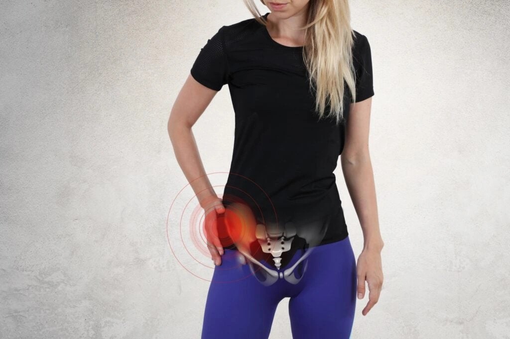
• Inner ankle pain without injury often signals flat feet problems – particularly posterior tibial tendon dysfunction, which causes pain behind the inner ankle bump and difficulty standing on tiptoes.
• Simple self-tests reveal flat feet connections – wet footprint tests, shoe wear patterns on inner edges, and visible toe count from behind indicate potential flat feet causing ankle issues.
• Conservative treatments provide significant relief for most people – targeted physiotherapy exercises, proper arch-supporting footwear, and custom orthotics address both symptoms and underlying biomechanics effectively.
• Prevention through strengthening prevents recurring problems – regular arch exercises, balance training, and early intervention for ankle sprains maintain stability and reduce future injury risk.
Most people with flat feet-related ankle pain find substantial improvement through conservative management, making early recognition and proper treatment essential for long-term comfort and mobility. Is your ankle pain actually coming from flat feet? Hidden foot mechanics might be the culprit behind your persistent discomfort. Many people experience ankle pain without realizing that their foot structure could be the root cause. In fact, flat feet affect approximately 25% of the general population, with higher prevalence among females and individuals with higher body mass index.
While most of us don’t think much about our foot arches, they significantly impact our entire lower body alignment. A recent study found that among 100 university students, 30 had flat feet, demonstrating how common this condition is even among younger adults. Importantly, the incidence of flat feet decreases with age, from 54% at age 3 to just 11.25% by ages 18-25. For those with symptomatic flexible flat feet, symptoms tend to be more severe when there’s greater forefoot abduction, creating a chain reaction of stress that travels up from the foot to the ankle.
In this guide, we’ll explore the hidden connections between flat feet and ankle pain, how to determine if your foot structure is causing your discomfort, and effective treatment options to finally find relief.
Understanding the Connection Between Flat Feet and Ankle Pain
The relationship between your foot structure and ankle discomfort runs deeper than most realize. Flat feet don’t just affect how your shoes fit—they can fundamentally alter the mechanics of your entire lower limb.
What Are Flat Feet (Pes Planus)?
Pes planus, commonly called “flat feet,” is characterized by the loss of the medial longitudinal arch of the foot. This condition affects approximately 25% of the general population, with women experiencing it more frequently than men. When someone with flat feet stands, the arches on the inside of their feet flatten under pressure, causing the feet to point outward with the entire sole touching the floor.
Flat feet typically appear in two forms:
- Flexible flat feet – The arch disappears during weight-bearing but returns when weight is removed. Most children naturally have flat feet, with arches developing as they begin walking. Nevertheless, this condition may persist into adulthood for some individuals.
- Rigid flat feet – Present in only about 1% of cases, these flat feet don’t form an arch even when not bearing weight.
The foot’s arches serve crucial biomechanical functions. They act as natural shock absorbers, adapt to uneven surfaces, store and release energy during walking, and protect the nerves and blood vessels in your feet. When these arches collapse or fail to develop properly, the entire mechanical foundation of your lower limbs becomes compromised.
How Flat Feet Affect Ankle Alignment
Flat feet fundamentally alter the alignment of your ankle and foot. Due to the collapse of the medial arch, people with pes planus typically experience heel valgus (outward pointing heel) and medial talar prominence. This misalignment doesn’t stay isolated to the foot.
The flattened arch causes your heel to point outward while simultaneously forcing your ankle to roll inward. This creates an imbalance between the invertor and evertor muscles surrounding your ankle. Studies using electromyography have confirmed that patients with flat feet show higher tibialis posterior activity and lower peroneus longus activity during midstance and propulsion phases of walking.
Furthermore, these muscle imbalances directly contribute to ankle instability. As the calcaneal pitch (heel angle) decreases, the tibialis anterior ratio increases while the peroneus longus ratio decreases—essentially creating an uneven pull across the ankle joint. This mechanical disadvantage explains why people with flat feet often experience ankle pain, especially after prolonged standing or activity.
The Kinetic Chain: Foot to Ankle Stress
The concept of the kinetic chain helps explain how foot problems create issues elsewhere in the body. Your foot serves as the foundation of your entire lower quarter kinetic chain. Therefore, when flat feet alter your normal biomechanics, the effects cascade upward.
The term “kinetic” refers to movement, while “chain” describes how individual joint movements coordinate to create functional movements like walking and running. Misalignment at any point in this chain—particularly at its foundation—can trigger problems throughout the system.
For individuals with flat feet, excessive flexibility of the subtalar joint increases pronation, creating an unstable base of support. This instability forces your body to make compensations:
- Your foot hyper-pronates, causing abnormal rear-foot eversion
- This creates abnormal loading patterns on both the subtalar and ankle joints
- The muscles surrounding your ankle work harder to maintain stability
- Your gait pattern changes to accommodate these mechanical disadvantages
Additionally, flat feet affect the somatosensory input from the foot to the brain. The anatomical changes alter how mechanoreceptors on your plantar surface transmit signals to your central nervous system. This disrupted communication further compounds movement inefficiencies and increases stress on ankle structures.
For many people, this connection between flat feet and ankle pain remains hidden until symptoms become severe. However, understanding this relationship is essential for proper diagnosis and effective treatment.
Common Types of Ankle Pain Caused by Flat Feet
Flat feet often manifest in specific ankle pain patterns that can drastically affect your daily mobility. Understanding these distinct pain presentations helps identify the root cause of your discomfort and guides appropriate treatment.
Medial Ankle Pain and Posterior Tibial Tendon Dysfunction
The inner ankle area commonly becomes painful for people with flat feet, typically due to Posterior Tibial Tendon Dysfunction (PTTD). This critical tendon runs from your calf muscle across the inside of your ankle to support your arch. In flat feet, this tendon undergoes excessive stress, leading to inflammation, weakness, and eventually degeneration.
PTTD progression follows a predictable pattern. Initially, you’ll notice pain and swelling along the tendon path behind your inner ankle bump. Over time, the tendon weakens, making it difficult to stand on tiptoes or “push off” while walking. Eventually, the arch collapses further, causing visible changes in foot structure – including a more pronounced flat foot appearance and outward turning of the heel and toes.
Without intervention, this painful cycle continues as inflammation weakens the tendon, making it more susceptible to injury. Each new injury further weakens the structure, creating ongoing stress on surrounding tissues.
Lateral Ankle Pain from Overpronation
The outer (lateral) ankle isn’t spared from flat feet complications. As the arch flattens, your ankle typically rolls inward excessively – a condition called overpronation. Subsequently, this misalignment forces your feet to point outward during walking.
Overpronation creates several issues that contribute to lateral ankle pain:
- The heel bone shifts outward relative to the ankle, creating impingement against the outer ankle bone
- Excessive pressure develops along the outer foot edge
- Increased risk of ankle instability and repeated sprains
People with flat feet are markedly more inclined to develop overpronation, which connects to numerous painful conditions including Achilles tendinitis, plantar fasciitis, shin splints, and both knee and hip pain.
Chronic Ankle Tendonitis in Flat Feet
Several tendons surrounding the ankle become vulnerable to inflammation in people with fallen arches. The most commonly affected include:
- Posterior Tibial Tendon: As mentioned earlier, this inner ankle tendon bears tremendous stress in flat feet, leading to chronic inflammation and potential tearing.
- Achilles Tendon: Connecting your calf muscle to your heel bone, this tendon undergoes increased strain with flat feet. The lack of proper arches forces greater range of motion in your feet, causing them to slide inward and forcing your Achilles tendons to work harder, becoming inflamed.
- Extensor Tendons: These tendons run along the top of your foot and can become inflamed with altered biomechanics.
Tendonitis symptoms generally include pain along the affected tendon, swelling, redness, stiffness (especially after inactivity), and gradually worsening discomfort with activity.
Peroneal Tendon Pain and Hindfoot Valgus
The peroneal tendons run along your outer ankle and provide crucial stability. Flat feet create conditions that frequently lead to peroneal tendon inflammation through several mechanisms:
First, the outward tilting of the heel (hindfoot valgus) places abnormal stress on these stabilizing tendons. Second, as your arch collapses, your foot’s biomechanics change, requiring these tendons to work harder to maintain balance.
Peroneal tendonitis symptoms include outer ankle pain, swelling, and a feeling of ankle instability. Left untreated, this condition can progress to tendon tears or subluxation (displacement from normal position).
Unlike many ankle injuries, peroneal tendon issues often develop gradually rather than from sudden trauma, making them harder to diagnose. In fact, about 60% of peroneal tendonitis cases are initially misdiagnosed as other conditions like sprains.
Why Does My Ankle Hurt with Flat Feet: Hidden Causes
Behind every painful step lies a complex mechanism that connects flat feet to ankle discomfort. Even minor changes in foot structure can trigger a cascade of biomechanical issues that affect your entire lower limb. Let’s uncover the hidden culprits behind your persistent ankle pain.
Subtalar Joint Overpronation
The subtalar joint—located just below your ankle—plays a crucial role in foot mechanics yet rarely gets the attention it deserves. In people with flat feet, this joint experiences excessive motion during walking or running.
Pronation requires a combination of dorsiflexion, abduction, and eversion movements. Though some pronation is normal and necessary, those with flat feet often develop overpronation, where the foot rolls inward excessively. This abnormal subtalar joint movement becomes a primary source of ankle pain.
Overpronation happens when your gait eventually causes the arches of your feet to flatten more than they would normally. This creates a vicious cycle: the flattened arch increases overpronation, which then stresses the ankle joint further.
The consequences of subtalar joint overpronation extend beyond mere discomfort. It increases the risk of foot and leg injuries as your body struggles to maintain proper alignment. Moreover, the soles of your shoes might reveal the first clue—if the inside section shows more wear than the outside, overpronation might be occurring.
Deltoid Ligament Stress and Ankle Ligament Strain
On the inner side of your ankle lies the deltoid ligament—a strong fibrous structure that prevents excessive outward movement of the foot. For those with flat feet, this critical ligament faces constant stress.
The deltoid ligament stabilizes the ankle joint, yet in flat-footed individuals, it remains perpetually stretched. Though deltoid ligament injuries are less common than outer ankle sprains, they’re typically more severe and require longer recovery periods.
Flat feet create the perfect conditions for deltoid ligament strain through several mechanisms:
- Excessive stress on the medial ankle due to arch collapse
- Poor foot biomechanics, specifically overpronation
- Repetitive strain from everyday activities
Consequently, neglected deltoid ligament injuries can lead to progressive deformity, posterior tibial tendon dysfunction, and worsening flat-foot conditions. This creates a dangerous feedback loop—flat feet strain the ligament, which then weakens, allowing the foot to flatten further.
Gait Abnormalities That Increase Ankle Load
Your walking pattern changes substantially with flat feet. These alterations might seem minor, yet they dramatically increase stress on your ankle joint.
Children with flat feet display a less functional gait pattern in terms of ankle kinetics than children without flat feet. Research shows that the higher the arch index value (flatter the foot), the lower the peak of ankle moment and generated ankle power during terminal stance.
People with flat feet typically demonstrate:
- Reduced ankle plantarflexion moment during push-off phase
- Lower power generation at the ankle
- Less efficient energy transfer through the foot
- Antalgic (pain-avoiding) walking patterns
These biomechanical changes result in weaker push-off ability, leading to less functional walking. Additionally, examiners often notice overpronation with ambulation in flat-footed individuals.
Ankle Pain After Standing Long Hours
Standing for extended periods puts your feet and ankles under sustained pressure. For those with flat feet, this becomes particularly problematic.
Having flat feet can cause ankle instability, leading to pain and other problems when you walk. Over time, this instability transfers too much stress to parts of your lower body that aren’t prepared to bear it.
Early symptoms might include foot pain after walking and ankle pain from overpronation. Moreover, dysfunction of the arch complex usually begins asymptomatic but gradually alters the biomechanics of the lower limbs and lumbar spine, causing an increased risk of pain and injury.
Flat feet dramatically increase your risk for chronic “rolling of the ankle”. This instability becomes particularly noticeable after long periods of standing, when muscles fatigue and structural support diminishes.
How to Tell If Your Flat Feet Are Causing Ankle Pain
Recognizing the connection between your flat feet and ankle pain requires attention to specific signs and symptoms. Many people overlook this relationship, despite clear indicators that their foot structure might be causing their discomfort. Is your ankle pain actually coming from flat feet? Hidden foot mechanics often reveal themselves through predictable patterns.
Self-Assessment: Foot Posture Signs
Checking your feet at home can provide valuable insights into whether your arch structure might be contributing to ankle discomfort. First, try the wet footprint test—wet your feet and stand on a flat surface like paper or concrete. If you see a complete imprint of your foot with no visible arch, you likely have flat feet.
Another telling sign appears when examining your feet from behind. Look at your Achilles tendon—on a flat foot, this tendon will appear arched and curved inward, with the inside of your foot positioned quite close to the ground. Additionally, check how many toes are visible when looking at your feet from behind—if you can see more than one or two toes, this suggests potential flat feet.
Your shoes also tell a revealing story. Inspect the wear patterns on your footwear—excessive wear on the inner edges of your soles often indicates overpronation, a condition closely linked to flat feet. This uneven wear pattern demonstrates how your biomechanics shift weight distribution abnormally.
When Inner Ankle Pain Points to Flat Feet
Inner ankle pain combined with specific symptoms often signals flat feet as the underlying cause. Pay attention to tenderness along the inside of your ankle, especially after standing or walking for extended periods. This pain typically follows a pattern—starting as a dull, dragging ache along the inner ankle that worsens with activity.
The posterior tibial tendon runs from your calf, behind your inner ankle bone, and connects to your arch. When this tendon becomes overworked from supporting collapsed arches, it often signals distress through pain and swelling near the inside of the ankle. A simple diagnostic test involves attempting to rise onto the ball of one foot—if your heel can’t lift properly or your foot wobbles inward during this movement, that’s a significant indicator of posterior tibial tendon dysfunction related to flat feet.
Ankle Pain Without Injury: Could It Be Your Arches?
Ankle discomfort without obvious trauma frequently stems from flat feet. Consider these key indicators:
- Pain that develops gradually rather than suddenly following an incident
- Discomfort that increases after long periods of standing or activity
- Symptoms that improve with rest but consistently return with activity
- Difficulty standing on your toes or feeling unstable on uneven surfaces
Flat feet don’t always cause problems immediately. Often, symptoms follow a progression—starting with foot fatigue and progressing to more persistent ankle pain. According to Cleveland Clinic, early symptoms might include foot pain after walking and ankle pain from overpronation, while over time, this can develop into gait disorders and chronic pain even when you’re not walking.
Importantly, if you notice that your ankle seems to turn inward as you walk, with your feet pointing outward, this pattern of overpronation strongly suggests flat feet as a contributing factor. This abnormal walking pattern transfers excess stress to your ankles, creating pain cycles that persist without proper intervention.
Treatment Options for Flat Feet Ankle Pain Relief
Finding relief for ankle pain caused by flat feet begins with targeted treatments that address both symptoms and underlying biomechanics. Effective management typically involves a combination of approaches tailored to your specific condition.
Flat Feet Physiotherapy Exercises
Strengthening exercises remain the cornerstone of flat feet treatment, directly targeting the muscles supporting your arches. Regular heel stretches reduce pain and may help correct fallen arches. Effective exercises include:
- Heel stretches against a wall, holding for 30 seconds (4 times each side)
- Tennis ball rolls under your arch for 2-3 minutes per foot
- Arch lifts where you roll weight to outer edges while lifting arches
- Towel scrunches that strengthen foot muscles through resistance
These exercises strengthen the tibialis posterior muscle and other foot stabilizers while improving overall foot posture. Indeed, some research indicates that proper exercise programming can improve arch function even in chronic cases.
Can Orthotics Fix Ankle Pain?
Orthotic devices provide substantial relief by supporting arches and improving foot alignment. They effectively distribute body weight evenly, reducing stress on sensitive areas and preventing common foot issues.
Orthotics come in two primary forms:
- Ready-made devices that offer general support
- Custom-made insoles that address your specific foot structure
Notably, arch support insoles with cushioned heel cups protect heels from impact and absorb shock at its source. For severe cases, custom-made footwear may be necessary to properly support the foot.
Best Shoes for Flat Feet Ankle Pain
Proper footwear selection proves critical for managing flat feet discomfort. Stability shoes with motion control features help support your arches and provide essential cushioning.
Look for shoes with:
- Stiff heel counters for stability
- Firm midfoot that resists twisting
- Wide fit to accommodate foot spreading

Dr. Manu Mengi is a best orthopedic doctor in Mohali, specializing in joint pain, arthritis, and sports injuries. With qualifications in orthopedics and advanced training in joint replacement, he provides effective care for bone and joint conditions, helping patients improve mobility and manage pain with the right treatment approach.