Understanding why heel pain disappears after walking helps you manage this common condition that affects 1 in 10 people at some point in their lives.
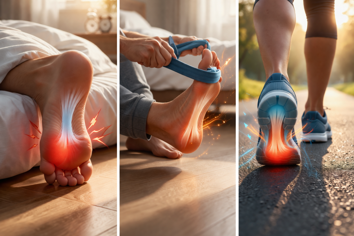
• Morning heel pain occurs because your plantar fascia tightens and cools during rest, then stretches suddenly when you stand
• Walking increases blood flow and warms the tissue, making it more flexible and reducing pain within minutes
• First-step pain that improves with movement is the hallmark symptom of plantar fasciitis, the most common cause of heel pain
• Simple morning stretches in bed, supportive footwear, and night splints can significantly reduce morning heel pain
• Pain that persists throughout the day or includes burning/tingling may indicate other conditions requiring different treatment
The key to managing heel pain lies in breaking the cycle of overnight tightening through proper stretching, supportive footwear, and maintaining tissue flexibility. Most cases improve with conservative treatments, making invasive procedures unnecessary for the majority of sufferers.
Have you ever wondered why your heel pain disappears after walking for a few minutes, even though those first steps in the morning feel unbearable? You’re not alone. Around 1 in 10 people will develop plantar fasciitis at some point throughout their life, and more than 2 million people in the U.S. are treated for it each year. This condition is the most common cause of heel pain, characterized by sharp discomfort when you first stand up. In fact, this distinctive pattern is known as first-step pain, and understanding why it happens can help you manage plantar fasciitis symptoms more effectively. We’ll explore what causes heel pain after rest and why movement brings relief.
The Morning Heel Pain Phenomenon: What You’re Experiencing
Heel pain first step in morning explained
That first step out of bed can feel like stepping on glass. The sharp, jolting sensation under your heel isn’t just typical stiffness. You might notice the pain while you’re still lying in bed, or it might strike the moment your foot touches the floor. The discomfort concentrates under the inner part of your heel, near where your arch attaches.
This sharp morning heel pain stands out from general foot aches in several distinct ways. The tenderness stays localized under the heel rather than spreading across your entire foot. Furthermore, the intensity peaks specifically during those first few steps after waking or after you’ve been sitting for extended periods. Many people describe the sensation as stabbing pain that catches them off guard each morning.
Why the pain feels worst after rest
Poor blood supply to your heel and foot area during rest explains why symptoms intensify in the morning. While you sleep, circulation to these areas becomes limited, which sets the stage for that brutal wake-up pain. The plantar fascia, that thick band of tissue running along your foot’s sole, shortens and tightens throughout the night. When you suddenly stand and stretch this tightened tissue, it responds with sharp discomfort.
Achilles tendinitis can produce similar morning symptoms. The band of tissue connecting your calf muscle to your heel bone can become inflamed, resulting in stiffness and pain in the heel area. In the light of restricted circulation during rest, these symptoms worsen overnight.
Other conditions mimic this pattern too. Stress fractures cause pain that develops gradually over days or weeks, often accompanied by swelling. Whereas plantar fasciitis pain eases after a few minutes of walking, stress fracture pain persists throughout the day. Hypothyroidism can also trigger morning heel pain through disrupted chemicals and hormones that lead to inflammation and swelling in your feet, ankles, and heels.
The pattern of pain throughout the day
The pain typically follows a predictable cycle. Those first few steps hurt the most. After walking around for several minutes, the discomfort subsides. Most people find relief within just a few minutes of activity.
The pain may return, though. After prolonged standing or extended activity, that familiar ache can creep back. Climbing stairs or working out might trigger increased pain afterward, even if you didn’t feel discomfort during the actual activity. This creates a frustrating pattern where rest brings stiffness, movement provides relief, but too much activity reignites the problem.
Stiffness in your foot or heel can make walking a challenge, particularly after waking up or sitting for long stretches. The tissue needs those initial minutes of movement to warm up and regain flexibility. Once warmed up, your heel might feel relatively normal until your next period of inactivity.
Why Heel Pain Disappears After Walking
The relief you feel after those first painful steps isn’t just in your head. Several biological processes kick in the moment you start moving, and they work together to ease your discomfort.
The plantar fascia tightens during rest
Your plantar fascia doesn’t maintain the same length throughout the day. While you sleep or sit for extended periods, this thick band of tissue becomes still and cool. The ligament shortens and contracts during these inactive hours, similar to how a rubber band left in cold temperatures loses its stretch.
The discomfort you feel stems from tiny tears in the plantar fascia that develop when tension or stress on the soft tissue becomes too great. When you suddenly stand after hours of rest, you’re forcing this tightened, cool tissue to stretch instantly. In effect, you’re pulling on tissue that hasn’t prepared for the load, which triggers that sharp, stabbing sensation.
Blood flow increases with movement
Movement changes everything about how blood reaches your feet. Walking gets blood flowing to your tendons, ligaments, and muscles. This increased circulation delivers oxygen and nutrients to the affected tissue, which helps reduce inflammation and supports the healing process.
The contrast between rest and activity makes a noticeable difference. During sleep, circulation to your heel area becomes limited. Once you start walking, your heart pumps blood more vigorously to your lower extremities. This surge in blood flow brings warmth and essential nutrients to the plantar fascia.
The tissue warms up and becomes more flexible
As you begin to walk around, you’ll notice that your heel pain decreases because the ligament has had a chance to warm up. Think of your plantar fascia like cold taffy. When it’s cool, it’s stiff and prone to tearing. Warmth makes it pliable and able to stretch without damage.
The warming process happens gradually with each step. Your muscles generate heat through contraction, and increased blood flow delivers additional warmth to the tissue. This combination makes the plantar fascia more flexible and better able to handle the stress of supporting your body weight. Hence, the pain subsides as the tissue reaches a more optimal temperature for function.
How long does the relief typically last
The pain usually goes away after walking for a few minutes. For most people, this relief comes quickly. As you get up and move, the pain normally decreases within just a few minutes of activity.
The relief isn’t permanent, though. You’ll find that the pain improves once you’ve walked around for a short time. However, the discomfort can return after you’ve walked for an extended amount of time. This creates a challenging balance where too little movement causes stiffness, but too much activity can reignite inflammation and pain.
Similarly, sitting down for another long stretch will restart the cycle. Your plantar fascia cools and tightens again during the next period of inactivity, which means you’ll likely experience that same sharp pain when you stand up. The pattern repeats throughout the day, with each period of rest followed by temporary relief through movement.
Is This First-Step Pain a Sign of Plantar Fasciitis?
Understanding plantar fasciitis symptoms
Plantar fasciitis stands as the most common cause of heel pain, affecting 10% of the general population at some point in their lives. The hallmark symptom is throbbing pain on the medial plantar heel that worsens with your first steps after rest. This pain typically decreases after further walking, though it can return with continued weight bearing.
Your doctor can identify plantar fasciitis through physical examination. Palpation of the medial calcaneal tuberosity, the bony prominence on the inner part of your heel, typically causes sharp, stabbing pain. Passive dorsiflexion, where your toes are gently pulled upward toward your shin, often elicits pain as well.
Heel spurs appear in approximately 50% of patients with plantar fasciitis, but they don’t correlate well with symptoms. These bony growths can also be found in people without plantar fasciitis. The spurs form as your bone’s response to traction forces from the plantar fascia and other foot muscles. Most people don’t feel pain from heel spurs, so their presence doesn’t necessarily explain your discomfort.
Other conditions that cause similar pain
Achilles tendinitis produces heel pain that shares similarities with plantar fasciitis but follows a different pattern. While both conditions worsen after rest due to limited circulation, Achilles tendinitis causes pain or discomfort throughout the day. In contrast, plantar fasciitis pain typically subsides after a few minutes of walking.
Stress fractures of the calcaneus develop from repetitive overload to your heel. Pain usually begins after increasing weight-bearing activities or switching to a harder walking surface. The discomfort initially occurs only during activity but can later appear at rest. You may notice swelling, and your doctor can perform a calcaneal squeeze test, which produces pain when the sides of your heel bone are compressed.
Nerve compression presents with burning, tingling, or numbness alongside heel pain. Tarsal tunnel syndrome results from compression of the posterior tibial nerve as it courses toward your heel. Patients with this condition describe pain with tingling sensations around the plantar and medial aspect of the heel. Lumbar radiculopathy at the L4-S2 levels can also cause heel pain, even without associated low back pain.
Heel pad syndrome produces a deep, bruise-like pain in the middle of your heel that can be reproduced with firm palpation. Pain intensifies when walking barefoot, on harder surfaces, or for prolonged periods.
Key differences to watch for
The anatomic location of your pain provides diagnostic clues. Plantar fasciitis pain concentrates at the medial calcaneal tubercle on the bottom inner part of your heel. Whereas tarsal tunnel syndrome typically feels most intense when standing and walking after long periods of rest, plantar fasciitis pain improves with activity.
Stress fracture pain persists throughout the day rather than improving with movement. You’ll experience point tenderness at the fracture site and pain when your heel is squeezed from side to side. Nerve-related heel pain brings additional symptoms like burning or tingling that plantar fasciitis doesn’t cause.
What Triggers Heel Pain After Sitting or Sleeping
Several specific factors trigger that sharp heel pain after sitting or sleeping. Understanding these triggers helps you identify patterns in your own discomfort and make adjustments to reduce morning stiffness.
Foot position during inactivity
The way you rest your feet in bed causes the plantar fascia ligament to tighten during sleep. Your foot naturally settles into a pointed position while you lie down, similar to how your foot would look when pressing a gas pedal. This shortened position allows the plantar fascia to contract and cool over several hours.
The muscles in your feet might feel tight while lying in bed. This isn’t just stiffness from being still. The tissue has literally shortened during your hours of rest. When you suddenly stand and force your foot into a flexed position, you’re stretching tissue that has spent the night in a relaxed, contracted state.
The role of the Achilles tendon
Tightness in the calf muscle increases pull on the tendon and the heel, which can irritate both the tendon and the fascia. Your Achilles tendon connects your calf muscle to your heel bone, handling about 93% of the plantar flexion force. Tight or weak calf muscles put you at higher risk for both Achilles tendinitis and heel pain.
Achilles tendinitis causes stiffness or tenderness in your tendon, along with leg weakness and swelling around the affected area. You may notice more discomfort after you’re active or the day after exercising, as you climb stairs or go uphill, and in the morning with improvement throughout the day.
Impact of footwear choices
Your shoe choices directly affect heel stress. Flat shoes with no support, ballet flats, flip-flops, and sandals provide little to no arch support or heel cushioning. Footwear without support causes the plantar fascia to stretch excessively with each step, increasing strain on the heel.
Worn-out shoes lose their supportive structure over time as cushioning thins and the heel counter weakens. High heels shift body weight forward, placing excessive pressure on the forefoot while tightening the Achilles tendon. When you switch back to flat shoes again, tension increases at the heel, triggering pain.
Weight and activity level factors
Patients with heel pain had a higher BMI (30.4) than those without heel pain (28.2). Excess body weight puts more pressure on your heels. Being on your feet all day for work, playing sports, or exercising on hard surfaces like warehouse floors or sidewalks can irritate your plantar fascia. Standing for long periods, especially on concrete floors, increases your likelihood of developing heel pain.
Managing Morning Heel Pain and Start-Up Pain
Most heel pain gets better over time with nonsurgical treatments that focus on easing discomfort, improving flexibility, and reducing stress on your heel.
Stretching exercises before getting out of bed
Before your feet hit the floor, spend a few minutes stretching while still in bed. Start with a point and flex warmup by sitting upright with legs extended, pointing your toes down, then flexing them back up 10 times. This warms up the soft tissues in your feet and prepares them for deeper stretches.
Next, wrap a belt or towel around the ball of one foot. Sit straight and pull the foot into a flexed position using your arm strength, holding for 20-30 seconds. Repeat 5-10 times per foot. Finish by massaging the bottom of your foot, working your thumbs from the inner arch to the outer edge for about five minutes.
Using night splints
Night splints keep your foot at a 90-degree angle overnight, preventing the plantar fascia and Achilles tendon from tightening. By maintaining this stretched position during sleep, night splints significantly reduce morning pain severity. Research shows consistent use over several weeks leads to noticeable improvements in mobility and pain relief.
Choosing proper footwear
Buy shoes with good arch support, thick soles, and extra cushioning. Avoid flats and replace worn athletic shoes before they stop supporting your feet. Over-the-counter or custom orthotics can take pressure off your heel. Proper footwear prevents excessive plantar fascia stretching with each step.
Ice and rest strategies
Apply ice twice daily for 15-20 minutes to reduce inflammation. Skip icing first thing in the morning; your feet need to warm up. Try rolling a frozen water bottle under your foot. Rest from high-impact activities like running and switch to low-impact options like swimming or cycling.
When over-the-counter treatments help
Nonsteroidal anti-inflammatory drugs like ibuprofen combined with ice packs ease pain and swelling. Don’t take NSAIDs for more than 10 days without consulting your doctor.
Conclusion
That sharp pain when you first step out of bed follows a predictable pattern. Your plantar fascia tightens during rest, circulation decreases, and the tissue cools. Once you start walking, blood flow increases, the ligament warms up, and flexibility returns. This explains why those first steps hurt so much while movement brings relief.
Above all, understanding this cycle helps you manage symptoms more effectively. Start with simple morning stretches before getting out of bed, invest in supportive footwear, and consider night splints to prevent overnight tightening. Most cases improve with these conservative treatments, so you won’t need invasive procedures. Pay attention to your body’s signals and adjust your routine accordingly for lasting relief.
FAQs
Q1. Why does heel pain feel better after walking for a few minutes?
When you start walking, blood flow increases to your heel area, delivering oxygen and nutrients that reduce inflammation. The plantar fascia tissue also warms up and becomes more flexible with movement, which is why the sharp pain you feel during those first steps gradually subsides after a few minutes of activity.
Q2. What causes the sharp heel pain when taking the first steps in the morning?
During sleep, the plantar fascia—the thick band of tissue along your foot’s sole—tightens and shortens while circulation to your heel decreases. When you suddenly stand and put weight on your foot, this cool, contracted tissue is forced to stretch quickly, causing that stabbing sensation under your heel.
Q3. Is heel pain that improves with walking always plantar fasciitis?
Not necessarily. While plantar fasciitis is the most common cause of heel pain that improves with movement, other conditions like Achilles tendinitis can produce similar symptoms. However, stress fractures cause pain that persists throughout the day rather than improving with activity, and nerve compression typically includes burning or tingling sensations alongside heel pain.
Q4. Can walking long distances make heel pain worse even if it initially feels better?
Yes. While a few minutes of walking provides temporary relief by warming up the tissue and increasing blood flow, prolonged walking or standing can reignite inflammation and pain. This creates a cycle where too little movement causes stiffness, but excessive activity can worsen symptoms and bring the discomfort back.
Q5. What can I do to reduce morning heel pain before getting out of bed?
Try stretching while still in bed by pointing and flexing your toes 10 times to warm up the tissue. Then use a belt or towel around the ball of your foot to gently pull it into a flexed position, holding for 20-30 seconds and repeating 5-10 times per foot. This prepares your plantar fascia for weight-bearing and can significantly reduce that first-step pain.
Consult with Dr. Manu Mengi for all type of Orthopedic problems he is the best Orthopedic doctor in Mohali visit for all kind of joint problems.

Dr. Manu Mengi is a best orthopedic doctor in Mohali, specializing in joint pain, arthritis, and sports injuries. With qualifications in orthopedics and advanced training in joint replacement, he provides effective care for bone and joint conditions, helping patients improve mobility and manage pain with the right treatment approach.


