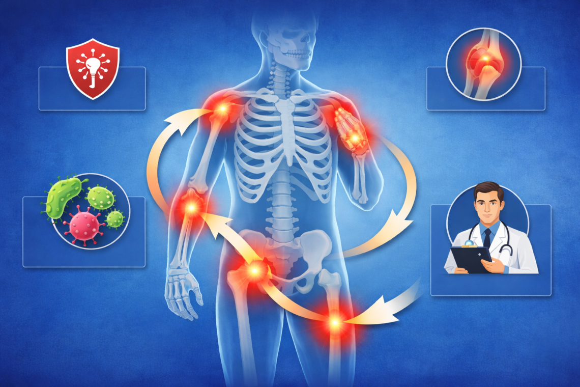
Why do I feel pain only when I start movement but it gets better after a few minutes? If you’ve experienced this pattern, you’re not alone. More than 100 million Americans are affected by chronic pain, and many deal with start-up pain in joints and muscles. Pain commonly peaks after sitting, sleeping, or long periods of rest, but fortunately, movement increases blood flow and delivers oxygen to stiff tissues. This initial movement pain doesn’t mean you should avoid activity. In fact, research shows that movement and exercise can significantly reduce pain and improve function. In this article, I’ll explain what causes this pain pattern and when you should be concerned.
What Start-Up Pain Actually Means
What Start-Up Pain Actually Means
Pain After Rest Is a Physical Response
Start-up pain refers to discomfort that occurs with your first few steps after getting up from bed or a chair and resolves after a minute or two of walking. This pattern isn’t random. Your body undergoes specific changes during periods of rest that directly affect how you feel when you begin moving again.
The term describes a protective mechanism more than a problem. When you’ve been still for hours, certain tissues in your body reach their tightest state. Muscles like the gastrocnemius become most restricted after prolonged sitting or sleeping. This tightness prevents your ankle from accommodating your body weight properly, forcing your heel to lift off the ground sooner and applying greater stress to various structures.
How Your Body Reacts to Inactivity
During sleep or extended rest, your joints remain relatively still. This lack of motion slows the circulation of synovial fluid, a gel-like substance that lubricates joints and allows for smooth, pain-free movement. When synovial fluid circulation reduces, it thickens. Upon waking, your bones receive less cushioning and may rub together, causing pain.
Blood flow to tissues decreases significantly when you’re sedentary. Reduced circulation slows healing and increases stiffness. Think of circulation as a delivery system: oxygen and nutrients can’t reach damaged tissues efficiently, and inflammatory waste products accumulate instead of being flushed away.
Physical inactivity triggers what experts call disuse syndrome, where your body’s systems deteriorate due to lack of use. Muscles begin atrophying, characterized by a reduction in muscle fiber area and overall muscle fiber count, leading to decreased muscle strength. Studies show that paraspinal muscles atrophy and increase in fat content as a result of physical inactivity.
Bones also respond to inactivity. Weight-bearing activity maintains bone density, whereas lack of movement leads to bone loss and osteoporosis development. Your cardiovascular system suffers too, with decreased oxygen intake and weakened heart function.
Why Movement Changes the Pain Signal
Once movement resumes, synovial fluid thins and circulates more efficiently. The warming up and stretching out of tight muscles during walking is the only thing that changes significantly to relieve start-up pain. This explains why pain typically disappears within minutes of activity.
Movement triggers healing processes that naturally reduce pain. Physical activity stimulates endorphin release, your body’s natural painkillers that are more potent than many medications without adverse side effects. This creates exercise-induced analgesia, essentially activating your own pain relief system.
Gentle movement helps regulate pain signals through gate control theory. When you move, you activate nerve pathways that block pain messages from reaching your brain, like closing a gate on those signals. Regular activity also helps regulate your body’s inflammatory response, reducing chronic inflammation that accompanies many pain conditions.
The dose matters as much as the exercise itself. Five minutes of daily activity beats an hour-long session once a week that triggers a flare-up. Consistency over intensity produces better results because your body needs regular stimulation to maintain fluid circulation, muscle length, and proper blood flow to painful areas.
Common Causes of Pain That Improves With Movement
Several medical conditions share this distinctive pain pattern where discomfort peaks after rest and eases with movement. Understanding which condition affects you helps determine the right approach to managing symptoms.
Early Osteoarthritis and Joint Wear
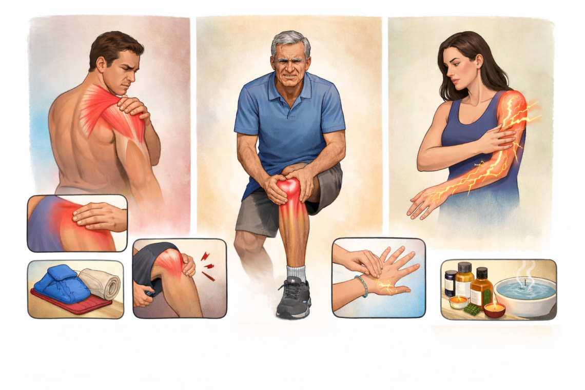
Osteoarthritis stiffness typically improves with movement. This happens when cartilage that cushions bone ends gradually wears away. Cartilage normally allows nearly frictionless joint motion, but as it breaks down, bone can eventually rub on bone. The condition affects knees, hips, hands, neck, and lower back most commonly.
Age increases your risk as normal wear and tear on joints contributes to cartilage breakdown. Joint injuries from sports or accidents years ago can also trigger osteoarthritis later. Repeated stress from certain jobs or activities places you at higher risk for developing this condition over time.
Mechanical Back Pain and Disk Stiffness
Roughly 80% of back problems are mechanical in nature. These respond best when you keep moving, even if pain persists. Mechanical back pain stems from arthritis in the spine, bulging disks, or strained muscles and ligaments.
Your spine contains over 30 segments, each with four joints and disks that can develop arthritis. These joints swell and enlarge, similar to arthritic knuckles. Disks between vertebrae act like shock absorbers, but they can bulge, rupture, or degenerate over time. Cartilage deterioration with age means bone-on-bone movement can cause pain initially, which eases as you move.
Muscle Tightness and Imbalances
Muscle imbalances develop from prolonged sitting, repetitive movements, poor posture, previous injuries, or one-sided activities. Office workers particularly face this issue. When opposing muscle groups develop unequal strength or flexibility, your body compensates in ways that lead to pain.
One side of a joint becomes stronger, shorter, and tighter while the other grows longer, looser, and weaker. This pulls joints out of position, straining surrounding structures. The resulting nerve irritation produces pain that often lessens once movement restores balance.
Plantar Fasciitis and Heel Pain
More than 2 million Americans receive treatment for plantar fasciitis each year. This condition causes inflammation in the thick tissue band running along your foot’s bottom. Pain when you stand after sleeping or sitting usually diminishes after walking for a few minutes.
Walking around after lying or sitting may ease plantar fasciitis symptoms as the ligament stretches out. The pain typically peaks first thing in the morning or when standing up after prolonged rest. Exercising or moving might temporarily relieve your pain, though it often worsens once you stop.
Mild Tendon Irritation
Tendonitis involves inflammation where connective tissues join muscles to bones. After age 40, tendons tolerate less stress, become less elastic, and tear more easily. Pain at the tendon site and surrounding area can worsen when you move, but mild cases often improve once tissues warm up and circulation increases.
Repetitive activities like gardening, painting, or sports cause most cases. Moving the affected area without pain indicates you should keep it mobile to prevent joint stiffness.
Why Your Joints and Muscles Hurt When You First Move
The mechanics behind start-up pain involve four interconnected systems that all respond poorly to rest. Understanding these mechanisms clarifies why those first steps hurt while later movement brings relief.
Joint Fluid Needs Movement to Work
Your body produces synovial fluid from blood plasma, and this thick, slippery substance contains proteins, enzymes, and high amounts of hyaluronic acid. As a matter of fact, research shows that hyaluronan secretion is directly coupled to movement. When you sit or sleep for hours, your joints produce less of this critical lubricant.
Synovial fluid serves multiple functions beyond lubrication. It transfers nutrients from your bloodstream to cartilage and other tissues it contacts. Production increases when exercise boosts circulation of fluid and nutrients to synovial membranes. The more you move, the more lubrication flows through your joints.
Without adequate movement, your joints lack the cushioning they need. Think of it like a car engine without enough oil. Parts rub together, creating friction and damage. Your joints experience similar stress when synovial fluid production drops during inactivity.
Muscles Shorten After Long Rest
Muscle fibers contract and seize up when they find it difficult to expand after prolonged inactivity. During extended periods without movement, muscles tighten due to restricted motion. Sitting too long causes hip flexors to tighten while gluteal muscles lengthen, creating an imbalance. The tightened muscles feel stiff, while lengthened muscles weaken.
Sleeping in awkward positions, sitting at a computer all day, or any period of inactivity leads to sore, tight muscles. Up to 1 in 4 people experience chronic muscle pain and stiffness. Dehydration and electrolyte imbalances worsen this tightening effect.
Inactivity leads to stiff muscles and decreased mobility and strength. Your musculoskeletal system suffers harm in the long run as muscles stiffen and weaken. Weak supporting muscles create more stress on bones and joints, ultimately worsening pain.
Blood Flow Drops During Inactivity
Blood flow and shear rate in the popliteal artery decline markedly after very short sitting periods of just 10 minutes. This rapid decrease in circulation means oxygen and nutrients can’t reach tissues efficiently. Waste products from cellular metabolism accumulate instead of being flushed away.
Reduced blood flow slows tissue healing and increases stiffness throughout your body. Sitting for prolonged periods puts you at higher risk of multiple health issues specifically because circulation drops so dramatically. Not exercising weakens bones and joints by reducing the delivery of essential nutrients needed for repair and maintenance.
Your Nervous System Stays on Alert
Nerve pain can occur if your nervous system malfunctions or sustains damage. After periods of rest, nerves may become sensitized and fire pain signals more readily when movement begins. This heightened state keeps your nervous system primed to detect potential threats to injured or irritated tissues.
The combination of reduced lubrication, shortened muscles, decreased circulation, and sensitized nerves creates the perfect storm for start-up pain. Each factor compounds the others, which explains why initial movement feels so uncomfortable before your body recalibrates.
When Start-Up Pain Is Normal vs. When to Worry
Signs This Pain Pattern Is Safe
Many who experience morning stiffness find it resolves on its own within 10 or 15 minutes. If your pain follows this timeline, you’re likely dealing with normal start-up stiffness rather than a serious condition. Safe pain typically rates between 2 and 4 out of 10 on a pain scale and decreases as you continue moving.
Pay attention to how your body responds during activity. If discomfort diminishes or disappears as you walk or move around, this indicates a mechanical issue rather than something more serious. New runners and people starting exercise programs often experience soreness the day after activity, which is normal. This type of achiness shouldn’t alarm you.
Red Flags That Need Medical Attention
Only about 1% of musculoskeletal cases involve serious pathology, but missing that small percentage can have significant consequences. Certain symptoms require immediate medical attention:
- Loss of bladder or bowel control combined with back pain
- Numbness in your groin, inner thighs, or buttocks (saddle anesthesia)
- Rapidly progressive weakness in your legs that affects walking
- Trouble breathing or dizziness alongside muscle pain
- Extreme muscle weakness that interferes with daily activities
- High fever and stiff neck
- Severe night pain that prevents sleep even after changing positions
Seek medical care if you notice signs of infection like redness and swelling around a sore muscle. A history of cancer, especially breast, prostate, lung, kidney, or thyroid cancer, combined with new pain warrants evaluation. Unexplained weight loss, fever, or night sweats alongside pain suggests possible infection or malignancy.
Age matters when assessing pain. Recent trauma combined with age over 50 increases fracture risk. A single red flag rarely signals emergency, but clusters of concerning symptoms require immediate assessment.
How Long Should Start-Up Pain Last
Start-up stiffness should dissipate within minutes of movement. If you’ve been resting for more than 7 to 10 days without significant improvement, or if pain returns every time you get active, reassess your recovery strategy. Pain that worsens during activity instead of improving signals a problem.
Muscle soreness from new exercise typically peaks around 36 to 48 hours after activity and generally resolves within 3 to 4 days. Anything persisting beyond a week deserves attention, especially if accompanied by localized, sharp bone tenderness that could indicate a stress fracture.
How to Reduce Morning Stiffness and First-Step Pain
Gentle Movement Before Getting Up
Stretching before your feet touch the floor helps wake up your body and improve circulation. While still lying down, flex your knees and feet in the air, then raise and lower your feet with knees elevated. Roll your ankles back and forth. Sit up in bed and slowly look left then right, roll your shoulders, work your elbows with biceps curls, flex your wrists, and open and close your hands several times.
Stretches That Help Joint Stiffness
Hold each stretch for 30 to 60 seconds without bouncing. A hamstring stretch involves lying on your back, bending one knee with foot flat, then lifting the other leg straight while pulling it toward your chest with your hands. Single knee to chest stretch requires pulling one bent knee toward your chest while keeping the other foot flat. Neck rolls, shoulder shrugs, and arm circles loosen upper body joints.
Heat vs. Cold for Start-Up Pain
Heat brings more blood to the affected area and reduces joint stiffness and muscle spasm. A warm shower relaxes muscles and joints, making movement easier. Alternating heat and cold therapy for 20 minutes at a time, several times daily, can relieve muscle stiffness.
Building Strength to Support Your Joints
Aim for two sessions per week that work major muscle groups. Choose low-impact options like walking, cycling, pool-based exercise, or using an elliptical machine. Daily range-of-motion exercises and stretching keep joints flexible. Water exercise classes reduce joint loading while allowing cardio and strengthening.
When to Consider Physical Therapy
Physical therapy helps when home remedies fail. A physical therapist assesses your range of motion and strength, then develops a tailored plan to improve joint mobility. Treatment may include stretching routines to break up contracted muscles, joint mobilization to improve range of motion, and guidance on improving body mechanics.
Conclusion
Start-up pain after rest is completely normal for millions of people. As a matter of fact, your body just needs a few minutes to increase blood flow, circulate synovial fluid, and warm up tight muscles. The discomfort you feel doesn’t mean you should avoid activity. Movement is your best medicine.
Pay attention to how your pain responds. If it eases within 10 to 15 minutes of walking, you’re dealing with mechanical stiffness rather than something serious. Daily stretching, gentle movement, and strengthening exercises will help reduce morning stiffness over time. However, if you notice red flags like progressive weakness, loss of bowel control, or pain that worsens with activity, seek medical attention right away.
Key Takeaways
Understanding why you feel pain when starting movement can help you manage it effectively and know when to seek medical attention.
• Start-up pain after rest is normal – it occurs because synovial fluid thickens, muscles tighten, and blood flow decreases during inactivity.
• Movement is medicine – gentle activity increases circulation, lubricates joints, and releases natural painkillers within 10-15 minutes.
• Stretch before getting up – perform ankle rolls, knee flexes, and gentle movements while still in bed to prepare your body.
• Seek help for red flags – progressive weakness, loss of bladder control, or pain that worsens with movement requires immediate medical attention.
• Build strength gradually – low-impact exercises like walking, swimming, and daily stretching prevent future stiffness and support joint health.
Remember, consistency beats intensity. Five minutes of daily movement is more beneficial than sporadic intense exercise sessions that may trigger pain flare-ups.
FAQs
Q1. Why does mechanical pain improve with movement?
Mechanical pain, which accounts for about 80% of back problems, responds best to continued movement. Your spine consists of multiple joints, and mechanical pain occurs when something restricts the movement of one or more of these joints. Keeping active helps maintain joint mobility and reduces stiffness, even when pain persists initially.
Q2. Can physical therapy make pain worse at first?
Yes, it’s normal to feel worse before getting better during physical therapy. Treatment involves mobilizing injured areas, breaking down scar tissue, and strengthening weak muscles, which can cause initial discomfort. This temporary increase in pain typically subsides after the first 2-3 weeks as your body adapts to the therapeutic exercises.
Q3. Why do I feel sore after trying a new activity even though I exercise regularly?
Soreness after a new activity occurs because you’re using your muscles in unfamiliar ways, causing tiny tears that lead to Delayed Onset Muscle Soreness (DOMS). Even if you’re fit, different movements engage muscles at varying capacities your body isn’t accustomed to. This is a normal part of building strength and adapting to new physical demands.
Q4. What symptoms should I never ignore when experiencing pain?
Seek immediate medical attention for severe abdominal pain, the worst headache you’ve ever experienced, low back pain with fever, chest pain, pain in one calf, or menstrual cramps that don’t improve with medication. Additionally, watch for loss of bladder or bowel control, numbness in the groin area, rapidly progressive leg weakness, or trouble breathing with muscle pain.
Q5. Why does my body ache after I stop moving?
Body aches after stopping movement can result from tiredness, exercise, or commonly occur with infections like the flu. However, persistent aches may also indicate underlying conditions such as fibromyalgia, arthritis, or lupus. If aches don’t resolve with rest and continue to worsen, it’s important to consult a healthcare provider for proper evaluation.
Q6. Why do I feel pain only when I start moving?
This is called start-up pain. It usually happens because joints or soft tissues become stiff after rest. Once you begin moving, lubrication improves, muscles activate, and the pain reduces.
Q7. Is it normal for pain to improve after movement?Yes, in many cases it is normal. Conditions like early osteoarthritis, muscle stiffness, or mild inflammation often feel worse at the start and improve as movement increases blood flow and flexibility.
Q8. What causes stiffness after sitting or resting?
During rest, joint fluid circulation slows and muscles tighten slightly. This leads to temporary stiffness, which improves once movement “warms up” the tissues.
Q9. Is start-up pain a sign of arthritis?
It can be. Early osteoarthritis commonly presents with pain during initial movement that improves with activity. However, it can also occur in muscle tightness or tendon issues.
Q10. Why does my knee hurt when I first stand up but then feels better?
This is often due to joint stiffness or early cartilage wear. As you start walking, the joint gets lubricated, reducing friction and easing the pain.
Q11. Why does heel pain improve after a few steps?
Classic example of plantar fasciitis. The fascia tightens during rest, causing pain with the first few steps, but loosens as you continue walking.
Q12. Can muscle problems cause start-up pain?
Yes. Tight or weak muscles can feel painful when first activated. Once they warm up and stretch slightly, the discomfort reduces.
Q13. When should I worry about this type of pain?
You should seek medical advice if:
- Pain is severe or worsening
- It does not improve with movement
- There is swelling, redness, or joint locking
- It interferes with daily activities
Q14. Does inflammation always get better with movement?
Not always. Mild inflammation may improve with movement, but active or severe inflammation (like in rheumatoid arthritis flare-ups) can worsen with activity.
Q15. How can I reduce start-up pain at home?
Simple measures include:
- Gentle warm-up exercises before activity
- Regular movement (avoid prolonged sitting)
- Stretching tight muscles
- Using heat therapy before activity
Consult for all types of joint problems and pain with Dr. Manu Manegi, best orthopedic doctor in Mohali.

Dr. Manu Mengi is a best orthopedic doctor in Mohali, specializing in joint pain, arthritis, and sports injuries. With qualifications in orthopedics and advanced training in joint replacement, he provides effective care for bone and joint conditions, helping patients improve mobility and manage pain with the right treatment approach.