Rotator Cuff Tear vs. Frozen Shoulder: A Doctor’s Guide to Spotting the Difference

Rotator Cuff Tear vs. Frozen Shoulder | Dr. Manu Mengi

Rotator Cuff Tear vs. Frozen Shoulder confusion is something I encounter frequently in my practice, with many patients mistakenly self-diagnosing their shoulder pain. These two conditions are often confused with each other, yet they present distinct symptoms and have significantly different recovery timelines.

In my experience, understanding the key differences can help you identify your condition more accurately. Rotator cuff tears typically have a sudden onset, particularly after activities like lifting something heavy or falling. At the same time, frozen shoulder develops gradually with pain that slowly worsens over weeks or months. The most telling difference? If you have a rotator cuff tear, you can likely lift your shoulder without assistance. However, with a frozen shoulder, you’ll face difficulty raising your arm by yourself.

Throughout this guide, I’ll walk you through the specific symptoms, causes, and treatments for both conditions. We’ll explore why frozen shoulder involves progressive stiffness accompanied by dull, aching pain (especially at night), while rotator cuff tears cause sharper, more localised pain when lifting or rotating the arm. By the end, you’ll understand why that stiffness in your shoulder joint might indicate one condition over the other.

This self-diagnosis often leads to anxiety. If you're in Mohali and struggling with shoulder pain, the most important step is to get a professional evaluation. Book an appointment with a specialist to get an accurate diagnosis and peace of mind.

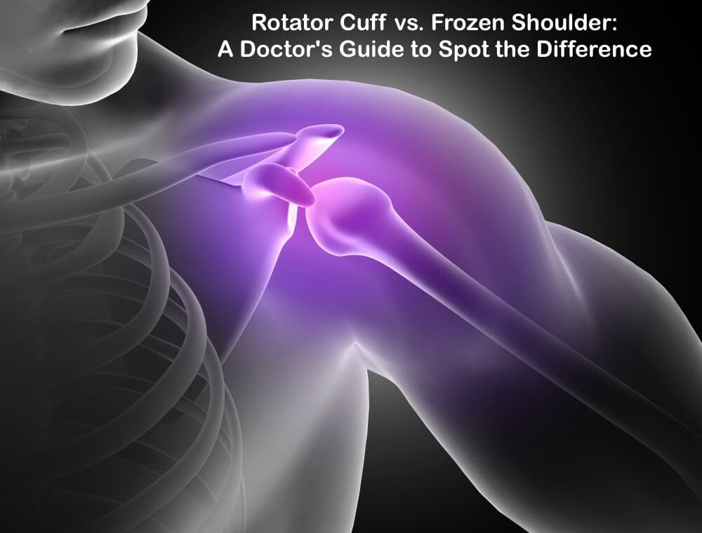
Understanding the Shoulder: Anatomy and Function

The shoulder represents one of the most complex and mobile joints in the human body. To fully understand the differences between rotator cuff injuries and frozen shoulder, we must first examine the intricate architecture that allows for this remarkable range of motion. Unlike other joints that prioritise stability, the shoulder sacrifices some stability for mobility, making it uniquely susceptible to certain conditions.

The role of the rotator cuff in shoulder movement

The rotator cuff forms the cornerstone of shoulder function – a group of four muscles and their tendons that surround the shoulder joint like a protective collar. This muscular quartet connects your shoulder blade (scapula) to your upper arm bone (humerus) and works together to enable precise movement while maintaining joint stability.

The four muscles that make up this critical structure include:

  • Supraspinatus: Initiates the first 15 degrees of arm abduction (lifting your arm away from your body)
  • Infraspinatus: Powers external rotation of the arm away from your body
  • Teres minor: Assists with external rotation and stabilisation
  • Subscapularis: Enables internal rotation (turning your arm toward your body)

A helpful mnemonic for remembering these muscles is “SITS” – Supraspinatus, Infraspinatus, Teres minor, and Subscapularis.

The primary biomechanical role of these muscles extends beyond their movement function. They compress the head of the humerus against the shallow glenoid cavity of the scapula, creating what doctors call “dynamic stability”. Without this muscular support, the disproportionate size between the humeral head and the glenoid fossa (a 4:1 surface area ratio) would make the joint highly unstable.

Furthermore, these muscles don’t work in isolation. For example, during abduction, the supraspinatus handles the initial 0-15 degrees of movement, then the deltoid muscle takes over from 15-90 degrees. Finally, the scapula must rotate to achieve full range of motion, which is accomplished by the trapezius and serratus anterior muscles.

What is the shoulder capsule, and why does it matter

Surrounding the glenohumeral joint is a fibrous sheath called the shoulder capsule. This structure extends from the anatomical neck of the humerus to the rim of the glenoid fossa, completely enveloping the joint. Unlike other joint capsules in your body, the shoulder capsule has a unique characteristic – it’s remarkably loose and lax, allowing the bones to separate by more than 2.5 cm.

This laxity isn’t a design flaw but rather a deliberate feature that permits the extensive freedom of movement characteristic of the shoulder joint. Nevertheless, this same feature also makes the joint more vulnerable to instability and injury.

The capsule doesn’t work alone in providing joint stability. It’s reinforced by:

  • The glenoid labrum – a fibrocartilaginous ring that deepens the glenoid cavity
  • Ligaments that connect bones to bones
  • Tendons that connect muscles to bones
  • The rotator cuff muscles themselves, which fuse with the capsule

Additionally, fluid-filled sacs called bursae surround the capsule, facilitating smooth movement by reducing friction between the structures. The synovial membrane inside the capsule produces lubricating fluid that further aids motion.

Interestingly, when the shoulder capsule becomes damaged or inflamed, it can lead to adhesive capsulitis – commonly known as frozen shoulder. In this condition, the capsule thickens and becomes stiff, significantly restricting movement and causing pain.

How shoulder mobility is affected by injuries

Given the complexity of the shoulder’s structure, it’s not surprising that injuries can substantially impact its function. Shoulder pain affects approximately 18 million Americans each year, with rotator cuff tears accounting for a significant number of these cases.

Rotator cuff tears can occur through trauma, overuse, or age-related degeneration. These tears affect one in every four adults aged 60 years and older, with the supraspinatus tendon being most commonly involved. Risk factors include smoking, high cholesterol, and family history.

When the rotator cuff is damaged, patients typically experience:

  • Pain during overhead activities
  • Limited range of motion
  • Weakness in the shoulder
  • Difficulty with daily activities

Studies using three-dimensional motion analysis have shown that individuals with symptomatic rotator cuff tears exhibit abnormal shoulder motion when performing common daily tasks compared to those without tears. This dysfunction occurs because rotator cuff injuries compromise the joint’s dynamic stability, creating muscle force imbalances.

Alternatively, when the shoulder capsule becomes inflamed and thickened in adhesive capsulitis (also known as frozen shoulder), patients experience progressive stiffness and significant pain. This condition not only limits one movement but can also restrict motion in multiple directions simultaneously.

Research shows that patients with rotator cuff-related shoulder pain demonstrate significantly less shoulder flexion, internal and external rotation range of motion compared to individuals without shoulder problems. This finding helps explain why these conditions have such a profound impact on everyday activities.

Both conditions can make simple tasks challenging – whether reaching for objects on high shelves, putting on a coat, or even combing your hair. Understanding the underlying anatomy helps explain why these different conditions can sometimes present with similar symptoms despite having distinct causes.

As we've seen, both conditions severely impact your daily life. The key to effective treatment is a correct diagnosis. If you're experiencing limited motion or pain, consider visiting a 
Specialised Shoulder Clinic in Mohali for a comprehensive assessment.

Rotator Cuff Tear: Causes, Symptoms, and Diagnosis

Rotator cuff tears represent one of the most common shoulder injuries I diagnose in my practice. These tears are the leading cause of shoulder pain and shoulder-related disability, affecting millions of people each year. In fact, more than 2 million Americans experience some rotator cuff problem annually. While these injuries can affect people of all ages, they’re particularly common in adults over 40, with prevalence increasing significantly with age.

As a physician, I find that many patients are surprised to learn that some individuals have rotator cuff tears without experiencing any symptoms whatsoever. This silent presentation can make diagnosis challenging in certain cases.

Common causes: trauma, overuse, and ageing

Rotator cuff tears typically fall into two distinct categories: acute traumatic tears and degenerative tears. Understanding this distinction is crucial for proper treatment.

Traumatic (Acute) Tears occur suddenly as a result of a specific injury. In my experience, patients often report:

  • Falling on an outstretched arm
  • Lifting something heavy with a jerking motion
  • Experiencing sudden force while pushing or pulling
  • Sustaining an injury during a shoulder dislocation

These traumatic tears account for approximately 8% of all rotator cuff injuries. They tend to stem from higher energy mechanisms and typically cause full-thickness tears, whereas degenerative tears often begin as partial-thickness injuries. Moreover, traumatic tears generally are larger and more likely to involve the subscapularis muscle.

Degenerative (Wear-Related) Tears develop gradually over time due to natural ageing processes. These constitute the majority of rotator cuff injuries I see in my practice. Several factors contribute to these tears:

  1. Repetitive stress – Certain activities that involve repeated shoulder motions put significant strain on the rotator cuff. Sports like baseball, tennis, rowing, and weightlifting carry an increased risk of overuse. Likewise, many occupations requiring repetitive overhead movements can lead to progressive damage.
  2. Decreased blood supply – As we age, blood flow to our rotator cuff tendons diminishes. Without adequate blood supply, the body’s natural ability to repair tendon damage becomes impaired, ultimately leading to tissue breakdown.
  3. Bone spurs, also known as age-related bone overgrowth (osteophytes), can develop on the acromion, creating friction against the rotator cuff tendons when you move your arm.

Essentially, the supraspinatus tendon bears the brunt of most rotator cuff injuries, as it endures the majority of shoulder-stabilising strain. Approximately 53% of patients with supraspinatus tears also have involvement of the infraspinatus.

Concerning symptoms, patients typically experience:

  • Pain localised to the anterior shoulder or greater tuberosity area
  • Discomfort when lying on the affected shoulder
  • Pain with overhead activities
  • Night pain that interrupts sleep
  • Weakness in the affected arm
  • Crackling or popping sensations during certain movements
  • A positive painful arc sign during clinical examination

Diagnosing rotator cuff tears involves several components. First, I take a detailed history, including the mechanism of injury and activities that aggravate or relieve symptoms. Next, I perform a thorough physical examination, checking for tenderness, range of motion, and specific clinical tests designed to stress the rotator cuff tendons. Additionally, diagnostic imaging plays a crucial role in confirming the diagnosis and determining the extent.

Specifically, when patients present with an inability to elevate their arm after a traumatic event, despite normal radiographs, I maintain a high index of suspicion for rotator cuff pathology, as these injuries can often be missed during initial assessment.

While this guide helps you understand the differences, only a physical examination can confirm your condition. For expert rotator cuff tear treatment in Mohali or frozen shoulder treatment in Mohali, seeking help from a qualified orthopaedic doctor is crucial. Learn more about your Shoulder Pain Treatment Options in Mohali.

Key Takeaways

Understanding the differences between rotator cuff tears and frozen shoulder can help you identify your condition and seek appropriate treatment more effectively.

• Onset patterns differ significantly: Rotator cuff tears typically occur suddenly after trauma or lifting, while frozen shoulder develops gradually over weeks to months with progressively worsening pain.

• Movement ability is the key differentiator: With rotator cuff tears, you can usually lift your arm without assistance, but frozen shoulder severely limits your ability to raise your arm independently.

• Pain characteristics vary between conditions: Rotator cuff tears cause sharp, localised pain during lifting or rotation, while frozen shoulder presents as dull, aching pain that’s particularly troublesome at night.

• Age and activity patterns matter for diagnosis: Rotator cuff tears affect over 25% of adults aged 60+, often from repetitive overhead activities or sudden trauma. At the same time, a frozen shoulder involves inflammation and stiffness of the capsule.

• The rotator cuff’s four muscles (SITS) – Supraspinatus, Infraspinatus, Teres minor, and Subscapularis – work together to provide dynamic shoulder stability, making tears particularly disabling for daily activities.

Both conditions significantly impact quality of life; however, accurate identification of symptoms, such as pain location, movement limitations, and onset patterns, enables more targeted treatment approaches and better outcomes.

FAQs

Q1. How can I differentiate between a frozen shoulder and a rotator cuff tear?

A1. The key difference lies in your ability to move your arm. If you can lift your arm independently, it’s more likely to be a rotator cuff tear. Frozen shoulder severely limits your ability to raise your arm without assistance. Additionally, rotator cuff tears often have a sudden onset, while frozen shoulder develops gradually over time.

Q2. Is it possible for a rotator cuff tear to be misdiagnosed as frozen shoulder?

A2. Yes, these conditions can be confused. Some partial rotator cuff tears may not cause pain and can mimic symptoms of a frozen shoulder. However, a thorough medical examination, including imaging tests, can help differentiate between the two conditions.

Q3. What are the most effective treatments for frozen shoulder?

A3. Most cases of frozen shoulder improve on their own within 12 to 18 months. For severe or persistent symptoms, treatments may include physical therapy, corticosteroid injections, and in some cases, surgery. Steroid injections can be particularly effective when administered early in the progression of the condition.

Q4. What other conditions can be mistaken for a rotator cuff tear?

A4. Several shoulder conditions can present similar symptoms to rotator cuff tears, including biceps tendinitis, shoulder impingement syndrome, labral tears, arthritis, bursitis, and tendinitis. Each of these conditions affects different parts of the shoulder structure but may cause similar pain or movement limitations.

Q5. How does age affect the likelihood of developing rotator cuff tears?

A5. The risk of developing a rotator cuff tear increases significantly with age. They are particularly common in adults over 40, with more than 25% of adults aged 60 and older experiencing some form of rotator cuff tear. This is often due to natural wear and tear, decreased blood supply to the tendons, and the development of bone spurs that can irritate the rotator cuff over time.

Still unsure about your shoulder pain? Don't let it limit your life any longer. If you're looking for an Affordable Shoulder Treatment in Mohali or simply want to consult with a trusted expert, reach out to us today. Schedule a consultation to get a clear diagnosis and start your journey to recovery.
📞 Call Us: +91 87250 11661 | 📍 Find Our Clinic: Google My Business

📖 Learn More About Shoulder Pain:

Dr. Manu Mengi – Shoulder Pain Specialist in Mohali

With extensive experience in diagnosing and treating complex shoulder conditions, Dr. Manu Mengi is a leading Shoulder Pain Specialist in the Mohali and Tricity region. He is dedicated to providing personalised care, utilising accurate diagnosis and evidence-based treatment plans to help patients regain pain-free movement and improve their quality of life.

Your Complete Patient Guide to Orthopedic Management of Achilles Tendon Tear

Despite being the largest tendon in your body and capable of withstanding forces up to 10 times your body weight, the Achilles tendon is surprisingly vulnerable to complications. Understanding the Orthopedic Management of Achilles Tendon Tear is the first step toward a successful recovery. Achilles tendon ruptures are very common sports injuries, occurring most frequently in people ages 30 to 40 and affecting men more often than women.

If you’re among the “weekend warriors” who exercise intensely without regular training, you face a higher risk of tearing your Achilles than younger, well-trained athletes. Additionally, these injuries can be missed in up to 25% of cases, making proper diagnosis crucial. Whether you’re dealing with a partial or complete tear, understanding your treatment options is essential for recovery. This comprehensive guide will walk you through everything you need to know about Achilles tendon tears—from identifying symptoms and risk factors to exploring both conservative and surgical treatment approaches that can help you return to your normal activities.

For expert diagnosis and a personalised treatment plan, 
Consult the Best Orthopedic Doctor in Mohali or call +91 87250 11661

Understanding Achilles Tendon Tear

The Achilles tendon is a critical structure that plays a significant role in your daily movements. Understanding its function and how it can tear will help you better recognise and manage this injury.

What is the Achilles tendon?

The Achilles tendon, also called the calcaneal tendon, is the thickest and strongest tendon in your body. This fibrous band of tissue connects your calf muscles (gastrocnemius and soleus) to your heel bone (calcaneus). Located at the back of your lower leg, this powerful tendon spans approximately 6 to 10 inches in adults.

What makes the Achilles tendon remarkable is its incredible strength—it can support forces up to four times your body weight. This impressive capacity allows you to perform essential movements like walking, running, and jumping. When your calf muscles contract, they pull on the Achilles tendon, causing your foot to point downward (plantarflexion), which helps lift your heel off the ground during physical activities.

Partial vs complete Achilles tendon tear

Achilles tendon tears exist on a spectrum from minor damage to complete rupture. Understanding the difference between partial and complete tears is crucial for proper treatment.

In a partial tear, only a portion of the tendon fibres is damaged. Think of this like a frayed rope where some strands remain intact while others are torn. With a partial tear, you’ll experience:

  • Localised soreness around the tendon
  • Some swelling that gradually decreases
  • Full function of the ankle despite pain

A complete tear occurs when the tendon ruptures entirely, separating into two distinct parts. This severe injury results in:

  • A noticeable “pop” or snapping sound at the moment of injury
  • Complete loss of strength and function in the ankle
  • Extreme difficulty walking or exercising
  • Significant swelling around the ankle
  • In some cases, visible displacement of calf muscles

The Thompson test is commonly used by doctors to diagnose a complete rupture—when squeezing the calf fails to produce normal foot movement, it indicates a tear.

Common causes and overuse injuries

Most Achilles tendon tears happen during recreational sports or athletic activities. The tendon typically ruptures when exposed to sudden, powerful forces, especially during activities requiring quick stops, starts, and pivots—such as soccer, football, basketball, tennis, or squash.

Several specific scenarios commonly lead to tears:

  • Landing awkwardly from a jump
  • Cutting movements during sports
  • Sudden acceleration or forceful pushing off with the foot
  • Direct trauma to the tendon
  • Tripping or falling, particularly when the foot is forced upward[18]

Certain factors increase your risk of experiencing an Achilles tendon tear. As you age, the tendon naturally becomes stiffer and weaker. “Weekend warriors”—adults who exercise intensely without regular training—face a higher risk than consistently trained athletes.

Furthermore, medical conditions like inflammatory diseases, diabetes, obesity, and certain medications (including fluoroquinolone antibiotics and corticosteroids) can weaken the tendon structure. Mechanical issues such as tight calf muscles, improper footwear, and training errors also contribute to vulnerability.

Overall, Achilles tendon ruptures affect approximately 12 per 100,000 individuals, most commonly occurring between ages 40 and 50, with men experiencing them 2 to 12 times more frequently than women.

If you're active and experiencing heel pain, visit a Sports Injury Clinic Mohali for a risk evaluation. Book your appointment or call +91 87250 11661

Recognising Symptoms and Risk Factors

Recognising an Achilles tendon tear promptly can make a significant difference in your treatment outcomes. By understanding the tell-tale signs and knowing your risk profile, you might prevent a delayed diagnosis that occurs in up to 25% of cases.

Sudden pop in the back of the ankle

The most distinctive sign of an Achilles tendon rupture is experiencing (and sometimes hearing) a sudden “pop” or “snap” at the back of your ankle. This sensation is so pronounced that many mistake it for being struck from behind. The sound represents the actual moment when your tendon fibres separate.

After this characteristic pop, you’ll likely feel immediate, intense pain. The sensation resembles what would happen if someone kicked you forcefully in the lower leg. Unlike gradual onset injuries, this moment is unmistakable and marks a clear point when damage occurred.

Call your doctor immediately if you experience this sudden snap during physical activity. This symptom alone strongly suggests a complete tear rather than a minor strain, consequently requiring proper medical evaluation.

Heel pain & swelling

Following a tear, sharp, sudden pain typically develops near your heel. Initially, this pain might be unbearable, though it sometimes subsides slightly after the acute injury. The area around your Achilles tendon will swell noticeably, often accompanied by bruising along the back of the ankle.

The discomfort pattern differs from other foot conditions. With an Achilles tendon tear, pain tends to be localised specifically to the back of the ankle where the tendon attaches to your heel bone. Throughout the day, this pain might intensify with activity.

Morning stiffness presents another common symptom, where the affected area feels particularly tight and sore when you first get up. As you move around, this stiffness sometimes improves temporarily.

Calf pain after injury

Beyond the immediate heel area, pain often radiates upward into your calf muscle. This happens because the calf muscles connect directly to the Achilles tendon, creating a continuous pain pathway.

The discomfort in your calf typically worsens during activities that engage these muscles, such as climbing stairs or walking uphill. Furthermore, weakness in the leg becomes apparent when attempting to push off with the affected foot.

For partial tears, you might maintain some function despite the pain. However, with complete ruptures, standing on tiptoes or performing basic foot movements becomes nearly impossible.

Risk factors for Achilles tendon tear

Several factors increase your vulnerability to Achilles tendon tears:

Age and Gender: The peak age for ruptures occurs between 30-40 years, though some sources identify 40-50 as the highest risk period. Men experience these injuries four to five times more frequently than women.

Activity Patterns: “Weekend warriors” face significantly higher risk than regularly trained athletes. Sports involving quick stops, starts, and pivots present the greatest danger—particularly soccer, basketball, tennis, football, and racquet games.

Medical Factors: Certain conditions predispose you to tendon injuries:

  • Previous Achilles tendinopathy
  • Diabetes
  • High cholesterol or blood pressure
  • Psoriasis
  • End-stage kidney disease
  • Inflammatory arthritis

Medication Usage: Some medications weaken tendon structure, notably:

  • Fluoroquinolone antibiotics[28]
  • Corticosteroid injections
  • Oral bisphosphonates

Biomechanical Issues: Physical characteristics matter too. Having tight calf muscles, flat arches, overpronation (ankles rolling inward), or legs of different lengths all increase your risk[30].

Understanding these symptoms and risk factors helps you identify potential problems early and seek appropriate orthopaedic management for Achilles tendon tears before complications develop.

Experienced a pop or snap in your ankle? Seek immediate consultation with an Orthopedic Doctor in Mohali for Achilles Tear, call +91 87250 11661

How Achilles Tendon Tears Are Diagnosed

Getting an accurate diagnosis is essential for proper orthopaedic management of an Achilles tendon tear. Medical professionals use several examination techniques combined with imaging to confirm your injury.

Physical exam and Thompson test

Your doctor will begin by asking about your symptoms and medical history, followed by a thorough physical examination of your lower leg. During this examination, they’ll check for tenderness, swelling, and potentially feel for a gap in your tendon if it has ruptured completely.

The Thompson test (sometimes called the calf squeeze test) is a key diagnostic procedure with 96-100% sensitivity and 93-100% specificity for detecting Achilles ruptures. Here’s how it works:

  • You’ll lie face down with your feet hanging over the edge of the exam table
  • Your doctor will gently squeeze your calf muscle
  • In a healthy tendon, this causes your foot to point downward naturally
  • If your foot doesn’t move during the squeeze, it indicates a likely rupture

To confirm the diagnosis, doctors often use additional clinical signs like checking for a palpable gap in the tendon (typically 3-6cm above the heel) and assessing plantar flexion strength.

MRI vs Ultrasound in Achilles tendon tear

Both MRI and ultrasound provide valuable diagnostic information, albeit with different strengths:

Ultrasound shows the tendon in real-time and demonstrates how it responds to movement. It’s highly accurate with 95% sensitivity and 99% specificity for detecting full-thickness tears. Ultrasound is generally:

  • More cost-effective
  • Readily available
  • Excellent for detecting tendinopathy and complete ruptures

MRI creates detailed images of soft tissues and is particularly valuable for:

  • Detecting partial tears (superior to ultrasound)
  • Assessing the distance between torn tendon ends
  • Postoperative evaluation
  • Ruling out other injuries with similar symptoms

Most specialists recommend ultrasound over MRI for initial diagnosis and monitoring, though your doctor may order both depending on your specific situation.

When to see a doctor

Seek immediate medical attention if you experience:

  • A popping or snapping sound at the time of injury
  • Suddenly, severe pain in the back of your ankle
  • Difficulty walking or standing on tiptoes
  • Visible swelling around the heel area

Even if you can walk with a ruptured Achilles (which many people can), it’s crucial to see a healthcare provider promptly. Using your ankle and putting full weight on it before diagnosis can worsen the injury. Importantly, up to 20% of Achilles tendon ruptures are initially misdiagnosed, often confused with ankle sprains, making proper medical evaluation essential for effective treatment.

For advanced diagnostic imaging and expert interpretation, visit the Best Ortho Doctor in Mohali. Schedule your visit.

Treatment Options: Conservative and Surgical

Treatment decisions for Achilles tendon tears depend on several factors, including your age, activity level, and the severity of your injury. Both non-surgical and surgical approaches offer viable pathways to recovery, each with distinct advantages.

Achilles tendon tear – conservative management

Conservative treatment involves non-surgical approaches that typically include rest, immobilisation, and controlled rehabilitation. This option is often suitable for older patients, those with limited activity goals, or individuals with health conditions that increase surgical risks.

For partial tears with less than 5mm gap between ruptured tendon edges, conservative management can be particularly effective. The traditional approach involves wearing a below-knee cast in an equinus (pointed down) position for four weeks without weight-bearing, followed by a neutral position cast with weight-bearing for another four weeks.

Surgical treatment of Achilles tendon tear

Surgical intervention appears to be the preferred method for athletes and younger, active individuals. The primary benefit of surgery is a lower re-rupture rate compared to non-surgical treatment.

The procedure typically involves making an incision in the back of your leg and stitching the torn tendon together. In cases of severe degeneration, surgeons may remove damaged portions and repair the remaining healthy tendon.

Minimally invasive Achilles tendon tear surgery

This advanced technique involves a small 3-4cm incision instead of the traditional 10cm cut. Through this smaller opening, specialised instruments guide sutures into the tendon to complete the repair.

The minimally invasive approach offers several advantages:

  • Reduced wound healing issues
  • Lower infection rates
  • Less scar tissue formation
  • Faster return to normal activities

Immobilisation vs early mobilisation in Achilles tendon tear

Historically, rigid cast immobilisation for six weeks was standard practice. Nevertheless, recent research strongly supports early functional rehabilitation and mobilisation.

Studies demonstrate that early mobilisation doesn’t increase re-rupture rates. Moreover, it offers superior benefits:

  • Decreases excessive adhesion formation
  • Improves the biomechanical properties of healing tissue
  • Enhances tendon gliding function
  • Reduces joint stiffness and muscle atrophy

Medication for tendon inflammation

Pain management typically begins with over-the-counter options like ibuprofen or naproxen sodium. For persistent discomfort, prescription medications might include COX-2 inhibitors, which potentially cause fewer gastrointestinal side effects than traditional NSAIDs.

PRP Achilles tendon tear therapy

Platelet-rich plasma (PRP) therapy involves injecting a concentrated solution of your own platelets into the injured area. These platelets contain growth factors that may promote tissue repair and regeneration.

Currently, evidence regarding PRP effectiveness remains mixed. Some studies show improvements in ankle dorsiflexion angle and calf circumference, whereas others found no significant differences in patient-reported outcomes at two years post-injury.

Explore all treatment options, including Minimally Invasive Achilles Surgery in Mohali, with the Best Orthopedician in Mohali. Discuss your choices at https://jointpaindoctor.in/ or 
Call *+91 87250 11661* to learn more about the Achilles Tear Surgery Cost Mohali.

Recovery, Rehab, and Return to Activity

Full healing from an Achilles tendon tear requires a comprehensive rehabilitation approach tailored to your specific needs. The recovery journey typically spans four to six months, regardless of whether you underwent surgical or non-surgical treatment.

Physical therapy and strengthening

Physical therapy serves as the cornerstone of Achilles tendon rehabilitation. The duration varies based on injury severity—from a few weeks to several months. Your therapist will focus on three primary goals: pain relief through various modalities, restoring proper movement patterns, and rebuilding muscle strength and balance.

Eccentric exercises stand out as the most evidence-based intervention for Achilles rehabilitation. This approach, typically performed twice daily for at least 11 weeks, has been shown to reduce pain by an average of 60% across multiple clinical trials. The Alfredson protocol remains the gold standard, gradually progressing from bilateral to single-leg heel raises.

For optimal recovery, maintain a consistent exercise regimen alongside gradually increasing weight-bearing activities. Initially, you’ll use a walking boot with progressively decreasing heel wedges until reaching a neutral position, usually around 6-8 weeks post-injury.

Custom orthotics post Achilles repair

Bespoke orthotics play a valuable role in recovery by providing proper foot alignment, enhancing shock absorption, and correcting biomechanical issues that might stress your healing Achilles tendon. These devices primarily keep your heel raised, reducing the workload on the tendon while protecting against re-rupture.

Studies have demonstrated that custom foot orthoses can significantly improve symptoms in athletes with Achilles tendinopathy, with participants reporting an average 92% improvement when using high-density EVA orthotics.

Equinus contracture after Achilles tendon tear

Equinus contracture—excessive tightness limiting ankle dorsiflexion—often develops following Achilles injuries. Conservative management through physical therapy, stretching, and night splints should be attempted first. For refractory cases, surgical options include gastrocnemius lengthening, soleus fascial release, or Achilles tendon lengthening procedures.

Return to sports after Achilles tendon tear

Returning to sports requires patience—full athletic activities should be avoided for at least 6 months post-injury. The return process follows a carefully structured progression: controlled strengthening, followed by plyometric training, and finally sport-specific movements.

Before resuming competitive activities, you should achieve specific milestones: single-leg heel raise at 90% height compared to your uninjured side, normal gait mechanics, and pain-free performance of sport-specific movements. Even with optimal rehabilitation, expect some persistent strength deficits (10-30%) in the affected leg beyond the one-year mark.

Access comprehensive rehabilitation programs at our Orthopedic Hospital in Mohali for the Achilles Rupture facility. Start your recovery journey with the Best Achilles Tendon Surgeon in Mohali by calling +91 87250 11661

Conclusion

Achilles tendon tears represent serious injuries that require prompt diagnosis and appropriate treatment for optimal recovery. Throughout this guide, we’ve explored how these tears happen, their symptoms, and the available treatment approaches. Whether you choose conservative management or surgical intervention, your recovery journey demands patience and commitment to rehabilitation protocols.

Most patients can expect a full recovery period of four to six months, though some strength deficits might persist beyond the one-year mark. During this time, physical therapy will become your essential ally, particularly through eccentric strengthening exercises that have proven highly effective for tendon healing.

Remember that each case differs based on factors like age, activity level, and tear severity. Therefore, working closely with healthcare professionals remains crucial for developing a personalised treatment plan. Custom orthotics might benefit your recovery by improving foot alignment and reducing stress on your healing tendon.

Though returning to sports and normal activities takes time, a structured approach to rehabilitation significantly improves your outcomes. Above all, don’t rush this process. Your body needs adequate time to rebuild the strongest tendon in your body.

Armed with this knowledge about Achilles tendon tears, you can now make informed decisions about your care if faced with this injury. Early recognition of symptoms, prompt medical attention, and dedication to your rehabilitation program will ultimately determine your successful return to the activities you enjoy.

For a successful recovery under expert guidance, book your final consultation at https://jointpaindoctor.in/ or call +91 87250 11661

Key Takeaways

Understanding Achilles tendon tears and their proper management can significantly impact your recovery outcomes and help you make informed treatment decisions.

• Recognise the warning signs early: A sudden “pop” sound, severe heel pain, and inability to stand on tiptoes indicate a potential Achilles rupture requiring immediate medical attention.

• Both surgical and conservative treatments work: Your age, activity level, and tear severity determine the best approach—athletes often benefit from surgery while older patients may succeed with non-surgical management.

• Early mobilisation beats prolonged immobilisation: Modern rehabilitation emphasises controlled movement over extended casting, leading to better outcomes and faster functional recovery.

• Recovery takes 4-6 months minimum: Patience is crucial as rushing back to activities increases re-rupture risk—expect some strength deficits even after one year.

• Physical therapy is non-negotiable: Eccentric strengthening exercises, particularly the Alfredson protocol, form the foundation of successful rehabilitation regardless of treatment method chosen.

The key to successful Achilles tendon recovery lies in prompt diagnosis, appropriate treatment selection, and unwavering commitment to structured rehabilitation. Don’t underestimate this injury—proper management now prevents long-term complications and ensures your return to normal activities.

Ready to start your treatment? Contact the Best Orthopedic Doctor Mohali today 
or call +91 87250 11661

FAQs

Q1. What are the main symptoms of an Achilles tendon tear?

A1. The primary symptoms include a sudden “pop” or snapping sensation in the back of the ankle, intense heel pain, swelling around the affected area, and difficulty walking or standing on tiptoes.

Q2. How long does it typically take to recover from an Achilles tendon tear?

A2. Recovery usually takes 4-6 months, regardless of whether surgical or non-surgical treatment is chosen. However, some strength deficits may persist for over a year.

Q3. Is surgery always necessary for an Achilles tendon tear?

A3. Not always. The decision between surgical and conservative treatment depends on factors like age, activity level, and tear severity. Athletes often benefit from surgery, while older patients may succeed with non-surgical management.

Q4. What role does physical therapy play in Achilles tendon tear recovery?

A4. Physical therapy is crucial for recovery, focusing on pain relief, restoring proper movement, and rebuilding strength. Eccentric exercises, particularly the Alfredson protocol, are considered highly effective for rehabilitation.

Q5. When can I return to sports after an Achilles tendon tear?

A5. Full athletic activities should be avoided for at least 6 months post-injury. Before returning to competitive sports, you should achieve specific milestones like single-leg heel raises at 90% height compared to the uninjured side and pain-free performance of sport-specific movements.

About the Doctor – Dr. Manu Mengi

Dr. Manu Mengi is a highly skilled and renowned Orthopedic Surgeon in Mohali, specialising in the management and treatment of sports injuries, particularly complex Achilles tendon tears. With extensive experience and a commitment to adopting the latest surgical techniques, including minimally invasive procedures, Dr. Mengi provides personalised care to each patient. He leads a state-of-the-art Ortho Clinic in Mohali that is equipped with advanced diagnostic technology to ensure accurate assessments and the most effective treatment plans. Dedicated to helping patients return to their active lifestyles, Dr. Mengi is considered one of the best orthopedic doctors in the region for Achilles tendon repair and rehabilitation.


Book an Appointment today - 
📱 +91 87250 11661 | 🌐 Read Reviews | 📍 Get Directions

The Hidden Link Between Your Sleeping Posture and Back Pain

Ever woken up with a tingling arm or an achy neck? Your sleeping posture & back pain might be more connected than you realise. Lower back pain is the leading cause of disability worldwide, with up to eight out of ten people experiencing at least one episode during their lifetime. If you’re struggling with persistent discomfort, consulting the Best Doctor for Back Pain Treatment in Mohali, Dr. Manu Mengi, can help diagnose and treat underlying issues effectively.

The relationship between sleep and pain works both ways. While back pain can make finding a comfortable sleeping position difficult, poor sleeping posture can cause or worsen your existing discomfort. Research clearly shows that there’s a significant correlation between pain and sleep quality. Consequently, making simple changes to your sleeping posture for back pain can lead to better spine alignment while sleeping and significantly reduce your discomfort.

Throughout this article, you’ll discover which positions support your spine and which ones might be contributing to your morning stiffness. Furthermore, you’ll learn practical tips for creating a sleep environment that helps alleviate back pain rather than causing it. Whether you’re dealing with chronic issues or occasional discomfort, understanding the hidden link between how you sleep and how you feel could be the key to waking up pain-free.

How sleep position affects your spine

Your spine is designed with specific natural curves that distribute weight and pressure throughout your body. During sleep, maintaining these curves becomes particularly important for preventing discomfort and injury.

Why spine alignment matters during sleep

The spine serves as the central support system for your body, protecting the spinal cord and helping distribute weight across muscles and bones. Good sleeping posture maintains these natural curves even while lying down. When your head, neck, or hips aren’t properly supported, the spine becomes misaligned, potentially leading to both immediate discomfort and long-term problems.

Misalignment during sleep puts uneven pressure on different parts of your spine. Sleeping on your back, for instance, places approximately 50 pounds of pressure on your spine. This pressure, when distributed unevenly due to poor posture, can cause muscle imbalances and strain that manifest as morning pain.

The role of gravity and pressure points

Gravity is a fundamental yet often overlooked factor affecting your sleep quality and spinal health. Research shows that gravitational forces continue to act on your body during sleep, influencing everything from muscle relaxation to breathing patterns.

Different sleeping positions interact with gravity in unique ways. When lying flat, gravity can cause soft tissues in your upper airway to relax and potentially obstruct breathing. Moreover, your mattress creates pressure points where your body makes the most contact with the surface.

A soft mattress allows the hips to sink too deeply, creating an unnatural spinal curve that may trigger lower back pain. Conversely, a mattress that’s too firm creates excessive pressure on contact points like shoulders and hips, potentially causing skin sores and muscle soreness.

Common signs your sleep posture is causing pain

Your body often signals when sleep posture is problematic. Watch for these warning signs:

  • Morning neck stiffness or soreness, especially if you sleep on your stomach with your head twisted
  • Waking with numbness or tingling in the arms or legs
  • Lower back pain that improves once you’ve been up and moving
  • Persistent shoulder pain on one side (usually the side you sleep on)
  • Headaches that begin shortly after waking

Sleep allows muscles surrounding the spine to fully relax through a process called muscle atonia. This relaxation is essential for recovery, but only works properly when your spine remains in neutral alignment throughout the night.

🔹 Struggling with chronic back pain? Visit the Best Back Pain Relief Clinics in Mohali for personalised treatment! Book an appointment now.

Best and worst sleep positions for back pain

Finding the right sleeping position can make a world of difference for your back. Each posture offers unique benefits and drawbacks depending on your specific condition.

Back sleeping: pros, cons, and pillow tips

Sleeping flat on your back is often considered the gold standard for spine health. This position distributes weight evenly across your body’s widest surface area, helping maintain the spine’s natural curve. Back sleeping keeps your head, neck and spine in a neutral position, reducing pressure that contributes to aches and pains.

To optimise this position, place a pillow underneath your knees. This simple adjustment helps reduce pressure on your lumbar spine. Additionally, using a thin pillow for your head prevents your neck from arching unnaturally.

Though ideal for spinal alignment, back sleeping isn’t perfect for everyone. This position may worsen snoring and sleep apnea as gravity causes the tongue or loose tissues in the throat to collapse and block the airway.

Side sleeping: how to do it right

Side sleeping ranks as the second-best position for avoiding back pain. This position helps keep your spine elongated and can significantly reduce pressure points along your back.

For optimal side sleeping, stretch your legs out straight rather than curling them. Place a firm pillow between your knees to maintain proper spinal alignment and prevent your top leg from pulling your spine out of position. Keep your ears aligned with your shoulders and your chin in a neutral position.

Fetal position: when it helps and when it hurts

The fetal position—lying on your side with knees drawn up toward your chest—is among the most popular sleeping positions despite its mixed benefits. This position can help open the space between your spinal vertebrae and reduce tension on your disks.

Nevertheless, curling too tightly can restrict movement and place excessive pressure on joints, particularly in the knees, hips, and spine. Over time, this may lead to stiffness or discomfort, especially without adequate mattress support.

Stomach sleeping: why it’s usually a bad idea

Stomach sleeping is generally considered the worst position for your spine. This position puts the most pressure on your spine’s muscles and joints because it flattens the natural curve of your spine. Additionally, it forces you to turn your neck to breathe, which can cause neck and upper back pain.

If you absolutely must sleep on your stomach, place a thin pillow under your pelvis and lower abdomen to reduce strain on your back.

Reclined sleeping: when it’s beneficial

For some people, particularly those with specific back conditions, reclined sleeping offers notable benefits. This position helps reduce pressure on your spine by creating an angle between your trunk and thighs.

Reclined positions can be especially helpful after surgery or for people with conditions like degenerative disk disease. The slightly elevated position also improves circulation and can reduce inflammation associated with back pain.

🔹 Looking for a Lower Back Pain Specialist in Mohali? Dr. Manu Mengi provides expert care tailored to your needs. Call +91 87250 11661 today!

How to improve your sleep setup

Creating an optimal sleep environment goes beyond just finding the right position. Your mattress, pillows, and supporting accessories play crucial roles in spinal health.

Choosing the right mattress for back pain relief

Medium-firm mattresses consistently show the most significant benefits for relieving back pain. According to research, 75% of orthopaedic surgeons recommend firmer surfaces for back pain sufferers. Memory foam and latex options provide excellent support by contouring to your body’s natural curves, distributing weight evenly and helping maintain proper spinal alignment. However, avoid mattresses that are too soft as they allow your body to sink excessively, causing your spine to curve unnaturally.

Pillow support for the lower back and neck

Proper pillow support can dramatically improve spine alignment. Lumbar support pillows placed beneath your lower back help maintain the natural curve of your spine. For neck support, cervical pillows with an S-shape design reduce pain by providing the perfect balance between softness and firmness. Side sleepers should use higher loft pillows to fill the gap between the shoulder and head, whereas back sleepers benefit from medium loft options. Interestingly, water pillows offer customizable support and pressure relief.

Using props like towels and body pillows

Simple household items can transform your sleep setup. Rolling up a towel or sheet and placing it where your lumbar area rests provides targeted support. For side sleepers, placing a pillow between your knees aligns your hips and reduces pressure on your lower back. Body pillows offer comprehensive support for side sleepers by simultaneously aligning the spine and reducing pressure on hips and shoulders.

Sleeping on the floor vs bed: does it help?

The evidence regarding floor-sleeping is primarily anecdotal. Some report back pain relief from firmer surfaces, yet scientific proof remains limited. The potential benefits include improved posture since softer surfaces allow your spine to curve while hard surfaces provide better support. Nevertheless, studies indicate medium-firm surfaces, not extremely firm ones, offer optimal pain relief.

Signs your mattress or pillow needs replacing

Replace your mattress every 6-8 years. Watch for visible indentations deeper than 1.5 inches, which indicate inadequate support. Other signs include waking with unexplained stiffness or pain, increased allergies from accumulated dust mites, and sleeping better elsewhere than in your bed. Your pillow needs replacing if it’s too thin, causing your head to rest lower than your chest.

🔹 Is your mattress worsening your back pain? Consult the Best Orthopedic Doctor in Mohali for Back Pain, Dr. Manu Mengi, for professional guidance.

Tips for long-term sleep posture correction

Correcting poor sleeping posture isn’t a one-night fix but requires consistent effort and proper techniques. Long-term improvement demands both behavioural changes and physical adjustments that train your body over time.

Stretching and mobility routines before bed

Gentle stretching before sleep can significantly improve the quality of your rest by relieving muscle tension that accumulates during the day. Consider your day like running a marathon—regardless of whether you’ve been walking, standing, or sitting, your muscles need relaxation to recover properly. Nighttime stretches for your neck, shoulders, and back help release tension and prevent muscle spasms that might wake you up. Ideally, perform these stretches 30-60 minutes before bedtime, not immediately before falling asleep.

Morning stiffness & sleep posture connection

Initially, many people dismiss morning back pain as “just part of ageing,” yet research reveals a direct link between sleep posture and waking discomfort. Studies show that people with cervical and lumbar pain spend approximately twice as long in provocative side-lying positions compared to those without pain. Furthermore, those experiencing spinal symptoms change sleep positions more frequently throughout the night. This correlation indicates that correcting sleep posture may effectively reduce morning stiffness.

How to sleep with scoliosis or herniated disks

For scoliosis patients, back sleeping typically offers the most relief as it helps maintain spine elongation without twisting. Alternatively, side sleeping with properly positioned pillows between knees and ankles can minimise asymmetrical tension. Those with herniated disks often find relief in the fetal position, which opens vertebral spaces and reduces pressure on disks. Sleeping in a reclined position creates a beneficial angle between your trunk and legs, allowing core muscles to rest.

Sleep advice for pregnancy-related back pain

Pregnant women experiencing back pain should avoid sleeping as it puts pressure on the aorta and vena cava. Instead, side sleeping with one or both knees bent provides optimal support. Place pillows between bent knees, under your abdomen, and behind your back. Consider using full-body pregnancy pillows specifically designed to support your changing body shape.

Training your body to adopt better positions

Changing sleep habits requires patience and consistency. The “tennis ball technique” effectively prevents rolling onto problematic positions—simply sew a tennis ball into your pyjamas to discourage certain positions. Electronic devices that vibrate when you roll into poor positions offer gentler feedback and better long-term compliance. Finally, gradually transition to new positions using strategic pillow placement, including supports under your knees for back sleeping or between your knees for side sleeping.

🔹 For expert Back Pain Treatment in Mohali, trust Dr. Manu Mengi’s proven methods. Schedule a visit today! Call +91 87250 11661.

Conclusion

The connection between your sleep position and back pain runs deeper than most people realise. Your nightly posture significantly impacts spinal health, potentially creating or relieving pain depending on your choices. Back sleeping generally offers the best support for your spine’s natural curves, while side sleeping with proper pillow support between your knees serves as an excellent alternative. Conversely, stomach sleeping typically causes more harm than good due to the unnatural neck twist and flattened spinal curve it creates.

Beyond position alone, your sleep setup plays a crucial role in back health. A medium-firm mattress paired with appropriate pillows creates the foundation for proper alignment throughout the night. Additionally, simple props like rolled towels or body pillows can transform your sleep experience by providing targeted support exactly where needed.

Changing long-established sleep habits certainly requires patience. Still, the benefits of proper sleep posture extend far beyond just your nights—they directly influence how you feel each morning and throughout your day. Persistent morning stiffness often signals the need for sleep posture adjustments rather than representing an inevitable part of ageing.

Your specific health conditions also factor into optimal sleeping arrangements. Whether dealing with scoliosis, herniated disks, or pregnancy-related discomfort, tailored approaches to sleep positioning can make a substantial difference in your pain levels.

Remember that small, consistent changes to your sleep habits can yield significant results over time. Therefore, implementing the recommended positions and support techniques gradually helps train your body toward better alignment. The effort you invest in improving your sleep posture now will reward you with more restful nights and pain-free mornings for years to come.

🔹 Ready to say goodbye to back pain? Consult Dr. Manu Mengi, the Best Doctor for Back Pain in Mohali, for lasting relief. Call +91 87250 11661.

Key Takeaways

Understanding the connection between sleep position and back pain can transform your nights and mornings, leading to better spinal health and reduced discomfort.

• Back sleeping is optimal for spine health – distributes weight evenly and maintains natural spinal curves, but add a pillow under the knees for best results.

• Medium-firm mattresses provide the best pain relief – 75% of orthopedic surgeons recommend firmer surfaces, as they support natural alignment without excessive sinking.

• Side sleeping requires proper pillow placement – place a firm pillow between the knees to maintain spinal alignment and prevent hip rotation.

• Stomach sleeping should be avoided – this position flattens natural spinal curves and forces neck twisting, creating the most pressure on spine muscles.

• Simple props can dramatically improve sleep quality – rolled towels for lumbar support and body pillows for side sleepers provide targeted relief at minimal cost.

Poor sleep posture affects 8 out of 10 people who experience back pain during their lifetime. The good news is that making these evidence-based adjustments to your sleep setup can break the cycle of pain and poor sleep quality, helping you wake up refreshed rather than stiff.

FAQs

Q1. How does sleeping position affect back pain?

A1. Different sleeping positions can either alleviate or exacerbate back pain. Back sleeping is generally considered the best for spine health as it maintains natural spinal curves. Side sleeping with proper pillow support is also beneficial. Stomach sleeping is usually the worst position as it flattens the spine’s natural curve and forces neck twisting.

Q2. What type of mattress is best for reducing back pain?

A2. Medium-firm mattresses are typically recommended for back pain relief. They provide adequate support to maintain proper spinal alignment without being too hard. Memory foam and latex options can be particularly effective as they contour to your body’s natural curves and distribute weight evenly.

Q3. Can sleeping with a partner contribute to back pain?

A3. Yes, sleeping with a partner can potentially lead to back pain if it causes you to adopt poor sleeping postures. Compromising your natural sleeping position to accommodate your partner or using an unsuitable mattress for two people can create compressions or twists in your body, potentially leading to discomfort.

Q4. Are there any props that can help improve sleep posture?

A4. Yes, several props can enhance sleep posture. Placing a pillow between your knees when side sleeping can help maintain spinal alignment. For back sleepers, a pillow under the knees can reduce pressure on the lower back. Lumbar support pillows and body pillows can also provide targeted support and improve overall sleep posture.

Q5. How often should I replace my mattress to prevent back pain?

A5. It’s generally recommended to replace your mattress every 6-8 years. However, if you notice visible indentations deeper than 1.5 inches, experience unexplained morning stiffness or pain, or find you sleep better elsewhere than in your bed, it might be time for a replacement sooner. A worn-out mattress can contribute to poor sleep posture and back pain.

About Best Doctor for Back Pain Treatment in Mohali – Dr. Manu Mengi

Dr. Manu Mengi is a renowned Back Pain Specialist in Mohali, offering advanced and personalised treatment for chronic and acute back pain. With years of experience in orthopedic care, he provides the most effective Lower Back Pain Treatment in Mohali, ensuring patients regain mobility and live pain-free lives. Whether you suffer from posture-related discomfort or severe spinal conditions, Dr. Mengi’s expertise ensures the highest standard of care.


📍 Visit his clinic today or check out his Google My Business profile for patient reviews!

How Excess Weight Damages Your Joints: Expert Advice from Mohali’s Top Doctor – Dr. Manu Mengi

Weight Management & Joint Health might be more connected than you realise. Did you know that being just 10 pounds overweight increases the force on your knees by 30-60 pounds with each step? This staggering multiplier effect explains why excess weight so dramatically impacts your joint health. If you’re struggling with joint pain, consulting the Best Joint Pain Specialist in Mohali, like Dr. Manu Mengi, can help you address both weight and joint issues effectively.

When you walk across level ground, the force on your knees already equals 1½ times your body weight. In fact, for every extra kilogram you carry, your joints—particularly the knees—experience about 3 to 5 kilograms of additional pressure. This mechanical stress doesn’t just affect your knees; it extends to all weight-bearing joints, making them more susceptible to pain and damage.

Even modest weight loss can significantly improve your joint comfort and mobility. Losing just 5% to 10% of your body weight can substantially reduce knee pain, while a sustained 10-15 pound weight loss in younger years can translate to a much lower risk of osteoarthritis later in life. Throughout this guide, you’ll discover the science behind how your joints work, why excess weight causes problems, and practical, evidence-based strategies to protect your joints for years to come.

How joints work and why weight matters

Joints are the remarkable connections between bones that allow your body to move with flexibility and precision. Understanding their structure helps explain why excess weight can be so damaging to these vital mechanisms.

What are joints, and how do they function

Your body contains various types of joints, but the most important for movement are synovial joints—the freely movable connections found in your knees, hips, ankles, shoulders, and wrists. These joints are surrounded by a fibrous capsule and lined with a specialised membrane called the synovium. Unlike other tissues, synovial joints contain a joint cavity that enables a wide range of motion. This remarkable design allows you to walk, run, climb, and perform countless daily activities with ease.

Why does joint health and mobility decline with stress

When you carry excess weight, your joints bear a surprisingly disproportionate burden. For every pound of body weight, your knees experience the equivalent of 1½ times that force when walking on level ground. This pressure multiplies dramatically during everyday activities—the force increases to 2-3 times your body weight when climbing stairs and 4-5 times when squatting to tie your shoelaces.

Over time, this additional pressure accelerates wear and tear on your joints. The mechanical stress creates an imbalanced weight distribution and can reduce your range of motion. Consequently, joints become stiff and painful, making everyday tasks increasingly difficult. This explains why being overweight is strongly associated with joint problems, especially in weight-bearing joints like knees and hips.

The role of cartilage and synovial fluid

Your joints rely on two critical components for smooth, pain-free movement. First, articular cartilage—a tough, slippery layer that covers the ends of bones—acts as a shock absorber during high-impact activities. This cartilage endures tremendous stress, especially during walking or running when impact frequency reaches 0.5 to 10 Hz.

Second, synovial fluid serves as your body’s natural joint lubricant. This specialised fluid contains hyaluronic acid (1-4 mg/ml) and lubricin, the most lubricating molecule in the human body. Together, these components reduce friction between moving joint surfaces and provide essential nutrients to the cartilage.

Excess weight accelerates the breakdown of this protective cartilage, resulting in conditions like osteoarthritis, where the cushioning wears away, leading to pain, swelling, and stiffness. Additionally, the degradation of these components means less protection and lubrication, creating a painful cycle of joint deterioration.

👉 Want a personalised joint-friendly workout plan? Visit the Obesity and Joint Pain Doctor in Mohali for expert guidance!

Mechanical stress: how extra weight strains your joints

The mechanical burden of excess weight on your joints creates a ripple effect throughout your body. Every extra pound multiplies stress on weight-bearing joints, accelerating wear and tear far beyond what many realise.

Knee pain due to overweight: the pressure multiplier effect

Excess weight places extraordinary stress on your knees—a relationship with dramatic mathematical consequences. For every pound lost, you remove four pounds of pressure from your knees with each step. This multiplier effect makes even modest weight loss profoundly beneficial. For instance, losing just 5 pounds removes 20 pounds of pressure from your knees, while a 10-pound loss eliminates a remarkable 40 pounds of pressure.

Furthermore, being overweight or obese significantly increases your risk of developing osteoarthritis. Studies show that for every 5kg (11 pounds) of weight gained, your risk of developing osteoarthritis increases by 36%. This progressive joint damage creates a challenging cycle—pain leads to reduced activity, which often results in additional weight gain.

Hip and ankle joint pain from excess weight

Beyond knees, your hips and ankles bear tremendous pressure from excess weight. These weight-bearing joints absorb shock with each step and movement. As your weight increases, both hips and ankles experience accelerated cartilage breakdown and inflammation. Specifically, the feet and ankles remain under constant pressure during standing or walking, potentially leading to conditions like plantar fasciitis and an increased risk of ankle sprains.

Body weight & joint stress during daily movement

Daily activities dramatically multiply joint pressure. Notably, when walking on level ground, the force on your knees equals approximately 1½ times your body weight. This means a 200-pound person places 300 pounds of pressure on their knees with each step. This pressure intensifies with:

    • 2-3 times your body weight when climbing stairs

    • 4-5 times your body weight when squatting to tie shoelaces

Additionally, inadequate lower limb strength in overweight individuals means less absorption of impact forces on weight-bearing joints. As a result, repetitive forces damage articular cartilage over time, potentially leading to permanent joint damage.

👉 Struggling with knee pain? Book a consultation with Dr. Manu Mengi, Mohali’s Top Joint Specialist, today!

Biological effects: inflammation and cartilage damage

Beyond the physical pressure of excess pounds lies a hidden aspect of joint damage. Your body fat actively contributes to joint deterioration through complex biological pathways that can affect every joint in your body.

Role of fat in joint inflammation

Fat tissue isn’t merely storage—it’s an active endocrine organ that secretes inflammatory substances called adipokines. These chemical messengers create a “chronic low-grade inflammatory state” throughout your body. When you carry excess weight, your adipose tissue expands and begins producing higher levels of pro-inflammatory compounds, including tumour necrosis factor-alpha (TNF-α), interleukin-1 beta (IL-1β), and interleukin-6 (IL-6).

Leptin, predominantly expressed by subcutaneous adipose tissue, stands out as particularly problematic. This adipokine increases levels of degradative enzymes and pro-inflammatory cytokines that directly attack joint tissues. Moreover, studies show that chondrocytes (cartilage cells) from obese individuals respond differently to leptin compared to those from normal-weight people, suggesting that obesity creates a biochemical environment where cartilage cells cannot properly protect themselves.

Cartilage damage due to obesity

Inflammation systematically undermines joint integrity. The inflammatory cytokines and proteases present in joints actively degrade cartilage, initiating a destructive cycle. As cartilage fragments release into the synovial space, they trigger further inflammation—synovial macrophages respond by producing more proteases and cytokines. This vicious feedback loop perpetuates joint deterioration regardless of mechanical stress.

Equally significant, obesity doubles your lifetime risk of symptomatic osteoarthritis compared to individuals with a normal BMI. This elevated risk applies even to non-weight-bearing joints, confirming that metabolic factors—not just mechanical pressure—drive joint damage.

Obesity & musculoskeletal disorders beyond weight-bearing joints

Remarkably, excess weight affects joints that bear little to no body weight. Studies demonstrate that obesity increases osteoarthritis risk in hands, shoulders, and other non-weight-bearing joints. This connection underscores how systemic inflammation from adipose tissue impacts your entire musculoskeletal system.

Obesity likewise contributes to numerous soft tissue injuries. Rotator cuff tendonitis, shoulder impingement, and carpal tunnel syndrome all occur more frequently in overweight individuals. Furthermore, surgical outcomes for these conditions typically show inferior results compared to those achieved in normal-weight patients.

The evidence overwhelmingly indicates that obesity represents more than just excess mechanical loading—it creates a body-wide inflammatory environment that attacks joints from within, regardless of their location or weight-bearing status.

Science-backed ways to protect your joints

Protecting your joints requires a multi-faceted approach that goes beyond understanding the problem. With proper strategies, you can minimise joint stress and maintain mobility for years to come.

Weight management through portion control and smart snacking

Losing weight significantly reduces joint pressure—every pound lost removes four pounds of pressure from your knees. Even a modest 5-10% reduction in body weight can substantially decrease joint pain.

Start by pre-portioning snacks into smaller containers instead of eating directly from large packages. Using smaller plates naturally reduces portion sizes, while the “plate method” helps balance meals: fill half your plate with vegetables, one-quarter with protein, and one-quarter with carbohydrates. Before reaching for a snack, drink water first—thirst is often mistaken for hunger.

Low-impact exercises for joint support

Regular exercise strengthens muscles supporting joints while decreasing inflammation. Low-impact activities provide benefits without adding stress to painful joints. Swimming offers an ideal option as water’s buoyancy reduces pressure while providing resistance for strength building. Walking on softer surfaces like tracks rather than concrete minimises impact. Additionally, cycling improves aerobic capacity, blood pressure, and body composition without dieting.

Daily routines to protect your joints

Start each day with gentle stretching to increase flexibility and decrease stiffness. Avoid sitting too long—stand or walk every 30 minutes to keep joint fluid moving. Apply heat before exercise to relax joints, then ice afterwards for up to 20 minutes to reduce inflammation. Listen to your body—if pain lasts more than two hours after activity, you’re likely pushing too hard.

Weight management in post menopausal joint pain

Estrogen helps protect joints and reduce inflammation, making menopausal women particularly vulnerable to joint pain. Combat this through regular exercise—both aerobic and resistance training improve mental health and reduce inflammatory markers. Incorporate omega-3 rich foods (salmon, walnuts), fruits, vegetables, and whole grains to fight inflammation. Stay well-hydrated to keep joints lubricated.

When to seek professional help

Consult a doctor if joint pain persists for more than three days or if you experience several episodes within a month. Seek immediate attention if joints become warm, red, or significantly limit your range of motion. Rheumatologists specialise in arthritis diagnoses, while orthopedists address degenerative conditions. Remember, early intervention can prevent permanent joint damage.

👉  For personalised weight loss plans that protect your joints, visit a trusted Weight Loss Clinic in Mohali. Combining diet, exercise, and expert guidance can significantly reduce knee and hip stress.
⭐ Rated 5/5 on Google: Read patient reviews here.

Conclusion

Protecting your joints requires both awareness and action. Throughout this guide, you’ve seen how excess weight creates a double threat to joint health—mechanical pressure multiplies force on weight-bearing joints while body fat actively promotes inflammation throughout your entire musculoskeletal system. Certainly, the relationship between weight and joint health proves more complex than most people realise.

Weight management stands as your most powerful tool for joint protection. Remember, losing just 10 pounds removes approximately 40 pounds of pressure from your knees with each step. Additionally, this weight loss reduces inflammatory compounds that attack cartilage regardless of joint location.

Low-impact exercises simultaneously strengthen supporting muscles and decrease inflammation without adding stress to painful joints. Meanwhile, simple daily habits like proper stretching, avoiding prolonged sitting, and alternating heat and ice therapy provide significant protection.

The evidence remains clear—joint health depends significantly on maintaining a healthy weight. Your efforts toward weight management today will determine your mobility and comfort for years ahead. Most importantly, addressing excess weight early prevents permanent joint damage that might otherwise limit your activities and independence later in life.

Your joints deserve expert care. Whether you need Affordable Joint Pain Treatment in Mohali or science-backed weight management tips for joint health, take action today. Even a 5% weight loss can transform your mobility.”

Listen to your body and seek professional help when needed. After all, your joints deserve the same care and attention as any other aspect of your health. Take these science-backed strategies and put them into practice—your future self will undoubtedly thank you.

FAQs

Q1. How does excess weight impact joint health?

A1. Excess weight significantly increases pressure on joints, particularly weight-bearing ones like knees and hips. For every extra pound of body weight, the force on your knees can increase by 1.5 to 4 times during various activities, accelerating wear and tear on cartilage and potentially leading to conditions like osteoarthritis.

Q2. Can losing weight improve joint pain?

A2. Yes, weight loss can substantially reduce joint pain. Even a modest weight loss of 5-10% of body weight can significantly decrease joint discomfort. For instance, losing just 10 pounds can remove approximately 40 pounds of pressure from your knees with each step you take.

Q3. Are non-weight-bearing joints affected by obesity?

A3. Surprisingly, yes. Obesity affects joints beyond those that bear weight, such as hands and shoulders. This is due to the systemic inflammation caused by excess fat tissue, which produces inflammatory compounds that can damage cartilage throughout the body.

Q4. What are some low-impact exercises that can help protect joints?

A4. Swimming, cycling, and walking on softer surfaces are excellent low-impact exercises that can strengthen muscles supporting joints without adding excessive stress. These activities improve overall fitness while minimising the risk of joint damage.

Q5. When should I seek professional help for joint pain?

A5. You should consult a doctor if joint pain persists for more than three days or if you experience several episodes within a month. Seek immediate attention if your joints become warm, red, or significantly limit your range of motion. Early intervention can prevent permanent joint damage.

Still have questions? Talk with our Best Joint Pain Specialist in Mohali today!  📱  +91 87250 11661 | 📍 SCO-41, Sector 80

⭐ Rated 5/5 on Google: Read patient reviews here.

About the Joint Pain Specialist

Dr. Manu Mengi is a renowned Joint Pain Specialist in Mohali, specialising in obesity-related joint disorders and non-surgical treatments. With over 15 years of experience, he has helped hundreds of patients reduce knee pain, improve mobility, and achieve sustainable weight loss. If you’re looking for the Best Doctors for Osteoarthritis in Mohali, Dr. Manu Mengi’s holistic approach combines physiotherapy, dietary guidance, and advanced pain management techniques.

Why Is My Neck Stuck? Understanding Wry Neck (Torticollis)

Have you ever woken up unable to turn your head? Understanding wry neck (torticollis) begins with recognising this alarming symptom that affects more people than you might think. Approximately 3 in every 100 babies are born with this condition, while many adults develop it suddenly and without warning. If you’re experiencing these symptoms, consult a Wry Neck Specialist Doctor in Mohali like Dr. Manu Mengi for prompt diagnosis.

Wry neck, also called torticollis, causes your neck to painfully twist and tilt to one side, often resulting in significant discomfort. This neck muscle spasm can make simple daily activities challenging, and in severe cases, torticollis neck symptoms can lead to chronic pain, social isolation, and even depression. The characteristic neck tilted to one side is not just uncomfortable—it’s a medical condition that requires proper attention.

In this comprehensive guide, we’ll explore everything you need to know about wry neck in 2025—from identifying early signs to understanding the various types and most effective treatment options. Whether you’re dealing with this condition yourself or are concerned about neck stiffness in your newborn, we’ll provide clear insights to help you navigate this challenging condition. For personalised care, visit the Best Neck Pain Doctor in Mohali.

What is Wry Neck (Torticollis)?


Torticollis, from the Latin words “tortus” (twisted) and “collum” (neck), is a condition where your neck muscles cause your head to twist and tilt at an odd angle. This painful condition affects millions worldwide, with studies suggesting that approximately 90% of people will experience at least one episode of torticollis during their lifetime.

Common signs like the neck tilted to one side

The most noticeable sign of torticollis is an abnormal positioning of the head and neck. Your head might tilt to one side while your chin rotates in the opposite direction. This isn’t just a simple stiffness—it’s a specific muscular issue causing:

  • Limited movement of the head and neck
  • Neck muscle tightness or stiffness
  • One shoulder appears higher than the other
  • Swollen neck muscles
  • Severe neck pain or pain radiating down the spine
  • Headaches in many cases

Depending on how your muscles are affected, torticollis can manifest in different positions: laterocollis (head tipped toward shoulder), rotational torticollis (head rotated along the longitudinal axis), anterocollis (chin towards chest), or retrocollis (head tilted backwards).

Difference between torticollis and general neck pain

Unlike general neck pain, torticollis creates a sensation that your neck is “stuck” in one position. Furthermore, any attempt to move it results in sharp, painful spasms. General neck pain typically allows some movement in all directions, albeit painful.

Another key difference is the sudden onset. Many people with torticollis report waking up with the condition, often describing a feeling that their neck is locked in place. The pain is typically more severe and localised than general neck stiffness, accompanied by visible muscle spasm and an abnormal head position.

Why is it called ‘wry neck’

The term “wry neck” has historical roots dating back to the 1520s, describing something “abnormally bent, distorted, or somewhat twisted to one side”. Initially, “wryneck” referred to a European bird with an unusual ability to twist its neck.

By 1753, the term was adopted in pathology to describe a person with a twisted, distorted, or stiff neck. Today, healthcare professionals use both terms interchangeably, with “torticollis” being the medical designation and “wry neck” serving as the common name for this distinctive condition.

——————————————————————————————————–

Types of Torticollis You Should Know

Torticollis manifests in several distinct forms, each with unique characteristics and treatment approaches. Recognising which type you’re dealing with is crucial for effective management.

Temporary torticollis

This form typically resolves within one or two days without specific medical treatment. Causes include swollen lymph nodes, ear infections, colds, or minor head and neck injuries resulting in swelling. Rest and keeping the neck still often helps during recovery.

Fixed torticollis

Also known as acute or permanent torticollis, this type stems from underlying muscular or bone structure problems. In children, it may cause facial asymmetry or a flattened appearance on one side. Fixed torticollis can also develop if tumours grow in the spinal cord, putting pressure on nerves.

Muscular torticollis

Representing the most common form of fixed torticollis, this condition occurs when muscles on one side of the neck are particularly tight or when scar tissue limits mobility. In congenital cases, the sternocleidomastoid (SCM) muscle becomes shortened and contracted, often presenting with a characteristic pea-sized lump.

Cervical dystonia

This rarer disorder (also called spasmodic torticollis) involves involuntary muscle contractions causing your head to twist painfully to one side. It primarily affects adults between 40-60 years old, with women more susceptible than men. Despite being the most common focal dystonia, affecting approximately 60,000 Americans, there’s no cure, though botulinum toxin injections often provide relief.

Klippel-Feil syndrome

This rare congenital condition (occurring in approximately 1 in 40,000 births) involves abnormal fusion of two or more cervical vertebrae. Beyond neck tilting, patients frequently present with a short neck, low hairline, and limited mobility. Many patients also experience scoliosis, kidney abnormalities, and hearing issues.

Benign paroxysmal torticollis in infants

This uncommon condition affects babies, causing episodic head tilting accompanied by vomiting, irritability, pallor, and sometimes ataxia. Episodes typically begin within the first year of life, can last from minutes to days, and generally resolve spontaneously by age 3-5. Interestingly, many children later develop migraine headaches, suggesting a potential link.

Struggling with persistent neck tilt or pain? Don’t wait—book an appointment with a Torticollis Specialist in Mohali for expert evaluation.

——————————————————————————————————–

What Causes Wry Neck in Adults and Children

The triggers behind wry neck vary significantly between adults and children, with causes ranging from minor injuries to complex medical conditions. Understanding these causes is essential for proper diagnosis and treatment.

Injury or trauma to the neck muscles

Neck trauma is a primary cause of acquired torticollis in both adults and children. Even minor injuries that you might not remember can trigger this condition. Cervical dystonia, a severe form of torticollis, often develops following head or neck injuries. Consequently, neck burns or scars may restrict muscle movement, leading to stiffness and torticollis symptoms.

Infections and inflammation

Infections play a significant role in developing temporary torticollis, especially in children. Swelling in the throat can cause tissues surrounding the upper spine to loosen, allowing vertebrae to shift and neck muscles to spasm. This can result from infections in the head, neck, ears, sinuses, jaw, teeth, or scalp. Notably, both bacterial and viral infections can trigger this inflammatory response.

Congenital causes in babies

Congenital torticollis affects 0.3% to 19.7% of newborns and typically results from shortening of the sternocleidomastoid muscle (SCM). Several factors contribute to this condition:

  • Intrauterine positioning (limited space in the womb)
  • Decreased amniotic fluid volume
  • Birth trauma or injury during delivery
  • Blood collection in the neck muscles (hematoma)
  • Abnormal muscle tissue thickening (fibrosis)

Surprisingly, up to 20% of babies with congenital torticollis also have hip dysplasia, and approximately 30% experience gastroesophageal reflux disease (GERD).

Medication side effects

Certain medications can induce torticollis as a side effect. Dopamine receptor antagonists like metoclopramide (used for gastrointestinal disorders) cause extrapyramidal side effects in 4% to 25% of cases. Additionally, phenothiazines and some depression medications can trigger temporary torticollis, particularly in children who are more susceptible to these reactions.

Poor posture and sleep habits

Inadequate ergonomics and poor sleeping positions frequently contribute to adult-onset torticollis. Sitting at a computer with poor posture or sleeping without proper neck support compresses the neck joints. Subsequently, stiffness builds up gradually over time until the joint becomes “stuck,” resulting in the characteristic painful twist of wry neck.

If poor posture or sudden neck spasms are disrupting your life, explore Neck Stiffness Treatment in Mohali with proven methods like heat therapy and targeted exercises.

——————————————————————————————————–

How Wry Neck is Diagnosed and Treated

Diagnosing wry neck requires a systematic approach from healthcare providers who rely on both physical assessment and advanced imaging to develop effective treatment plans.

Physical exam and imaging tests

Physicians typically confirm torticollis through a thorough medical history and physical examination, assessing your head position, neck mobility, and muscle tension. For deeper investigation, they may order X-rays to check for bone abnormalities, MRI scans to examine soft tissues, or CT scans to identify degenerative arthritis. In certain cases, an electromyogram (EMG) helps determine which specific muscles are affected.

Home remedies: heat, massage, rest

For temporary torticollis, several at-home treatments often provide relief. Applying heat packs relaxes tight muscles, whereas ice packs reduce inflammation during the first 24-48 hours. Getting adequate rest, particularly in positions that don’t aggravate symptoms, can be beneficial since torticollis symptoms may diminish during sleep. Gentle massage and stress reduction techniques frequently help manage persistent symptoms.

Medications: pain relief and muscle relaxants

Over-the-counter painkillers like acetaminophen or ibuprofen reduce discomfort, yet severe cases might necessitate prescription medications. Muscle relaxants alleviate spasms, whereas anticholinergics (such as trihexyphenidyl) sometimes help with dystonic torticollis. For certain types, doctors occasionally prescribe medications used for Parkinson’s disease symptoms.

Physical therapy and stretching

Physical therapy represents a cornerstone of torticollis treatment, with 95% of infants achieving acceptable neck movement when therapy starts early. Therapists typically recommend gentle stretching exercises, neck traction, and posture correction. For congenital muscular torticollis, structured protocols involving daily stretching exercises (minimum 3 minutes each time, 3 times daily) have shown significant improvements.

Surgical options for severe cases

When conservative treatments fail, surgical interventions become necessary. Options include sternocleidomastoid muscle release, selective denervation, and vertebral fusion. For congenital muscular torticollis, approximately 10% of children eventually require surgery to lengthen the affected neck muscle, typically performed around preschool age.

Botox and brain stimulation for cervical dystonia

Botulinum toxin injections effectively treat cervical dystonia by temporarily preventing muscle contractions. Studies show an 18.4% improvement in symptoms at four weeks after injection. For severe, medication-resistant cases, deep brain stimulation—involving electrodes implanted in the globus pallidus or subthalamic nucleus—provides another advanced treatment option.

Early intervention is key! Contact the Neck Pain Clinic in Mohali for advanced options like Botox or physical therapy tailored to your condition.

——————————————————————————————————–

Conclusion

Wry neck might seem like a simple muscle cramp at first, but as we’ve seen throughout this guide, torticollis represents a complex condition with numerous causes and manifestations. Whether you wake up with a suddenly stiff neck or notice your baby consistently tilting their head to one side, recognising the signs early can significantly improve treatment outcomes.

Understanding the specific type of torticollis affecting you or your loved one undoubtedly helps determine the most effective treatment approach. From temporary cases caused by minor injuries to more serious forms like cervical dystonia, each variation requires a tailored response. Consequently, professional medical evaluation remains essential for proper diagnosis and treatment planning.

Most people with torticollis find relief through a combination of treatments rather than a single solution. Heat therapy, gentle stretching, and over-the-counter pain medications often address mild cases effectively. Nevertheless, persistent symptoms might require physical therapy, prescription medications, or even surgical intervention in severe instances.

Parents of infants with congenital torticollis should take heart—early intervention through physical therapy resolves approximately 95% of cases without surgery. Likewise, adults suffering from cervical dystonia can find substantial relief through botulinum toxin injections and specialised physical therapy techniques.

Living with wry neck certainly presents challenges, yet modern treatment approaches continue improving quality of life for those affected. Though painful and sometimes frightening when first experienced, torticollis symptoms typically respond well to appropriate care. Therefore, if you recognise the telltale signs of a neck “stuck” in an abnormal position, seek help from the Wry Neck Treatment in Mohali team for timely relief.

FAQs

Q1. How long does torticollis typically last?

A1. Torticollis often resolves within 1-2 days for most people. However, some cases may take up to 6 weeks to fully settle. In rare instances, patients might require prolonged rehabilitation beyond 6 weeks.

Q2. Can wry neck become a permanent condition?

A2. While temporary wry neck caused by minor injuries or illnesses is usually treatable, some forms of torticollis can be long-lasting. Congenital and severe cases may lead to chronic issues if left untreated, potentially causing complications like swollen neck muscles.

Q3. What are effective home remedies for torticollis?

A3. Common home treatments for torticollis include applying heat packs to relax tight muscles, gentle massage, and getting adequate rest. Over-the-counter pain relievers like acetaminophen or ibuprofen can also help manage discomfort.

Q4. When should I seek medical attention for a wry neck?

A4. If you experience persistent neck stiffness, severe pain, or difficulty moving your head, it’s advisable to consult a healthcare professional. Early diagnosis and treatment can prevent potential complications and improve outcomes.

Q5. What are the treatment options for severe cases of torticollis?

A5. For severe or persistent cases of torticollis, treatment options may include physical therapy, prescription medications, botulinum toxin injections, and in some instances, surgical interventions. The specific treatment plan depends on the underlying cause and type of torticollis.

About the Best Neck Pain Doctor in Mohali

Dr. Manu Mengi is a renowned Wry Neck Specialist Doctor in Mohali, offering expert care for torticollis, cervical dystonia, and chronic neck pain. With years of experience, Dr. Manu Mengi provides personalised treatments ranging from non-invasive therapies to advanced interventions like Botox and corrective exercises. Patients from across the region—not just Mohali—trust his clinic for compassionate, evidence-based care. For emergency neck pain relief or long-term solutions, schedule a consultation today.