Gout Treatment in Mohali | Dr. Manu Mengi Explains

Have you ever woken up in the middle of the night with your big toe feeling like it’s on fire? This intense pain is often a hallmark of gout, a painful form of arthritis that affects about 1-2% of adults. Finding effective Gout Treatment in Mohali is the first step toward lasting relief. Historically known as “the disease of kings,” gout has actually become more common in recent decades.

Gout occurs when urate crystals accumulate in your joints, causing inflammation and intense pain during an attack. This condition most commonly affects your big toe joint, though it can target other joints as well. High uric acid levels in your body are the primary culprit, often resulting from eating or drinking foods high in purines. Additionally, certain risk factors increase your likelihood of developing gout, including being overweight, having high blood pressure, diabetes, kidney disease, or heart failure. Men are three times more likely to develop this condition than women, though anyone can be affected.

Fortunately, with early diagnosis, appropriate treatment, and lifestyle changes, gout is one of the most controllable forms of arthritis. In this comprehensive guide, Dr. Manu Mengi explains everything you need to know about managing and preventing gout flares.

> > > CTA: Is your big toe swollen and red? Don't ignore the signs. Consult with Dr. Manu Mengi, a leading Red Painful Toe Specialist in Mohali, for an accurate diagnosis. Book your appointment today: +91 87250 11661

Understanding Gout: What It Is and How It Starts

Gout represents a complex form of inflammatory arthritis that occurs when sharp, needle-shaped crystals form in and around your joints. These crystals cause intense inflammation, resulting in sudden attacks of severe pain, swelling, and tenderness.

What is gout and how it affect joints

Gout is characterised by the formation of monosodium urate (MSU) crystals in joints and surrounding tissues. These crystals trigger highly inflammatory responses in the affected areas. The inflammation doesn’t just cause pain—it can eventually lead to joint damage if left untreated. Furthermore, gout progresses through several stages, beginning with asymptomatic hyperuricemia, followed by acute gout flares, intervals between attacks, and eventually, in some cases, chronic gout with tophi (crystal deposits under the skin).

The role of uric acid and purines

At the heart of gout is uric acid, the final product of purine metabolism. Purines are chemicals naturally found in your body and certain foods. Normally, uric acid dissolves in your blood and passes through your kidneys into urine. However, problems arise in two scenarios: either your body produces too much uric acid, or your kidneys don’t excrete enough.

Consequently, uric acid can build up in your bloodstream—a condition called hyperuricemia. When levels exceed 6.8 mg/dL (the saturation threshold), uric acid can form those painful MSU crystals. Essentially, these crystals are what cause the inflammation and intense pain of a gout attack.

Why the big toe is often the first target

The first metatarsophalangeal joint (your big toe joint) is ground zero for approximately 50% of initial gout attacks, with about 90% of patients experiencing at least one attack in this location. This preference for the big toe isn’t random—there are scientific reasons behind it.

Temperature plays a significant role in crystal formation. A reduction of even 2°C is sufficient to lower the solubility point of urate from 6.8 to 6.0 mg/dL. The big toe, being farther from your body’s core and having less tissue coverage, naturally maintains a slightly lower temperature. Additionally, this joint experiences repetitive trauma from walking, creating an ideal environment for crystal formation.

Unlike other forms of arthritis, gout is largely controllable with proper management of uric acid levels. Understanding this connection between uric acid and gout symptoms is the first step toward effective treatment.

> > > CTA: Struggling with high uric acid? Get personalised advice from the Best Gout Doctor in Mohali. Dr. Mengi creates tailored management plans to prevent crystal formation and stop the pain. Call now: +91 87250 11661

Recognising the Signs: Symptoms and Triggers

The sudden, excruciating pain of a gout attack often strikes without warning, leaving you searching for relief in the middle of the night. Recognising the symptoms early can help you seek treatment promptly and potentially reduce the severity of flares.

Gout symptoms in the foot and other joints

While the big toe is gout’s favourite target, the condition doesn’t limit itself to just one location. Gout can also affect your ankles, knees, elbows, wrists, and fingers. The pain is typically most severe within the first 4-12 hours after it begins. As gout progresses, you might experience a limited range of motion in the affected joints.

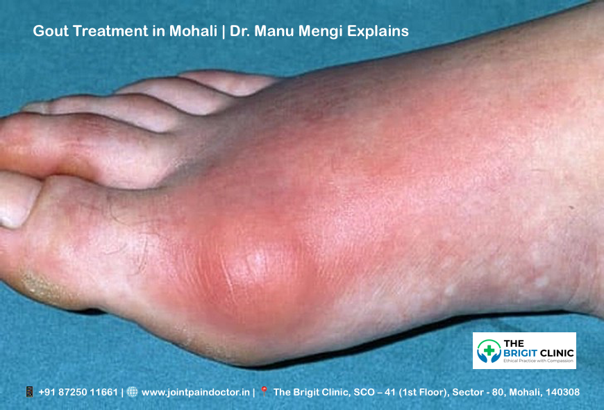
Swollen big toe joint and severe tenderness

The joint at the base of your big toe often bears the brunt of a gout attack. During a flare, this joint becomes so tender that even the light touch of a bedsheet can cause excruciating pain. You’ll notice significant swelling along with this tenderness, making it difficult to wear shoes or walk normally.

Redness & heat in joints during a flare

Beyond pain and swelling, visual cues help identify a gout attack. The affected joint typically becomes noticeably red or discoloured. Moreover, you’ll feel warmth or even a sensation like the joint is “on fire”. This inflammation is your body’s response to the sharp urate crystals irritating the joint.

Gout attack signs and how long they last

Gout flares often begin suddenly at night, with pain intense enough to wake you from sleep. A typical gout attack lasts between one and two weeks. Between flares, you usually won’t experience any symptoms. Nevertheless, without proper treatment, attacks may become more frequent and last longer over time.

Alcohol binge and other common triggers

Research shows alcohol consumption is the most common trigger for gout attacks, responsible for 14% of flares in one study. Specifically, drinking more than one alcoholic beverage in a day raises your risk of experiencing a gout attack. Beer appears to pose a greater risk than spirits, due to its high purine content.

Other common triggers include:

  • Consuming red meat or seafood
  • Dehydration
  • Physical injury or excessive activity
  • Extremely warm or cold weather

For those with gout, experts recommend avoiding alcoholic beverages entirely, despite the potential benefits of moderate consumption for other health conditions.

> > > CTA: Woken up by a sudden, fiery joint pain? You don't have to suffer through a gout flare alone. Contact Dr. Manu Mengi's Clinic in Mohali for urgent consultation and effective pain relief strategies.

What Causes Gout: Risk Factors and Underlying Conditions

Understanding what causes gout begins with identifying its root mechanism – disrupted uric acid balance in your body. Let’s explore the factors that put you at risk.

High uric acid levels and hyperuricemia

Hyperuricemia occurs when uric acid levels exceed 6.8 mg/dL in your blood. This condition happens when your body either produces too much uric acid or your kidneys don’t excrete enough. Although one-quarter of people with hyperuricemia develop gout, most remain symptom-free.

Risk factors for gout – diet, alcohol, obesity

Your dietary choices significantly impact gout risk. Foods high in purines, such as red meat, organ meats, and certain seafood, can raise uric acid levels. Beer consumption poses a greater risk than other alcoholic beverages because of its high guanosine content. Weight gain is another major factor – obesity increases your risk of gout by 3.26 times compared to a normal weight.

Genetics & gout: family history matters

Gout runs in families, with heritability estimated at approximately 60%. About 20% of people with gout have a relative with the condition. Research shows that an additive genetic score of high-risk alleles can increase gout risk up to 41-fold.

Medications that trigger gout

Certain medications raise uric acid levels in your blood. Diuretics increase the relative risk of gout by almost 80%. Other culprits include low-dose aspirin, cyclosporine, and some transplant medications.

Hormonal changes in gout (e.g., menopause)

For women, menopause significantly increases gout risk. The hormone estrogen helps eliminate uric acid through urine. After menopause, estrogen production declines, causing uric acid levels to rise.

Gout & kidney disease: a two-way link

A bidirectional relationship exists between gout and kidney disease. Kidney disease impairs uric acid excretion, raising gout risk. Conversely, high uric acid can damage the kidneys by forming stones and causing scarring.

> > > CTA: Confused about how your diet or medications affect your gout? Get a personalised Uric Acid Diet Consultation in Mohali with Dr. Mengi to understand your triggers and build a prevention plan.

Diagnosis and Treatment Options Explained

Getting an accurate diagnosis is crucial for effective gout management. Modern diagnostic techniques have improved physicians’ ability to detect and monitor this painful condition.

Ultrasound for gout detection

Ultrasound has emerged as a valuable tool for diagnosing gout, offering high-resolution imaging without radiation. The characteristic “double contour sign” (an irregular hyperechoic line over cartilage) has 43.7% sensitivity but 99% specificity for gout. Ultrasound can also identify tophaceous deposits in soft tissues and joints that might not be evident during clinical examination.

X-ray changes in chronic gout

X-rays typically show normal findings early in the disease. In chronic gout, hallmark features include marginal erosions with overhanging edges and sclerotic margins. Soft-tissue nodules (tophi) may appear as dense masses with higher attenuation than surrounding tissues.

Corticosteroids in gout treatment

Corticosteroids effectively treat acute gout flares, especially for patients who cannot tolerate NSAIDs. Options include oral prednisone (30-40mg daily for 5 days), intravenous administration, or direct joint injections. For single-joint gout, intra-articular injections offer fewer systemic side effects.

Gout treatment without medications

Non-pharmaceutical approaches include applying ice to affected joints, resting the inflamed area, and drinking plenty of water. Maintaining a healthy weight through regular exercise helps prevent flares, with low-impact activities like walking or swimming being ideal.

Managing chronic gout vs acute gout

Acute gout requires rapid inflammation control within 24 hours of symptom onset. Chronic gout management focuses on lowering serum uric acid below 6 mg/dL through long-term therapy. This distinction is essential—treating inflammation during flares differs from preventing future attacks through urate-lowering strategies.

> > > CTA: Need a precise diagnosis? As a leading Uric Acid Treatment Doctor in Mohali, Dr. Mengi uses advanced ultrasound imaging to accurately detect gout crystals and create a targeted treatment strategy for both acute and chronic gout. Visit our clinic to know more.

Conclusion

Gout, while extremely painful, remains one of the most manageable forms of arthritis when properly addressed. Understanding the connection between uric acid levels and gout attacks enables you to take control of this condition. Certainly, the buildup of urate crystals causes those excruciating flares, but knowledge about triggers like alcohol consumption, purine-rich foods, and certain medications gives you the power to prevent attacks.

Therefore, working closely with healthcare providers becomes essential for proper diagnosis and treatment. Advanced diagnostic tools, such as ultrasound, can detect gout even before visible symptoms appear. After diagnosis, treatment options range from medications that reduce inflammation during acute attacks to long-term strategies for lowering uric acid levels below the crystal-forming threshold.

Besides medical interventions, lifestyle modifications play a crucial role in gout management. Weight loss, adequate hydration, and avoiding high-purine foods significantly reduce your risk of future flares. Additionally, limiting alcohol consumption, particularly beer, helps maintain lower uric acid levels.

Remember that gout management requires both immediate relief during attacks and ongoing prevention between episodes. Though historically called “the disease of kings,” gout does not have to rule your life. With early detection, appropriate treatment, and consistent lifestyle adjustments, you can effectively manage this condition and maintain joint health for years to come.

Key Takeaways

Understanding gout empowers you to manage this painful but controllable form of arthritis effectively through proper diagnosis, treatment, and lifestyle modifications.

• Gout occurs when uric acid crystals form in joints, most commonly affecting the big toe first due to lower temperature and repetitive trauma from walking.

• Alcohol consumption is the top trigger for gout attacks, with beer posing a higher risk than other alcoholic beverages due to its purine content.

• Early diagnosis using ultrasound and X-rays enables targeted treatment, distinguishing between acute flare management and chronic prevention strategies.

• Lifestyle changes like weight loss, hydration, and avoiding high-purine foods significantly reduce attack frequency and severity.

• Gout is highly manageable when uric acid levels stay below 6 mg/dL through medication, diet modifications, and consistent medical monitoring.

With proper understanding of triggers, symptoms, and treatment options, gout doesn’t have to control your life. The key lies in working with healthcare providers to develop a comprehensive management plan that addresses both immediate pain relief and long-term prevention strategies.

> > > CTA: Ready to take control of your gout? Partner with Dr. Manu Mengi Gout Specialist Mohali, for a comprehensive management plan that combines advanced treatment with sustainable lifestyle changes. Your journey to a pain-free life starts here.

FAQs

Q1. What exactly is gout, and how does it affect the body?

A1. Gout is a form of inflammatory arthritis caused by the buildup of uric acid crystals in joints, most commonly in the big toe. It results in sudden attacks of severe pain, swelling, and tenderness. Gout occurs when the body either produces too much uric acid or the kidneys don’t excrete enough, leading to crystal formation in joints.

Q2. What are the most common triggers for a gout attack?

A2. The most common trigger for gout attacks is alcohol consumption, particularly beer, due to its high purine content. Other triggers include consuming red meat or seafood, dehydration, physical injury, and extreme weather conditions. Avoiding these triggers, especially limiting alcohol intake, can help reduce the frequency of gout flares.

Q3. How can I quickly relieve gout symptoms at home?

A3. To quickly relieve gout symptoms at home, apply ice to the affected joint, rest the inflamed area, and drink plenty of water. Some people find cherry juice helpful in reducing uric acid levels. However, it’s important to consult with a healthcare provider for proper treatment, as these methods provide temporary relief but don’t address the underlying cause.

Q4. Are there any non-medication approaches to managing gout?

A4. Yes, there are several non-medication approaches to managing gout. These include maintaining a healthy weight through regular low-impact exercise like walking or swimming, staying well-hydrated, and avoiding foods high in purines. Lifestyle modifications play a crucial role in preventing gout flares and managing the condition long-term.

Q5. How is gout diagnosed, and what treatment options are available?

A5. Gout is typically diagnosed through a combination of clinical symptoms, blood tests to measure uric acid levels, and imaging techniques like ultrasound or X-rays. Treatment options include medications to reduce inflammation during acute attacks, such as NSAIDs or corticosteroids, and long-term therapies to lower uric acid levels. The goal is to bring serum uric acid below 6 mg/dL to prevent future attacks and complications.

Dr. Manu Mengi – Gout Specialist Mohali

If you are looking for expert and compassionate care for gout in Mohali, look no further than Dr. Manu Mengi. Renowned as one of the Best Orthopedic Doctors in Chandigarh Mohali, Dr. Manu Mengi specialises in the diagnosis and management of gout and other joint pain disorders. His patient-centric approach combines cutting-edge diagnostic technology with evidence-based treatment protocols to provide both immediate relief and long-term prevention strategies. Whether you’re experiencing your first painful flare or struggling with chronic gout, Dr. Mengi and his team are dedicated to helping you regain your mobility and live a pain-free life. Schedule a consultation at his Mohali clinic to begin your journey toward effective gout management.

Book Appointment - Call Us: +91 87250 11661 | Find Our Clinic: Get Directions


Rotator Cuff Tear vs. Frozen Shoulder: A Doctor’s Guide to Spotting the Difference

Rotator Cuff Tear vs. Frozen Shoulder | Dr. Manu Mengi

Rotator Cuff Tear vs. Frozen Shoulder confusion is something I encounter frequently in my practice, with many patients mistakenly self-diagnosing their shoulder pain. These two conditions are often confused with each other, yet they present distinct symptoms and have significantly different recovery timelines.

In my experience, understanding the key differences can help you identify your condition more accurately. Rotator cuff tears typically have a sudden onset, particularly after activities like lifting something heavy or falling. At the same time, frozen shoulder develops gradually with pain that slowly worsens over weeks or months. The most telling difference? If you have a rotator cuff tear, you can likely lift your shoulder without assistance. However, with a frozen shoulder, you’ll face difficulty raising your arm by yourself.

Throughout this guide, I’ll walk you through the specific symptoms, causes, and treatments for both conditions. We’ll explore why frozen shoulder involves progressive stiffness accompanied by dull, aching pain (especially at night), while rotator cuff tears cause sharper, more localised pain when lifting or rotating the arm. By the end, you’ll understand why that stiffness in your shoulder joint might indicate one condition over the other.

This self-diagnosis often leads to anxiety. If you're in Mohali and struggling with shoulder pain, the most important step is to get a professional evaluation. Book an appointment with a specialist to get an accurate diagnosis and peace of mind.

Understanding the Shoulder: Anatomy and Function

The shoulder represents one of the most complex and mobile joints in the human body. To fully understand the differences between rotator cuff injuries and frozen shoulder, we must first examine the intricate architecture that allows for this remarkable range of motion. Unlike other joints that prioritise stability, the shoulder sacrifices some stability for mobility, making it uniquely susceptible to certain conditions.

The role of the rotator cuff in shoulder movement

The rotator cuff forms the cornerstone of shoulder function – a group of four muscles and their tendons that surround the shoulder joint like a protective collar. This muscular quartet connects your shoulder blade (scapula) to your upper arm bone (humerus) and works together to enable precise movement while maintaining joint stability.

The four muscles that make up this critical structure include:

  • Supraspinatus: Initiates the first 15 degrees of arm abduction (lifting your arm away from your body)
  • Infraspinatus: Powers external rotation of the arm away from your body
  • Teres minor: Assists with external rotation and stabilisation
  • Subscapularis: Enables internal rotation (turning your arm toward your body)

A helpful mnemonic for remembering these muscles is “SITS” – Supraspinatus, Infraspinatus, Teres minor, and Subscapularis.

The primary biomechanical role of these muscles extends beyond their movement function. They compress the head of the humerus against the shallow glenoid cavity of the scapula, creating what doctors call “dynamic stability”. Without this muscular support, the disproportionate size between the humeral head and the glenoid fossa (a 4:1 surface area ratio) would make the joint highly unstable.

Furthermore, these muscles don’t work in isolation. For example, during abduction, the supraspinatus handles the initial 0-15 degrees of movement, then the deltoid muscle takes over from 15-90 degrees. Finally, the scapula must rotate to achieve full range of motion, which is accomplished by the trapezius and serratus anterior muscles.

What is the shoulder capsule, and why does it matter

Surrounding the glenohumeral joint is a fibrous sheath called the shoulder capsule. This structure extends from the anatomical neck of the humerus to the rim of the glenoid fossa, completely enveloping the joint. Unlike other joint capsules in your body, the shoulder capsule has a unique characteristic – it’s remarkably loose and lax, allowing the bones to separate by more than 2.5 cm.

This laxity isn’t a design flaw but rather a deliberate feature that permits the extensive freedom of movement characteristic of the shoulder joint. Nevertheless, this same feature also makes the joint more vulnerable to instability and injury.

The capsule doesn’t work alone in providing joint stability. It’s reinforced by:

  • The glenoid labrum – a fibrocartilaginous ring that deepens the glenoid cavity
  • Ligaments that connect bones to bones
  • Tendons that connect muscles to bones
  • The rotator cuff muscles themselves, which fuse with the capsule

Additionally, fluid-filled sacs called bursae surround the capsule, facilitating smooth movement by reducing friction between the structures. The synovial membrane inside the capsule produces lubricating fluid that further aids motion.

Interestingly, when the shoulder capsule becomes damaged or inflamed, it can lead to adhesive capsulitis – commonly known as frozen shoulder. In this condition, the capsule thickens and becomes stiff, significantly restricting movement and causing pain.

How shoulder mobility is affected by injuries

Given the complexity of the shoulder’s structure, it’s not surprising that injuries can substantially impact its function. Shoulder pain affects approximately 18 million Americans each year, with rotator cuff tears accounting for a significant number of these cases.

Rotator cuff tears can occur through trauma, overuse, or age-related degeneration. These tears affect one in every four adults aged 60 years and older, with the supraspinatus tendon being most commonly involved. Risk factors include smoking, high cholesterol, and family history.

When the rotator cuff is damaged, patients typically experience:

  • Pain during overhead activities
  • Limited range of motion
  • Weakness in the shoulder
  • Difficulty with daily activities

Studies using three-dimensional motion analysis have shown that individuals with symptomatic rotator cuff tears exhibit abnormal shoulder motion when performing common daily tasks compared to those without tears. This dysfunction occurs because rotator cuff injuries compromise the joint’s dynamic stability, creating muscle force imbalances.

Alternatively, when the shoulder capsule becomes inflamed and thickened in adhesive capsulitis (also known as frozen shoulder), patients experience progressive stiffness and significant pain. This condition not only limits one movement but can also restrict motion in multiple directions simultaneously.

Research shows that patients with rotator cuff-related shoulder pain demonstrate significantly less shoulder flexion, internal and external rotation range of motion compared to individuals without shoulder problems. This finding helps explain why these conditions have such a profound impact on everyday activities.

Both conditions can make simple tasks challenging – whether reaching for objects on high shelves, putting on a coat, or even combing your hair. Understanding the underlying anatomy helps explain why these different conditions can sometimes present with similar symptoms despite having distinct causes.

As we've seen, both conditions severely impact your daily life. The key to effective treatment is a correct diagnosis. If you're experiencing limited motion or pain, consider visiting a 
Specialised Shoulder Clinic in Mohali for a comprehensive assessment.

Rotator Cuff Tear: Causes, Symptoms, and Diagnosis

Rotator cuff tears represent one of the most common shoulder injuries I diagnose in my practice. These tears are the leading cause of shoulder pain and shoulder-related disability, affecting millions of people each year. In fact, more than 2 million Americans experience some rotator cuff problem annually. While these injuries can affect people of all ages, they’re particularly common in adults over 40, with prevalence increasing significantly with age.

As a physician, I find that many patients are surprised to learn that some individuals have rotator cuff tears without experiencing any symptoms whatsoever. This silent presentation can make diagnosis challenging in certain cases.

Common causes: trauma, overuse, and ageing

Rotator cuff tears typically fall into two distinct categories: acute traumatic tears and degenerative tears. Understanding this distinction is crucial for proper treatment.

Traumatic (Acute) Tears occur suddenly as a result of a specific injury. In my experience, patients often report:

  • Falling on an outstretched arm
  • Lifting something heavy with a jerking motion
  • Experiencing sudden force while pushing or pulling
  • Sustaining an injury during a shoulder dislocation

These traumatic tears account for approximately 8% of all rotator cuff injuries. They tend to stem from higher energy mechanisms and typically cause full-thickness tears, whereas degenerative tears often begin as partial-thickness injuries. Moreover, traumatic tears generally are larger and more likely to involve the subscapularis muscle.

Degenerative (Wear-Related) Tears develop gradually over time due to natural ageing processes. These constitute the majority of rotator cuff injuries I see in my practice. Several factors contribute to these tears:

  1. Repetitive stress – Certain activities that involve repeated shoulder motions put significant strain on the rotator cuff. Sports like baseball, tennis, rowing, and weightlifting carry an increased risk of overuse. Likewise, many occupations requiring repetitive overhead movements can lead to progressive damage.
  2. Decreased blood supply – As we age, blood flow to our rotator cuff tendons diminishes. Without adequate blood supply, the body’s natural ability to repair tendon damage becomes impaired, ultimately leading to tissue breakdown.
  3. Bone spurs, also known as age-related bone overgrowth (osteophytes), can develop on the acromion, creating friction against the rotator cuff tendons when you move your arm.

Essentially, the supraspinatus tendon bears the brunt of most rotator cuff injuries, as it endures the majority of shoulder-stabilising strain. Approximately 53% of patients with supraspinatus tears also have involvement of the infraspinatus.

Concerning symptoms, patients typically experience:

  • Pain localised to the anterior shoulder or greater tuberosity area
  • Discomfort when lying on the affected shoulder
  • Pain with overhead activities
  • Night pain that interrupts sleep
  • Weakness in the affected arm
  • Crackling or popping sensations during certain movements
  • A positive painful arc sign during clinical examination

Diagnosing rotator cuff tears involves several components. First, I take a detailed history, including the mechanism of injury and activities that aggravate or relieve symptoms. Next, I perform a thorough physical examination, checking for tenderness, range of motion, and specific clinical tests designed to stress the rotator cuff tendons. Additionally, diagnostic imaging plays a crucial role in confirming the diagnosis and determining the extent.

Specifically, when patients present with an inability to elevate their arm after a traumatic event, despite normal radiographs, I maintain a high index of suspicion for rotator cuff pathology, as these injuries can often be missed during initial assessment.

While this guide helps you understand the differences, only a physical examination can confirm your condition. For expert rotator cuff tear treatment in Mohali or frozen shoulder treatment in Mohali, seeking help from a qualified orthopaedic doctor is crucial. Learn more about your Shoulder Pain Treatment Options in Mohali.

Key Takeaways

Understanding the differences between rotator cuff tears and frozen shoulder can help you identify your condition and seek appropriate treatment more effectively.

• Onset patterns differ significantly: Rotator cuff tears typically occur suddenly after trauma or lifting, while frozen shoulder develops gradually over weeks to months with progressively worsening pain.

• Movement ability is the key differentiator: With rotator cuff tears, you can usually lift your arm without assistance, but frozen shoulder severely limits your ability to raise your arm independently.

• Pain characteristics vary between conditions: Rotator cuff tears cause sharp, localised pain during lifting or rotation, while frozen shoulder presents as dull, aching pain that’s particularly troublesome at night.

• Age and activity patterns matter for diagnosis: Rotator cuff tears affect over 25% of adults aged 60+, often from repetitive overhead activities or sudden trauma. At the same time, a frozen shoulder involves inflammation and stiffness of the capsule.

• The rotator cuff’s four muscles (SITS) – Supraspinatus, Infraspinatus, Teres minor, and Subscapularis – work together to provide dynamic shoulder stability, making tears particularly disabling for daily activities.

Both conditions significantly impact quality of life; however, accurate identification of symptoms, such as pain location, movement limitations, and onset patterns, enables more targeted treatment approaches and better outcomes.

FAQs

Q1. How can I differentiate between a frozen shoulder and a rotator cuff tear?

A1. The key difference lies in your ability to move your arm. If you can lift your arm independently, it’s more likely to be a rotator cuff tear. Frozen shoulder severely limits your ability to raise your arm without assistance. Additionally, rotator cuff tears often have a sudden onset, while frozen shoulder develops gradually over time.

Q2. Is it possible for a rotator cuff tear to be misdiagnosed as frozen shoulder?

A2. Yes, these conditions can be confused. Some partial rotator cuff tears may not cause pain and can mimic symptoms of a frozen shoulder. However, a thorough medical examination, including imaging tests, can help differentiate between the two conditions.

Q3. What are the most effective treatments for frozen shoulder?

A3. Most cases of frozen shoulder improve on their own within 12 to 18 months. For severe or persistent symptoms, treatments may include physical therapy, corticosteroid injections, and in some cases, surgery. Steroid injections can be particularly effective when administered early in the progression of the condition.

Q4. What other conditions can be mistaken for a rotator cuff tear?

A4. Several shoulder conditions can present similar symptoms to rotator cuff tears, including biceps tendinitis, shoulder impingement syndrome, labral tears, arthritis, bursitis, and tendinitis. Each of these conditions affects different parts of the shoulder structure but may cause similar pain or movement limitations.

Q5. How does age affect the likelihood of developing rotator cuff tears?

A5. The risk of developing a rotator cuff tear increases significantly with age. They are particularly common in adults over 40, with more than 25% of adults aged 60 and older experiencing some form of rotator cuff tear. This is often due to natural wear and tear, decreased blood supply to the tendons, and the development of bone spurs that can irritate the rotator cuff over time.

Still unsure about your shoulder pain? Don't let it limit your life any longer. If you're looking for an Affordable Shoulder Treatment in Mohali or simply want to consult with a trusted expert, reach out to us today. Schedule a consultation to get a clear diagnosis and start your journey to recovery.
📞 Call Us: +91 87250 11661 | 📍 Find Our Clinic: Google My Business

📖 Learn More About Shoulder Pain:

Dr. Manu Mengi – Shoulder Pain Specialist in Mohali

With extensive experience in diagnosing and treating complex shoulder conditions, Dr. Manu Mengi is a leading Shoulder Pain Specialist in the Mohali and Tricity region. He is dedicated to providing personalised care, utilising accurate diagnosis and evidence-based treatment plans to help patients regain pain-free movement and improve their quality of life.

Your Complete Patient Guide to Orthopedic Management of Achilles Tendon Tear

Despite being the largest tendon in your body and capable of withstanding forces up to 10 times your body weight, the Achilles tendon is surprisingly vulnerable to complications. Understanding the Orthopedic Management of Achilles Tendon Tear is the first step toward a successful recovery. Achilles tendon ruptures are very common sports injuries, occurring most frequently in people ages 30 to 40 and affecting men more often than women.

If you’re among the “weekend warriors” who exercise intensely without regular training, you face a higher risk of tearing your Achilles than younger, well-trained athletes. Additionally, these injuries can be missed in up to 25% of cases, making proper diagnosis crucial. Whether you’re dealing with a partial or complete tear, understanding your treatment options is essential for recovery. This comprehensive guide will walk you through everything you need to know about Achilles tendon tears—from identifying symptoms and risk factors to exploring both conservative and surgical treatment approaches that can help you return to your normal activities.

For expert diagnosis and a personalised treatment plan, 
Consult the Best Orthopedic Doctor in Mohali or call +91 87250 11661

Understanding Achilles Tendon Tear

The Achilles tendon is a critical structure that plays a significant role in your daily movements. Understanding its function and how it can tear will help you better recognise and manage this injury.

What is the Achilles tendon?

The Achilles tendon, also called the calcaneal tendon, is the thickest and strongest tendon in your body. This fibrous band of tissue connects your calf muscles (gastrocnemius and soleus) to your heel bone (calcaneus). Located at the back of your lower leg, this powerful tendon spans approximately 6 to 10 inches in adults.

What makes the Achilles tendon remarkable is its incredible strength—it can support forces up to four times your body weight. This impressive capacity allows you to perform essential movements like walking, running, and jumping. When your calf muscles contract, they pull on the Achilles tendon, causing your foot to point downward (plantarflexion), which helps lift your heel off the ground during physical activities.

Partial vs complete Achilles tendon tear

Achilles tendon tears exist on a spectrum from minor damage to complete rupture. Understanding the difference between partial and complete tears is crucial for proper treatment.

In a partial tear, only a portion of the tendon fibres is damaged. Think of this like a frayed rope where some strands remain intact while others are torn. With a partial tear, you’ll experience:

  • Localised soreness around the tendon
  • Some swelling that gradually decreases
  • Full function of the ankle despite pain

A complete tear occurs when the tendon ruptures entirely, separating into two distinct parts. This severe injury results in:

  • A noticeable “pop” or snapping sound at the moment of injury
  • Complete loss of strength and function in the ankle
  • Extreme difficulty walking or exercising
  • Significant swelling around the ankle
  • In some cases, visible displacement of calf muscles

The Thompson test is commonly used by doctors to diagnose a complete rupture—when squeezing the calf fails to produce normal foot movement, it indicates a tear.

Common causes and overuse injuries

Most Achilles tendon tears happen during recreational sports or athletic activities. The tendon typically ruptures when exposed to sudden, powerful forces, especially during activities requiring quick stops, starts, and pivots—such as soccer, football, basketball, tennis, or squash.

Several specific scenarios commonly lead to tears:

  • Landing awkwardly from a jump
  • Cutting movements during sports
  • Sudden acceleration or forceful pushing off with the foot
  • Direct trauma to the tendon
  • Tripping or falling, particularly when the foot is forced upward[18]

Certain factors increase your risk of experiencing an Achilles tendon tear. As you age, the tendon naturally becomes stiffer and weaker. “Weekend warriors”—adults who exercise intensely without regular training—face a higher risk than consistently trained athletes.

Furthermore, medical conditions like inflammatory diseases, diabetes, obesity, and certain medications (including fluoroquinolone antibiotics and corticosteroids) can weaken the tendon structure. Mechanical issues such as tight calf muscles, improper footwear, and training errors also contribute to vulnerability.

Overall, Achilles tendon ruptures affect approximately 12 per 100,000 individuals, most commonly occurring between ages 40 and 50, with men experiencing them 2 to 12 times more frequently than women.

If you're active and experiencing heel pain, visit a Sports Injury Clinic Mohali for a risk evaluation. Book your appointment or call +91 87250 11661

Recognising Symptoms and Risk Factors

Recognising an Achilles tendon tear promptly can make a significant difference in your treatment outcomes. By understanding the tell-tale signs and knowing your risk profile, you might prevent a delayed diagnosis that occurs in up to 25% of cases.

Sudden pop in the back of the ankle

The most distinctive sign of an Achilles tendon rupture is experiencing (and sometimes hearing) a sudden “pop” or “snap” at the back of your ankle. This sensation is so pronounced that many mistake it for being struck from behind. The sound represents the actual moment when your tendon fibres separate.

After this characteristic pop, you’ll likely feel immediate, intense pain. The sensation resembles what would happen if someone kicked you forcefully in the lower leg. Unlike gradual onset injuries, this moment is unmistakable and marks a clear point when damage occurred.

Call your doctor immediately if you experience this sudden snap during physical activity. This symptom alone strongly suggests a complete tear rather than a minor strain, consequently requiring proper medical evaluation.

Heel pain & swelling

Following a tear, sharp, sudden pain typically develops near your heel. Initially, this pain might be unbearable, though it sometimes subsides slightly after the acute injury. The area around your Achilles tendon will swell noticeably, often accompanied by bruising along the back of the ankle.

The discomfort pattern differs from other foot conditions. With an Achilles tendon tear, pain tends to be localised specifically to the back of the ankle where the tendon attaches to your heel bone. Throughout the day, this pain might intensify with activity.

Morning stiffness presents another common symptom, where the affected area feels particularly tight and sore when you first get up. As you move around, this stiffness sometimes improves temporarily.

Calf pain after injury

Beyond the immediate heel area, pain often radiates upward into your calf muscle. This happens because the calf muscles connect directly to the Achilles tendon, creating a continuous pain pathway.

The discomfort in your calf typically worsens during activities that engage these muscles, such as climbing stairs or walking uphill. Furthermore, weakness in the leg becomes apparent when attempting to push off with the affected foot.

For partial tears, you might maintain some function despite the pain. However, with complete ruptures, standing on tiptoes or performing basic foot movements becomes nearly impossible.

Risk factors for Achilles tendon tear

Several factors increase your vulnerability to Achilles tendon tears:

Age and Gender: The peak age for ruptures occurs between 30-40 years, though some sources identify 40-50 as the highest risk period. Men experience these injuries four to five times more frequently than women.

Activity Patterns: “Weekend warriors” face significantly higher risk than regularly trained athletes. Sports involving quick stops, starts, and pivots present the greatest danger—particularly soccer, basketball, tennis, football, and racquet games.

Medical Factors: Certain conditions predispose you to tendon injuries:

  • Previous Achilles tendinopathy
  • Diabetes
  • High cholesterol or blood pressure
  • Psoriasis
  • End-stage kidney disease
  • Inflammatory arthritis

Medication Usage: Some medications weaken tendon structure, notably:

  • Fluoroquinolone antibiotics[28]
  • Corticosteroid injections
  • Oral bisphosphonates

Biomechanical Issues: Physical characteristics matter too. Having tight calf muscles, flat arches, overpronation (ankles rolling inward), or legs of different lengths all increase your risk[30].

Understanding these symptoms and risk factors helps you identify potential problems early and seek appropriate orthopaedic management for Achilles tendon tears before complications develop.

Experienced a pop or snap in your ankle? Seek immediate consultation with an Orthopedic Doctor in Mohali for Achilles Tear, call +91 87250 11661

How Achilles Tendon Tears Are Diagnosed

Getting an accurate diagnosis is essential for proper orthopaedic management of an Achilles tendon tear. Medical professionals use several examination techniques combined with imaging to confirm your injury.

Physical exam and Thompson test

Your doctor will begin by asking about your symptoms and medical history, followed by a thorough physical examination of your lower leg. During this examination, they’ll check for tenderness, swelling, and potentially feel for a gap in your tendon if it has ruptured completely.

The Thompson test (sometimes called the calf squeeze test) is a key diagnostic procedure with 96-100% sensitivity and 93-100% specificity for detecting Achilles ruptures. Here’s how it works:

  • You’ll lie face down with your feet hanging over the edge of the exam table
  • Your doctor will gently squeeze your calf muscle
  • In a healthy tendon, this causes your foot to point downward naturally
  • If your foot doesn’t move during the squeeze, it indicates a likely rupture

To confirm the diagnosis, doctors often use additional clinical signs like checking for a palpable gap in the tendon (typically 3-6cm above the heel) and assessing plantar flexion strength.

MRI vs Ultrasound in Achilles tendon tear

Both MRI and ultrasound provide valuable diagnostic information, albeit with different strengths:

Ultrasound shows the tendon in real-time and demonstrates how it responds to movement. It’s highly accurate with 95% sensitivity and 99% specificity for detecting full-thickness tears. Ultrasound is generally:

  • More cost-effective
  • Readily available
  • Excellent for detecting tendinopathy and complete ruptures

MRI creates detailed images of soft tissues and is particularly valuable for:

  • Detecting partial tears (superior to ultrasound)
  • Assessing the distance between torn tendon ends
  • Postoperative evaluation
  • Ruling out other injuries with similar symptoms

Most specialists recommend ultrasound over MRI for initial diagnosis and monitoring, though your doctor may order both depending on your specific situation.

When to see a doctor

Seek immediate medical attention if you experience:

  • A popping or snapping sound at the time of injury
  • Suddenly, severe pain in the back of your ankle
  • Difficulty walking or standing on tiptoes
  • Visible swelling around the heel area

Even if you can walk with a ruptured Achilles (which many people can), it’s crucial to see a healthcare provider promptly. Using your ankle and putting full weight on it before diagnosis can worsen the injury. Importantly, up to 20% of Achilles tendon ruptures are initially misdiagnosed, often confused with ankle sprains, making proper medical evaluation essential for effective treatment.

For advanced diagnostic imaging and expert interpretation, visit the Best Ortho Doctor in Mohali. Schedule your visit.

Treatment Options: Conservative and Surgical

Treatment decisions for Achilles tendon tears depend on several factors, including your age, activity level, and the severity of your injury. Both non-surgical and surgical approaches offer viable pathways to recovery, each with distinct advantages.

Achilles tendon tear – conservative management

Conservative treatment involves non-surgical approaches that typically include rest, immobilisation, and controlled rehabilitation. This option is often suitable for older patients, those with limited activity goals, or individuals with health conditions that increase surgical risks.

For partial tears with less than 5mm gap between ruptured tendon edges, conservative management can be particularly effective. The traditional approach involves wearing a below-knee cast in an equinus (pointed down) position for four weeks without weight-bearing, followed by a neutral position cast with weight-bearing for another four weeks.

Surgical treatment of Achilles tendon tear

Surgical intervention appears to be the preferred method for athletes and younger, active individuals. The primary benefit of surgery is a lower re-rupture rate compared to non-surgical treatment.

The procedure typically involves making an incision in the back of your leg and stitching the torn tendon together. In cases of severe degeneration, surgeons may remove damaged portions and repair the remaining healthy tendon.

Minimally invasive Achilles tendon tear surgery

This advanced technique involves a small 3-4cm incision instead of the traditional 10cm cut. Through this smaller opening, specialised instruments guide sutures into the tendon to complete the repair.

The minimally invasive approach offers several advantages:

  • Reduced wound healing issues
  • Lower infection rates
  • Less scar tissue formation
  • Faster return to normal activities

Immobilisation vs early mobilisation in Achilles tendon tear

Historically, rigid cast immobilisation for six weeks was standard practice. Nevertheless, recent research strongly supports early functional rehabilitation and mobilisation.

Studies demonstrate that early mobilisation doesn’t increase re-rupture rates. Moreover, it offers superior benefits:

  • Decreases excessive adhesion formation
  • Improves the biomechanical properties of healing tissue
  • Enhances tendon gliding function
  • Reduces joint stiffness and muscle atrophy

Medication for tendon inflammation

Pain management typically begins with over-the-counter options like ibuprofen or naproxen sodium. For persistent discomfort, prescription medications might include COX-2 inhibitors, which potentially cause fewer gastrointestinal side effects than traditional NSAIDs.

PRP Achilles tendon tear therapy

Platelet-rich plasma (PRP) therapy involves injecting a concentrated solution of your own platelets into the injured area. These platelets contain growth factors that may promote tissue repair and regeneration.

Currently, evidence regarding PRP effectiveness remains mixed. Some studies show improvements in ankle dorsiflexion angle and calf circumference, whereas others found no significant differences in patient-reported outcomes at two years post-injury.

Explore all treatment options, including Minimally Invasive Achilles Surgery in Mohali, with the Best Orthopedician in Mohali. Discuss your choices at https://jointpaindoctor.in/ or 
Call *+91 87250 11661* to learn more about the Achilles Tear Surgery Cost Mohali.

Recovery, Rehab, and Return to Activity

Full healing from an Achilles tendon tear requires a comprehensive rehabilitation approach tailored to your specific needs. The recovery journey typically spans four to six months, regardless of whether you underwent surgical or non-surgical treatment.

Physical therapy and strengthening

Physical therapy serves as the cornerstone of Achilles tendon rehabilitation. The duration varies based on injury severity—from a few weeks to several months. Your therapist will focus on three primary goals: pain relief through various modalities, restoring proper movement patterns, and rebuilding muscle strength and balance.

Eccentric exercises stand out as the most evidence-based intervention for Achilles rehabilitation. This approach, typically performed twice daily for at least 11 weeks, has been shown to reduce pain by an average of 60% across multiple clinical trials. The Alfredson protocol remains the gold standard, gradually progressing from bilateral to single-leg heel raises.

For optimal recovery, maintain a consistent exercise regimen alongside gradually increasing weight-bearing activities. Initially, you’ll use a walking boot with progressively decreasing heel wedges until reaching a neutral position, usually around 6-8 weeks post-injury.

Custom orthotics post Achilles repair

Bespoke orthotics play a valuable role in recovery by providing proper foot alignment, enhancing shock absorption, and correcting biomechanical issues that might stress your healing Achilles tendon. These devices primarily keep your heel raised, reducing the workload on the tendon while protecting against re-rupture.

Studies have demonstrated that custom foot orthoses can significantly improve symptoms in athletes with Achilles tendinopathy, with participants reporting an average 92% improvement when using high-density EVA orthotics.

Equinus contracture after Achilles tendon tear

Equinus contracture—excessive tightness limiting ankle dorsiflexion—often develops following Achilles injuries. Conservative management through physical therapy, stretching, and night splints should be attempted first. For refractory cases, surgical options include gastrocnemius lengthening, soleus fascial release, or Achilles tendon lengthening procedures.

Return to sports after Achilles tendon tear

Returning to sports requires patience—full athletic activities should be avoided for at least 6 months post-injury. The return process follows a carefully structured progression: controlled strengthening, followed by plyometric training, and finally sport-specific movements.

Before resuming competitive activities, you should achieve specific milestones: single-leg heel raise at 90% height compared to your uninjured side, normal gait mechanics, and pain-free performance of sport-specific movements. Even with optimal rehabilitation, expect some persistent strength deficits (10-30%) in the affected leg beyond the one-year mark.

Access comprehensive rehabilitation programs at our Orthopedic Hospital in Mohali for the Achilles Rupture facility. Start your recovery journey with the Best Achilles Tendon Surgeon in Mohali by calling +91 87250 11661

Conclusion

Achilles tendon tears represent serious injuries that require prompt diagnosis and appropriate treatment for optimal recovery. Throughout this guide, we’ve explored how these tears happen, their symptoms, and the available treatment approaches. Whether you choose conservative management or surgical intervention, your recovery journey demands patience and commitment to rehabilitation protocols.

Most patients can expect a full recovery period of four to six months, though some strength deficits might persist beyond the one-year mark. During this time, physical therapy will become your essential ally, particularly through eccentric strengthening exercises that have proven highly effective for tendon healing.

Remember that each case differs based on factors like age, activity level, and tear severity. Therefore, working closely with healthcare professionals remains crucial for developing a personalised treatment plan. Custom orthotics might benefit your recovery by improving foot alignment and reducing stress on your healing tendon.

Though returning to sports and normal activities takes time, a structured approach to rehabilitation significantly improves your outcomes. Above all, don’t rush this process. Your body needs adequate time to rebuild the strongest tendon in your body.

Armed with this knowledge about Achilles tendon tears, you can now make informed decisions about your care if faced with this injury. Early recognition of symptoms, prompt medical attention, and dedication to your rehabilitation program will ultimately determine your successful return to the activities you enjoy.

For a successful recovery under expert guidance, book your final consultation at https://jointpaindoctor.in/ or call +91 87250 11661

Key Takeaways

Understanding Achilles tendon tears and their proper management can significantly impact your recovery outcomes and help you make informed treatment decisions.

• Recognise the warning signs early: A sudden “pop” sound, severe heel pain, and inability to stand on tiptoes indicate a potential Achilles rupture requiring immediate medical attention.

• Both surgical and conservative treatments work: Your age, activity level, and tear severity determine the best approach—athletes often benefit from surgery while older patients may succeed with non-surgical management.

• Early mobilisation beats prolonged immobilisation: Modern rehabilitation emphasises controlled movement over extended casting, leading to better outcomes and faster functional recovery.

• Recovery takes 4-6 months minimum: Patience is crucial as rushing back to activities increases re-rupture risk—expect some strength deficits even after one year.

• Physical therapy is non-negotiable: Eccentric strengthening exercises, particularly the Alfredson protocol, form the foundation of successful rehabilitation regardless of treatment method chosen.

The key to successful Achilles tendon recovery lies in prompt diagnosis, appropriate treatment selection, and unwavering commitment to structured rehabilitation. Don’t underestimate this injury—proper management now prevents long-term complications and ensures your return to normal activities.

Ready to start your treatment? Contact the Best Orthopedic Doctor Mohali today 
or call +91 87250 11661

FAQs

Q1. What are the main symptoms of an Achilles tendon tear?

A1. The primary symptoms include a sudden “pop” or snapping sensation in the back of the ankle, intense heel pain, swelling around the affected area, and difficulty walking or standing on tiptoes.

Q2. How long does it typically take to recover from an Achilles tendon tear?

A2. Recovery usually takes 4-6 months, regardless of whether surgical or non-surgical treatment is chosen. However, some strength deficits may persist for over a year.

Q3. Is surgery always necessary for an Achilles tendon tear?

A3. Not always. The decision between surgical and conservative treatment depends on factors like age, activity level, and tear severity. Athletes often benefit from surgery, while older patients may succeed with non-surgical management.

Q4. What role does physical therapy play in Achilles tendon tear recovery?

A4. Physical therapy is crucial for recovery, focusing on pain relief, restoring proper movement, and rebuilding strength. Eccentric exercises, particularly the Alfredson protocol, are considered highly effective for rehabilitation.

Q5. When can I return to sports after an Achilles tendon tear?

A5. Full athletic activities should be avoided for at least 6 months post-injury. Before returning to competitive sports, you should achieve specific milestones like single-leg heel raises at 90% height compared to the uninjured side and pain-free performance of sport-specific movements.

About the Doctor – Dr. Manu Mengi

Dr. Manu Mengi is a highly skilled and renowned Orthopedic Surgeon in Mohali, specialising in the management and treatment of sports injuries, particularly complex Achilles tendon tears. With extensive experience and a commitment to adopting the latest surgical techniques, including minimally invasive procedures, Dr. Mengi provides personalised care to each patient. He leads a state-of-the-art Ortho Clinic in Mohali that is equipped with advanced diagnostic technology to ensure accurate assessments and the most effective treatment plans. Dedicated to helping patients return to their active lifestyles, Dr. Mengi is considered one of the best orthopedic doctors in the region for Achilles tendon repair and rehabilitation.


Book an Appointment today - 
📱 +91 87250 11661 | 🌐 Read Reviews | 📍 Get Directions

The Hidden Link Between Your Sleeping Posture and Back Pain

Ever woken up with a tingling arm or an achy neck? Your sleeping posture & back pain might be more connected than you realise. Lower back pain is the leading cause of disability worldwide, with up to eight out of ten people experiencing at least one episode during their lifetime. If you’re struggling with persistent discomfort, consulting the Best Doctor for Back Pain Treatment in Mohali, Dr. Manu Mengi, can help diagnose and treat underlying issues effectively.

The relationship between sleep and pain works both ways. While back pain can make finding a comfortable sleeping position difficult, poor sleeping posture can cause or worsen your existing discomfort. Research clearly shows that there’s a significant correlation between pain and sleep quality. Consequently, making simple changes to your sleeping posture for back pain can lead to better spine alignment while sleeping and significantly reduce your discomfort.

Throughout this article, you’ll discover which positions support your spine and which ones might be contributing to your morning stiffness. Furthermore, you’ll learn practical tips for creating a sleep environment that helps alleviate back pain rather than causing it. Whether you’re dealing with chronic issues or occasional discomfort, understanding the hidden link between how you sleep and how you feel could be the key to waking up pain-free.

How sleep position affects your spine

Your spine is designed with specific natural curves that distribute weight and pressure throughout your body. During sleep, maintaining these curves becomes particularly important for preventing discomfort and injury.

Why spine alignment matters during sleep

The spine serves as the central support system for your body, protecting the spinal cord and helping distribute weight across muscles and bones. Good sleeping posture maintains these natural curves even while lying down. When your head, neck, or hips aren’t properly supported, the spine becomes misaligned, potentially leading to both immediate discomfort and long-term problems.

Misalignment during sleep puts uneven pressure on different parts of your spine. Sleeping on your back, for instance, places approximately 50 pounds of pressure on your spine. This pressure, when distributed unevenly due to poor posture, can cause muscle imbalances and strain that manifest as morning pain.

The role of gravity and pressure points

Gravity is a fundamental yet often overlooked factor affecting your sleep quality and spinal health. Research shows that gravitational forces continue to act on your body during sleep, influencing everything from muscle relaxation to breathing patterns.

Different sleeping positions interact with gravity in unique ways. When lying flat, gravity can cause soft tissues in your upper airway to relax and potentially obstruct breathing. Moreover, your mattress creates pressure points where your body makes the most contact with the surface.

A soft mattress allows the hips to sink too deeply, creating an unnatural spinal curve that may trigger lower back pain. Conversely, a mattress that’s too firm creates excessive pressure on contact points like shoulders and hips, potentially causing skin sores and muscle soreness.

Common signs your sleep posture is causing pain

Your body often signals when sleep posture is problematic. Watch for these warning signs:

  • Morning neck stiffness or soreness, especially if you sleep on your stomach with your head twisted
  • Waking with numbness or tingling in the arms or legs
  • Lower back pain that improves once you’ve been up and moving
  • Persistent shoulder pain on one side (usually the side you sleep on)
  • Headaches that begin shortly after waking

Sleep allows muscles surrounding the spine to fully relax through a process called muscle atonia. This relaxation is essential for recovery, but only works properly when your spine remains in neutral alignment throughout the night.

🔹 Struggling with chronic back pain? Visit the Best Back Pain Relief Clinics in Mohali for personalised treatment! Book an appointment now.

Best and worst sleep positions for back pain

Finding the right sleeping position can make a world of difference for your back. Each posture offers unique benefits and drawbacks depending on your specific condition.

Back sleeping: pros, cons, and pillow tips

Sleeping flat on your back is often considered the gold standard for spine health. This position distributes weight evenly across your body’s widest surface area, helping maintain the spine’s natural curve. Back sleeping keeps your head, neck and spine in a neutral position, reducing pressure that contributes to aches and pains.

To optimise this position, place a pillow underneath your knees. This simple adjustment helps reduce pressure on your lumbar spine. Additionally, using a thin pillow for your head prevents your neck from arching unnaturally.

Though ideal for spinal alignment, back sleeping isn’t perfect for everyone. This position may worsen snoring and sleep apnea as gravity causes the tongue or loose tissues in the throat to collapse and block the airway.

Side sleeping: how to do it right

Side sleeping ranks as the second-best position for avoiding back pain. This position helps keep your spine elongated and can significantly reduce pressure points along your back.

For optimal side sleeping, stretch your legs out straight rather than curling them. Place a firm pillow between your knees to maintain proper spinal alignment and prevent your top leg from pulling your spine out of position. Keep your ears aligned with your shoulders and your chin in a neutral position.

Fetal position: when it helps and when it hurts

The fetal position—lying on your side with knees drawn up toward your chest—is among the most popular sleeping positions despite its mixed benefits. This position can help open the space between your spinal vertebrae and reduce tension on your disks.

Nevertheless, curling too tightly can restrict movement and place excessive pressure on joints, particularly in the knees, hips, and spine. Over time, this may lead to stiffness or discomfort, especially without adequate mattress support.

Stomach sleeping: why it’s usually a bad idea

Stomach sleeping is generally considered the worst position for your spine. This position puts the most pressure on your spine’s muscles and joints because it flattens the natural curve of your spine. Additionally, it forces you to turn your neck to breathe, which can cause neck and upper back pain.

If you absolutely must sleep on your stomach, place a thin pillow under your pelvis and lower abdomen to reduce strain on your back.

Reclined sleeping: when it’s beneficial

For some people, particularly those with specific back conditions, reclined sleeping offers notable benefits. This position helps reduce pressure on your spine by creating an angle between your trunk and thighs.

Reclined positions can be especially helpful after surgery or for people with conditions like degenerative disk disease. The slightly elevated position also improves circulation and can reduce inflammation associated with back pain.

🔹 Looking for a Lower Back Pain Specialist in Mohali? Dr. Manu Mengi provides expert care tailored to your needs. Call +91 87250 11661 today!

How to improve your sleep setup

Creating an optimal sleep environment goes beyond just finding the right position. Your mattress, pillows, and supporting accessories play crucial roles in spinal health.

Choosing the right mattress for back pain relief

Medium-firm mattresses consistently show the most significant benefits for relieving back pain. According to research, 75% of orthopaedic surgeons recommend firmer surfaces for back pain sufferers. Memory foam and latex options provide excellent support by contouring to your body’s natural curves, distributing weight evenly and helping maintain proper spinal alignment. However, avoid mattresses that are too soft as they allow your body to sink excessively, causing your spine to curve unnaturally.

Pillow support for the lower back and neck

Proper pillow support can dramatically improve spine alignment. Lumbar support pillows placed beneath your lower back help maintain the natural curve of your spine. For neck support, cervical pillows with an S-shape design reduce pain by providing the perfect balance between softness and firmness. Side sleepers should use higher loft pillows to fill the gap between the shoulder and head, whereas back sleepers benefit from medium loft options. Interestingly, water pillows offer customizable support and pressure relief.

Using props like towels and body pillows

Simple household items can transform your sleep setup. Rolling up a towel or sheet and placing it where your lumbar area rests provides targeted support. For side sleepers, placing a pillow between your knees aligns your hips and reduces pressure on your lower back. Body pillows offer comprehensive support for side sleepers by simultaneously aligning the spine and reducing pressure on hips and shoulders.

Sleeping on the floor vs bed: does it help?

The evidence regarding floor-sleeping is primarily anecdotal. Some report back pain relief from firmer surfaces, yet scientific proof remains limited. The potential benefits include improved posture since softer surfaces allow your spine to curve while hard surfaces provide better support. Nevertheless, studies indicate medium-firm surfaces, not extremely firm ones, offer optimal pain relief.

Signs your mattress or pillow needs replacing

Replace your mattress every 6-8 years. Watch for visible indentations deeper than 1.5 inches, which indicate inadequate support. Other signs include waking with unexplained stiffness or pain, increased allergies from accumulated dust mites, and sleeping better elsewhere than in your bed. Your pillow needs replacing if it’s too thin, causing your head to rest lower than your chest.

🔹 Is your mattress worsening your back pain? Consult the Best Orthopedic Doctor in Mohali for Back Pain, Dr. Manu Mengi, for professional guidance.

Tips for long-term sleep posture correction

Correcting poor sleeping posture isn’t a one-night fix but requires consistent effort and proper techniques. Long-term improvement demands both behavioural changes and physical adjustments that train your body over time.

Stretching and mobility routines before bed

Gentle stretching before sleep can significantly improve the quality of your rest by relieving muscle tension that accumulates during the day. Consider your day like running a marathon—regardless of whether you’ve been walking, standing, or sitting, your muscles need relaxation to recover properly. Nighttime stretches for your neck, shoulders, and back help release tension and prevent muscle spasms that might wake you up. Ideally, perform these stretches 30-60 minutes before bedtime, not immediately before falling asleep.

Morning stiffness & sleep posture connection

Initially, many people dismiss morning back pain as “just part of ageing,” yet research reveals a direct link between sleep posture and waking discomfort. Studies show that people with cervical and lumbar pain spend approximately twice as long in provocative side-lying positions compared to those without pain. Furthermore, those experiencing spinal symptoms change sleep positions more frequently throughout the night. This correlation indicates that correcting sleep posture may effectively reduce morning stiffness.

How to sleep with scoliosis or herniated disks

For scoliosis patients, back sleeping typically offers the most relief as it helps maintain spine elongation without twisting. Alternatively, side sleeping with properly positioned pillows between knees and ankles can minimise asymmetrical tension. Those with herniated disks often find relief in the fetal position, which opens vertebral spaces and reduces pressure on disks. Sleeping in a reclined position creates a beneficial angle between your trunk and legs, allowing core muscles to rest.

Sleep advice for pregnancy-related back pain

Pregnant women experiencing back pain should avoid sleeping as it puts pressure on the aorta and vena cava. Instead, side sleeping with one or both knees bent provides optimal support. Place pillows between bent knees, under your abdomen, and behind your back. Consider using full-body pregnancy pillows specifically designed to support your changing body shape.

Training your body to adopt better positions

Changing sleep habits requires patience and consistency. The “tennis ball technique” effectively prevents rolling onto problematic positions—simply sew a tennis ball into your pyjamas to discourage certain positions. Electronic devices that vibrate when you roll into poor positions offer gentler feedback and better long-term compliance. Finally, gradually transition to new positions using strategic pillow placement, including supports under your knees for back sleeping or between your knees for side sleeping.

🔹 For expert Back Pain Treatment in Mohali, trust Dr. Manu Mengi’s proven methods. Schedule a visit today! Call +91 87250 11661.

Conclusion

The connection between your sleep position and back pain runs deeper than most people realise. Your nightly posture significantly impacts spinal health, potentially creating or relieving pain depending on your choices. Back sleeping generally offers the best support for your spine’s natural curves, while side sleeping with proper pillow support between your knees serves as an excellent alternative. Conversely, stomach sleeping typically causes more harm than good due to the unnatural neck twist and flattened spinal curve it creates.

Beyond position alone, your sleep setup plays a crucial role in back health. A medium-firm mattress paired with appropriate pillows creates the foundation for proper alignment throughout the night. Additionally, simple props like rolled towels or body pillows can transform your sleep experience by providing targeted support exactly where needed.

Changing long-established sleep habits certainly requires patience. Still, the benefits of proper sleep posture extend far beyond just your nights—they directly influence how you feel each morning and throughout your day. Persistent morning stiffness often signals the need for sleep posture adjustments rather than representing an inevitable part of ageing.

Your specific health conditions also factor into optimal sleeping arrangements. Whether dealing with scoliosis, herniated disks, or pregnancy-related discomfort, tailored approaches to sleep positioning can make a substantial difference in your pain levels.

Remember that small, consistent changes to your sleep habits can yield significant results over time. Therefore, implementing the recommended positions and support techniques gradually helps train your body toward better alignment. The effort you invest in improving your sleep posture now will reward you with more restful nights and pain-free mornings for years to come.

🔹 Ready to say goodbye to back pain? Consult Dr. Manu Mengi, the Best Doctor for Back Pain in Mohali, for lasting relief. Call +91 87250 11661.

Key Takeaways

Understanding the connection between sleep position and back pain can transform your nights and mornings, leading to better spinal health and reduced discomfort.

• Back sleeping is optimal for spine health – distributes weight evenly and maintains natural spinal curves, but add a pillow under the knees for best results.

• Medium-firm mattresses provide the best pain relief – 75% of orthopedic surgeons recommend firmer surfaces, as they support natural alignment without excessive sinking.

• Side sleeping requires proper pillow placement – place a firm pillow between the knees to maintain spinal alignment and prevent hip rotation.

• Stomach sleeping should be avoided – this position flattens natural spinal curves and forces neck twisting, creating the most pressure on spine muscles.

• Simple props can dramatically improve sleep quality – rolled towels for lumbar support and body pillows for side sleepers provide targeted relief at minimal cost.

Poor sleep posture affects 8 out of 10 people who experience back pain during their lifetime. The good news is that making these evidence-based adjustments to your sleep setup can break the cycle of pain and poor sleep quality, helping you wake up refreshed rather than stiff.

FAQs

Q1. How does sleeping position affect back pain?

A1. Different sleeping positions can either alleviate or exacerbate back pain. Back sleeping is generally considered the best for spine health as it maintains natural spinal curves. Side sleeping with proper pillow support is also beneficial. Stomach sleeping is usually the worst position as it flattens the spine’s natural curve and forces neck twisting.

Q2. What type of mattress is best for reducing back pain?

A2. Medium-firm mattresses are typically recommended for back pain relief. They provide adequate support to maintain proper spinal alignment without being too hard. Memory foam and latex options can be particularly effective as they contour to your body’s natural curves and distribute weight evenly.

Q3. Can sleeping with a partner contribute to back pain?

A3. Yes, sleeping with a partner can potentially lead to back pain if it causes you to adopt poor sleeping postures. Compromising your natural sleeping position to accommodate your partner or using an unsuitable mattress for two people can create compressions or twists in your body, potentially leading to discomfort.

Q4. Are there any props that can help improve sleep posture?

A4. Yes, several props can enhance sleep posture. Placing a pillow between your knees when side sleeping can help maintain spinal alignment. For back sleepers, a pillow under the knees can reduce pressure on the lower back. Lumbar support pillows and body pillows can also provide targeted support and improve overall sleep posture.

Q5. How often should I replace my mattress to prevent back pain?

A5. It’s generally recommended to replace your mattress every 6-8 years. However, if you notice visible indentations deeper than 1.5 inches, experience unexplained morning stiffness or pain, or find you sleep better elsewhere than in your bed, it might be time for a replacement sooner. A worn-out mattress can contribute to poor sleep posture and back pain.

About Best Doctor for Back Pain Treatment in Mohali – Dr. Manu Mengi

Dr. Manu Mengi is a renowned Back Pain Specialist in Mohali, offering advanced and personalised treatment for chronic and acute back pain. With years of experience in orthopedic care, he provides the most effective Lower Back Pain Treatment in Mohali, ensuring patients regain mobility and live pain-free lives. Whether you suffer from posture-related discomfort or severe spinal conditions, Dr. Mengi’s expertise ensures the highest standard of care.


📍 Visit his clinic today or check out his Google My Business profile for patient reviews!

Trigger Finger: A Doctor’s Complete Guide to Symptoms & Treatment

Trigger finger affects millions worldwide, ranking among the top 5 causes of disability in the hand. As an Orthopedic Specialist in Mohali, I’ve seen how this condition—medically known as stenosing tenosynovitis—causes significant discomfort through painful clicking, stiffness, and fingers stuck in a bent position. Women face this challenge six times more frequently than men, particularly between the ages of 40 to 60. Additionally, those with diabetes experience trigger finger at much higher rates—approximately 10% compared to just 2-3% of the general population.

If you’re seeking Trigger Finger Treatment in Mohali, early diagnosis and care can prevent long-term complications. Book a consultation today.

Hand stiffness and finger locking are especially troublesome during morning hours and often worsen after periods of heavy hand use. Certain occupations face greater risks, with farmers, industrial workers, and musicians developing trigger finger more frequently due to repetitive finger movements. The good news, however, is that effective treatments exist—from conservative approaches like splinting and steroid injections to surgical options when needed. In this comprehensive guide, I’ll walk you through everything you need to know about trigger finger, from recognising early symptoms to understanding your treatment options.

What is Trigger Finger?

Stenosing tenosynovitis, commonly known as trigger finger, occurs when a finger becomes stuck in a bent position before suddenly releasing, similar to pulling and releasing a trigger. This condition develops when inflammation narrows the space within the sheath surrounding the flexor tendons that control finger movement.

Trigger finger meaning and overview

Trigger finger happens when bands of tissue called pulleys (specifically the A1 pulley) become inflamed or thickened, making it difficult for tendons to slide smoothly. As your finger bends, the tendon may catch and then suddenly release, creating a distinctive clicking or popping sensation at the base of the affected digit.

Initially, you might notice mild discomfort that gradually worsens. Many patients report that their symptoms are most severe upon waking in the morning. Without treatment, the affected finger may eventually become locked in a flexed position, requiring force to straighten it, and in advanced cases, making it impossible to fully extend.

Difference between arthritis & trigger finger

Although both conditions cause hand pain and stiffness, trigger finger and arthritis are fundamentally different. Trigger finger affects the tendons and their sheaths rather than the joints themselves. Arthritis involves inflammation and breakdown of joint tissues, eventually causing bones to rub against each other across the 27 joints in your hands and fingers.

Furthermore, trigger finger presents with characteristic catching or locking sensations not typically seen in arthritis. You might also feel a small nodule in your palm from the enlarged tendon or thickened pulley – another distinctive feature of trigger finger.

Commonly affected fingers and demographics

Trigger finger shows clear patterns in which digits it affects. The ring finger is most commonly affected, followed by the middle finger, with the thumb close behind. When the thumb is involved, the condition is specifically called trigger thumb.

This condition displays interesting demographic patterns:

  • Women develop trigger finger approximately twice as often as men (68% vs. 32%)
  • Most cases occur between the ages of 40-60, with an average age of 63 years
  • The dominant hand is affected in 70% of cases
  • Right-handed individuals experience trigger finger in their right hand 60% of the time

While anyone can develop trigger finger, certain groups face higher risks. The condition affects 2-3% of the general population but jumps to 10-20% among people with diabetes. Other conditions like rheumatoid arthritis, gout, and thyroid disease also increase susceptibility.

Recognising the Symptoms Early

Identifying trigger finger early can help prevent progression to more severe symptoms. The condition typically begins gradually without any apparent injury and may follow periods of extensive hand use.

Early signs of trigger finger

The first indications of trigger finger often include a painful clicking or snapping sensation when bending or straightening your finger. You might notice mild stiffness, particularly in the morning, that gradually improves with movement throughout the day. Many patients report a strange feeling as if something is catching in their finger. These symptoms frequently start subtly and worsen over time if left untreated.

Finger locking and hand stiffness

As trigger finger progresses, the catching sensation becomes more pronounced. Your finger may temporarily lock in a bent position before suddenly popping straight—the characteristic “triggering” that gives this condition its name. Morning stiffness becomes more noticeable, making it difficult to make a fist or grasp objects firmly. In advanced cases, the affected digit may become completely locked in a bent position, requiring you to use your other hand to manually straighten it.

Swollen finger joint with pain

A key physical sign of trigger finger is tenderness or a noticeable bump (nodule) at the base of the affected finger in your palm. This nodule forms where the tendon has become swollen and thickened. Soreness typically intensifies when gripping objects or when pressure is applied to this area. The inflamed tendon struggles to glide smoothly through its sheath, consequently causing pain during finger movement.

When symptoms are worse during the day

Trigger finger symptoms follow distinct patterns throughout the day. The condition is notably worse after periods of inactivity, particularly first thing in the morning. Stiffness and locking tend to improve somewhat with gentle, consistent hand use as the day progresses. Nevertheless, symptoms often intensify during activities requiring firm grasping or pinching motions. Unlike some hand conditions that worsen with overuse, trigger finger is primarily troublesome after rest periods and upon initial movement.

Understanding Causes and Risk Factors

The root cause of trigger finger lies in the mechanical disruption of tendon movement. Understanding why and how this condition develops can help identify those at higher risk.

Inflamed tendon in finger: how it starts

Trigger finger begins when tendons that control your finger movements become irritated and swollen. These rope-like structures normally slide smoothly through a protective tunnel called the tendon sheath. With repeated use or certain health conditions, the tendon or its surrounding sheath becomes inflamed, creating friction during movement. Over time, this constant irritation forms a small lump (nodule) on the tendon. This nodule further complicates movement by getting caught on the A1 pulley (part of the sheath), causing the characteristic catching sensation.

Trigger finger in diabetes and thyroid conditions

People with diabetes face a significantly higher risk—approximately 10% of diabetics develop trigger finger compared to just 2-3% of the general population. Interestingly, research shows the length of time living with diabetes correlates more strongly with trigger finger risk than blood sugar levels. The theory is that chronic high blood glucose creates cross-links between collagen molecules, resulting in collagen buildup in tendon sheaths.

Thyroid conditions likewise increase vulnerability to trigger finger. Studies show trigger finger occurs in 10% of patients with subclinical hypothyroidism. Moreover, hypothyroid patients have the highest prevalence of Dupuytren’s contracture (21.7%), limited joint mobility (8.7%), and carpal tunnel syndrome (30.4%).

Hand pain in manual workers and repetitive strain

Occupations involving forceful, repetitive hand movements significantly increase trigger finger risk. Construction workers and manual labourers who regularly use vibrating power tools often develop this condition. Farmers, industrial workers, and anyone performing strenuous repetitive motions face similar risks. The constant strain causes microtrauma to the tendon-sheath complex, leading to inflammation and eventual tendon adhesion.

Trigger finger in computer users and musicians

Musicians face unique trigger finger risks due to their intensive, precise finger movements. Guitar players frequently develop multiple trigger fingers, sometimes affecting five or six digits simultaneously. One hand surgeon likens the condition to “a trumpet valve sticking,” noting that trumpet players must regularly oil their instrument valves to prevent similar mechanical issues. Computer users who spend hours typing or using a mouse also experience higher rates of trigger finger due to repetitive strain.

Diagnosis and Treatment Options

Diagnosing and treating trigger finger involves a structured approach that varies based on symptom severity and patient-specific factors. Let’s explore the complete treatment pathway from initial diagnosis to surgical intervention.

If you’ve been struggling with morning finger stiffness, a simple splint might solve your problem. Get a personalised assessment to find out.

Trigger finger diagnosis: what to expect

Physicians primarily diagnose trigger finger through a physical examination. During the exam, your doctor will check for tenderness over the flexor tendon, thickening of the tendon sheath, and the characteristic clicking sensation when you bend and straighten your digit. While X-rays are typically unnecessary, ultrasound may occasionally be used to evaluate tendon inflammation and to demonstrate the catching phenomenon during tendon movement.

Non-surgical treatment for trigger finger

Most cases begin with conservative approaches. Effective non-surgical options include:

  • Rest and activity modification: Avoiding repetitive gripping and using padded gloves for protection
  • Anti-inflammatory medications: Over-the-counter NSAIDs like ibuprofen to reduce pain and swelling
  • Gentle stretching exercises: To maintain mobility and decrease stiffness
  • Splinting: To immobilise and rest the affected digit

Splint for trigger finger: when and how to use

Splinting works by limiting tendon gliding and reducing inflammation. Research shows that wearing a splint for 6-10 weeks can be remarkably effective—one study found 87% of participants no longer required surgery or injections after completing a splinting regimen. Splints should typically position the metacarpophalangeal joint at 10-15 degrees of flexion. For milder cases, splinting alone may be the initial recommended treatment.

Steroid injections and their effectiveness

Corticosteroid injections into the tendon sheath represent a highly effective first-line intervention. Studies demonstrate an overall success rate of 66-69%. Interestingly, effectiveness varies by digit—the thumb responds best with a 92% success rate. Some patients may benefit from multiple injections, with efficacy increasing from 34% after one injection to 66% after three.

Trigger finger release surgery explained

When conservative treatments fail, surgical release of the A1 pulley becomes necessary. The procedure takes approximately 20 minutes under local anaesthesia on an outpatient basis. Two main approaches exist:

  1. Open release surgery: A small incision allows direct visualisation and release of the pulley
  2. Percutaneous release: Using a needle through the skin to divide the pulley, though this carries slightly higher risks

Most patients experience significant improvement after surgery, with a success rate exceeding 90%.

Trigger finger treatment in Mohali: what to know

Treatment protocols generally follow similar patterns globally, including in specialised centres. Regardless of location, early intervention yields better outcomes, with treatment plans tailored to individual needs and severity levels.

Is surgery your best option? Schedule a surgical consultation with Mohali’s Top Hand Surgeon.

Conclusion

Trigger finger, though painful and frustrating, remains a highly treatable condition when addressed properly. Throughout this guide, we’ve examined how this tendon disorder causes the characteristic catching and locking sensations that affect daily activities. Additionally, we’ve explored the demographic patterns showing that women and diabetic patients face significantly higher risks.

Early recognition of symptoms certainly makes a difference in treatment outcomes. Patients should watch for the initial signs—painful clicking, morning stiffness, and that distinctive catching sensation when bending fingers. Undoubtedly, understanding the underlying causes helps explain why certain groups experience higher rates of trigger finger. Manual workers, musicians, and computer users face elevated risks due to repetitive strain, while diabetes and thyroid conditions create physiological vulnerabilities.

The good news? Most patients respond well to conservative treatments. Splinting alone resolves many mild cases, while steroid injections prove effective for moderate symptoms, especially in the thumb. Nevertheless, surgical intervention remains a reliable option when other approaches fail, with success rates exceeding 90%.

As a physician who has treated countless trigger finger cases, I emphasise the importance of seeking treatment rather than enduring the discomfort. Though frustrating, this condition rarely causes permanent damage if addressed properly. Therefore, anyone experiencing symptoms should consult with a Hand Specialist in Mohali for Finger Pain to determine the most appropriate treatment path. With proper care, most patients return to normal hand function, free from the pain and limitations this condition can cause.

Don’t let finger pain limit your life—contact Dr. Manu Mengi’s clinic for same-week appointments.

FAQs

Q1. What is the most effective treatment for trigger finger?

A1. The most effective treatment for trigger finger depends on the severity of the condition. For mild cases, conservative approaches like splinting and anti-inflammatory medications may suffice. Corticosteroid injections are highly effective for moderate cases, especially in the thumb. When conservative treatments fail, surgical release of the A1 pulley is considered the gold standard, with success rates exceeding 90%.

Q2. Can vitamin deficiencies contribute to trigger finger?

A2. While vitamin deficiencies are not typically considered a primary cause of trigger finger, some research suggests that severe vitamin B6 deficiency may contribute to hand and finger issues. However, trigger finger is more commonly associated with factors like repetitive strain, diabetes, and certain occupations rather than specific vitamin deficiencies.

Q3. Are there any topical treatments that can help with trigger finger?

A3. Some topical treatments, such as diclofenac gel (a topical NSAID), may help reduce pain and inflammation associated with trigger finger. However, these are generally considered supplementary treatments and may not address the underlying mechanical issue causing the condition.

Q4. How long does it take for trigger finger to improve with treatment?

A4. The timeline for improvement varies depending on the treatment method. Splinting may show results after 6-10 weeks of consistent use. Steroid injections can provide relief within a few days to a few weeks. Surgical treatment often leads to immediate improvement in movement, with full recovery typically occurring within a few weeks to months.

Q5. Who is most at risk of developing trigger finger?

A5. Trigger finger is more common in women, particularly between the ages of 40-60. People with diabetes face a significantly higher risk, with about 10% developing the condition compared to 2-3% of the general population. Additionally, individuals in occupations involving repetitive hand movements, such as musicians, manual labourers, and frequent computer users, are at increased risk of developing trigger finger.

About the Doctor

Dr. Manu Mengi is a renowned Best Orthopedic Doctor for Trigger Finger in Mohali, specialising in hand and wrist disorders, including trigger finger, carpal tunnel syndrome, and arthritis. With years of experience in non-surgical and surgical trigger finger treatments, Dr. Manu Mengi has helped hundreds of patients regain pain-free hand movement. If you’re looking for Affordable Trigger Finger Surgery in Mohali or expert advice on managing finger stiffness, schedule a consultation today for personalised care.

Struggling to straighten your finger? WhatsApp [+91 87250 11661] us a photo of your hand for quick advice!