Rheumatoid Factor Positive But No Symptoms: What Your Blood Test Really Means – Dr. Manu Mengi

Why do your blood tests show rheumatoid factor positive when you feel perfectly fine? This question worries many people who receive unexpected lab results. Actually, rheumatoid factor has been found in up to 4% of young, healthy individuals, and it’s particularly common in older adults. A positive RF test doesn’t automatically mean rheumatoid arthritis. We’ll explain what rheumatoid factor positive meaning really is, why asymptomatic rheumatoid factor occurs, how it differs from anti-CCP testing, and when you should be concerned about your results.

What Does a Positive Rheumatoid Factor Test Mean?

What Does a Positive Rheumatoid Factor Test Mean?

Understanding Rheumatoid Factor and Its Normal Function

Rheumatoid factor is an antibody your immune system produces, but it behaves differently from the antibodies that protect you from illness. Most antibodies act as defenders, targeting germs, viruses, and other harmful invaders in your bloodstream. Rheumatoid factor, on the other hand, attacks healthy cells in your body.

Specifically, RF targets the Fc portion of immunoglobulin G (IgG), one of the most common antibodies your body makes to fight infections. Your immune system creates rheumatoid factor either by mistake or when it becomes overactive while fighting an infection. In reality, this protein doesn’t have a specific protective target like other antibodies, which is why it sometimes damages healthy tissue throughout your body.

The presence of RF isn’t always problematic. Many people have small amounts in their blood and remain perfectly healthy. RF can be part of your body’s normal response to various triggers, including bacterial toxins like lipopolysaccharides or viruses such as Epstein-Barr virus. In these cases, the RFs produced are low-affinity, transient antibodies that form immune complexes, which are then cleared by inflammatory cells. Their role could actually be protective in this context.

How RF Tests Work and What They Measure

An RF test measures the amount of rheumatoid factor in your bloodstream through a simple blood sample. A healthcare professional draws blood from a vein in your arm using a small needle, then sends the sample to a laboratory for analysis.

Labs typically measure rheumatoid factor in units per milliliter of blood (U/mL). Most assays detect IgM rheumatoid factors, although RF can occur in all immunoglobulin classes, including IgG and IgA. The test results come back either as a numerical value showing how many units of RF exist in each milliliter of your blood, or as a simple positive or negative designation.

A negative result means you have little or no rheumatoid factor in your blood. Correspondingly, a positive result indicates higher levels were detected. The test itself takes only a few minutes, though results may not return for several days.

Rheumatoid Factor Normal Range and Interpretation

A normal, healthy amount of rheumatoid factor is less than 20 units per milliliter (<20 U/mL)[7]. Some laboratories use slightly different thresholds, with results below 14 IU/mL considered normal, while others use higher cutoffs. Values may be slightly higher in older patients.

RF above 20 IU/mL is not considered enough to diagnose RA on its own, as other factors can elevate these levels. In fact, about 20% of people with confirmed RA will not have an abnormal RF test, while 5% of people who do not have RA will show elevated RF. This demonstrates why a positive result serves as a clue rather than a definitive diagnosis.

The sensitivity of RF testing in RA patients ranges from 60% to 90%, with a specificity of 85%. However, RF results are positive in less than 40% of patients with early RA. Higher RF levels generally indicate a greater likelihood of having a condition linked to rheumatoid factors, but people with rheumatoid arthritis can have little or no detectable RF. Similarly, people without rheumatoid arthritis can have substantial amounts of RF in their blood.

Your provider considers your RF levels alongside other test results, your symptoms, and your medical history to determine what’s causing any changes in your blood work.

Why Your RF Can Be Positive Without Any Symptoms

Rheumatoid Factor in Healthy Individuals

Perfectly healthy people can have elevated rheumatoid factor without any disease. In fact, RF appears in up to 4% of young, healthy individuals. Many people carry small amounts of RF in their blood and remain completely healthy throughout their lives.

Some people have high RF levels and never experience any symptoms. High amounts aren’t always a sign of a health condition. Your body might produce rheumatoid factor for no apparent reason or without a specific cause. This happens because RF can form as a normal immune response to various triggers, not just autoimmune conditions.

Most asymptomatic persons with a positive RF do not progress to RA. This bears repeating because it addresses the primary concern when you receive unexpected results. The presence of RF in your blood doesn’t predict that you’ll develop rheumatoid arthritis or any other condition. Your immune system sometimes produces these antibodies temporarily or maintains low levels indefinitely without causing tissue damage.

Age-Related Increase in Rheumatoid Factor

RF becomes increasingly common as you get older. The prevalence of RF can be as high as 14.1% in apparently healthy people aged 67-95, with a mean age of 81. This dramatic increase with age explains why many seniors receive positive results during routine blood work.

RF is also 3.5 times more common in healthy elderly subjects aged over 65 than in their younger counterparts. Your immune system changes as you age, producing more autoantibodies even when you’re healthy. These age-related changes don’t necessarily indicate disease development.

All these factors may alter the natural history of arthritis in elderly patients who have RF either in good health or in a non-arthritic presentation. Where you experience symptoms and their severity depends on where your immune system damages healthy tissue in your body.

The Timeline: When Symptoms May or May Not Develop

Some patients actually develop RF preceding the symptomatic disease. The earlier onset of RF in such patients has been associated with more severe disease. However, this represents only a subset of people with positive RF results.

Alternatively, there is a subset of patients in whom the appearance of RF follows symptoms. About 80% of all patients with rheumatoid arthritis will eventually be seropositive for rheumatoid factor, while only 40% are positive at clinical onset. This timeline variation demonstrates that RF positivity can occur at different stages or not at all.

People without rheumatoid arthritis can have substantial amounts of RF in their blood. Correspondingly, people with rheumatoid arthritis can have little or no rheumatoid factor. Your RF status today doesn’t determine your health status tomorrow.

Other Conditions That Cause Elevated Rheumatoid Factor

Other Conditions That Cause Elevated Rheumatoid Factor

Autoimmune Diseases Beyond Rheumatoid Arthritis

Rheumatoid arthritis isn’t the only autoimmune condition that triggers RF production. Several other autoimmune disorders cause elevated rheumatoid factor levels, sometimes even higher than those seen in RA patients.

Patients with Sjögren’s syndrome commonly show positive RF results. In effect, patients with primary Sjögren’s often have a positive RF test without having RA. Other rheumatic diseases that elevate RF include systemic lupus erythematosus (SLE), scleroderma, mixed connective tissue disease, and mixed cryoglobulinemia. Juvenile idiopathic arthritis also produces elevated RF in affected children.

These conditions share a common feature: uncontrolled immune system activity that becomes misdirected toward our own tissues rather than protecting us from infectious organisms. We believe some people inherit the tendency to develop these conditions, and that the diseases develop when something in the environment, perhaps an infection, stimulates the immune system to react.

Chronic Infections and RF Positivity

Chronic infections frequently cause RF elevation even when you don’t have any autoimmune disease. Hepatitis C stands out as a particularly common trigger. Hepatitis C could have very high levels of RFs, as high as 76%. Elevated rheumatoid factor levels and systemic immune activation are highly prevalent during chronic hepatitis C virus infection. This connection is so strong that it has been suggested that HCV status should be checked in all patients with increased RF levels.

The explanation lies in the nature of cryoglobulins produced during hepatitis C infection. They are cold precipitating IgM antibodies against IgG, which matches the basic definition of rheumatoid factor. By the same token, the positive rheumatoid factor could be from hepatitis C, particularly when you have arthritis and vasculitis.

Tuberculosis and subacute infective endocarditis also elevate RF. Infective endocarditis is associated with high inflammatory markers including rheumatoid factor. Actually, the latex test was positive in 45% of patients with infective endocarditis. More severe infection cases, as evidenced by duration of fever after initiation of antibiotic therapy, were more likely to develop rheumatoid factor.

Sarcoidosis and certain malignancies like primary sclerosing cholangitis also show RF elevation. Certain types of cancer, including leukemia, can cause high RF levels.

Temporary vs Persistent RF Elevation

The persistence of your elevated RF depends largely on what’s causing it. It’s not certain how a subset of chronic infections and inflammatory diseases lead to increased RF production. One explanation is chronic and persistent stimulation of the immune system by these disorders leading to a state of chronic inflammation.

Successful treatment of the underlying disorder frequently leads to the disappearance of these autoantibodies. Your RF levels can return to normal once the infection clears or the inflammatory condition resolves.

However, RF increases can be relatively stable in participants who have still not developed RA, especially in those with a combined increase of IgM and IgA RF at 74%. Unlike RA where no erosive joint changes occur, rheumatoid factor may be present in up to 50-85% of patients with certain chronic infections without causing joint damage.

Anti-CCP vs Rheumatoid Factor: Understanding the Difference

Anti-CCP vs Rheumatoid Factor: Understanding the Difference

Why Anti-CCP Is More Specific for RA

Anti-CCP stands for anti-cyclic citrullinated peptide, an autoantibody that appears early in RA development. The immune system produces anti-CCP when it mistakenly attacks certain peptides in joint linings, causing inflammation that leads to pain, swelling, and eventual joint damage. This antibody targets something fundamentally different from what RF targets, which explains why anti-CCP performs better as a diagnostic marker.

The specificity of anti-CCP reaches 95-98%, meaning it’s rarely positive in other conditions. Some studies report specificity at 96% with a positive likelihood ratio of about 14. In contrast, RF shows considerably lower specificity, ranging from 18% in some studies to 80-90% in early inflammatory arthritis. Given that RF appears in healthy individuals and numerous other conditions, anti-CCP delivers far more reliable results when diagnosing RA.

Anti-CCP antibodies may appear years before symptoms develop. Elevated levels indicate a more aggressive and destructive form of disease, serving as a marker for disease progression. In reality, anti-CCP remains negative in normal individuals, whereas RF is present in 5% of healthy people. This distinction matters when you’re trying to understand unexpected positive results without symptoms.

What It Means When RF Is Positive But Anti-CCP Is Negative

This specific combination suggests you’re unlikely to have RA, though clinical evaluation remains necessary. Studies show that 24.2% of anti-CCP-negative patients tested positive for RF. The interpretation changes based on your clinical presentation and other test results.

When anti-CCP is positive but RF is negative, you may be in the early stages of RA or could develop it in the future. This pattern appears in approximately 7.3% of cases. Correspondingly, when both tests come back negative, RA becomes highly unlikely, though seronegative RA still requires clinical evaluation to rule out.

Combining Both Tests for Better Accuracy

Doctors order both tests together, along with C-reactive protein and erythrocyte sedimentation rate, for complete diagnosis. Double positivity for RF and anti-CCP notably increases the positive likelihood ratio of RA. When both tests show positive results, you probably have rheumatoid arthritis, occurring in about 11.3% of cases.

Testing for both RF and anti-CCP is ideal for diagnosis. If testing for only one marker, anti-CCP is most recommended. The combined approach provides more accurate diagnosis than using RF testing results alone. Studies have shown that anti-CCP is positive even in RA patients who are RF negative.

When to Worry About Your Positive Rheumatoid Factor and What to Do Next

Warning Signs That Require Medical Attention

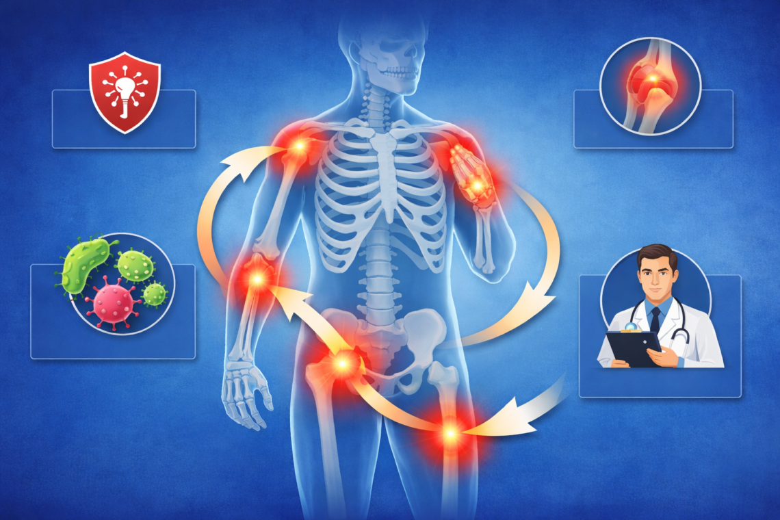
Joint symptoms warrant immediate medical attention. Pain, swelling, stiffness, and tenderness in more than one joint signal potential rheumatoid arthritis development. Morning stiffness or pain after sitting for long periods indicates inflammatory arthritis. RA typically affects joints on both sides of your body, so if one hand or knee hurts, watch the other.

Increased joint pain and swelling, new joint deformities, or feeling more tired and stiff for longer periods suggest your condition may be worsening. Fatigue, weakness, and low-grade fever accompanying joint problems require evaluation. Hard bumps under your skin near joints, called rheumatoid nodules, are red flags.

Patients with inflammatory joint disease should be referred to a rheumatology subspecialist, especially if symptoms last more than six weeks.

When to Repeat or Further Investigate Your Test Results

Inappropriate repeat testing of RF is common, and the results only rarely change. Patients who have clearly tested positive for RF or anti-CCP should not be retested routinely. RF and anti-CCP have no role in assessing disease activity.

Your clinician may check your RF level again if you were initially negative, but ongoing monitoring is based on symptoms, inflammation markers, and imaging. However, most asymptomatic persons with a positive RF do not progress to RA.

Do You Need Treatment If RF Is Positive But You Have No Joint Pain?

Treatment depends on the underlying cause. Without symptoms, you don’t need treatment for RA. Early diagnosis and effective treatment are important only when symptoms develop.

Working With Your Doctor to Monitor Your Condition

Track any new or worsening symptoms and share details with your clinician. This helps them adjust your treatment plan if needed. Regular monitoring focuses on symptom development rather than repeatedly checking RF levels.

Conclusion

We’ve demystified what rheumatoid factor positive really means when you have no symptoms. By and large, your positive RF test doesn’t predict rheumatoid arthritis development, particularly if your anti-CCP remains negative. Many healthy people carry RF in their blood without ever experiencing problems. Age-related increases, chronic infections, and various autoimmune conditions can all elevate your RF levels.

Focus on your symptoms rather than the numbers. You should worry only on the condition that you develop joint pain, swelling, or morning stiffness affecting multiple joints. Work with your doctor to monitor your health, but remember that most asymptomatic people with positive RF never progress to disease. Your blood test is just one piece of your overall health picture.

Key Takeaways

Understanding your positive rheumatoid factor test without symptoms can ease unnecessary worry and guide appropriate next steps.

• Positive RF doesn’t equal rheumatoid arthritis – Up to 4% of healthy young people and 14% of elderly individuals have elevated RF without any disease

• Anti-CCP testing is more reliable – Anti-CCP has 95-98% specificity for RA compared to RF’s lower accuracy, making it the better diagnostic marker

• Multiple conditions cause RF elevation – Chronic infections like hepatitis C, autoimmune diseases, and even normal aging can trigger positive results

• Symptoms matter more than numbers – Focus on joint pain, swelling, and morning stiffness rather than RF levels; most asymptomatic people never develop RA

• Monitor symptoms, not RF levels – Routine retesting of positive RF is unnecessary; work with your doctor to watch for actual disease signs instead

The key insight is that RF positivity without symptoms rarely indicates future disease development, especially when combined with negative anti-CCP results and absence of joint inflammation.

FAQs

Q1. Is it possible to have elevated rheumatoid factor without experiencing any symptoms?

Yes, it’s entirely possible to have high RF levels without any symptoms. Many healthy individuals have elevated rheumatoid factor in their blood and never develop any health problems. Up to 4% of young, healthy people and as many as 14% of elderly individuals have positive RF tests without any disease. The presence of RF alone doesn’t predict that you’ll develop rheumatoid arthritis or other conditions.

Q2. What does it mean if my rheumatoid factor is positive but my anti-CCP test is negative?

When RF is positive but anti-CCP is negative, it generally suggests you’re unlikely to have rheumatoid arthritis, though clinical evaluation is still important. Anti-CCP is much more specific for RA (95-98% specificity) compared to RF, which can be elevated in many other conditions including chronic infections, other autoimmune diseases, and normal aging. This combination is relatively common and doesn’t necessarily indicate disease.

Q3. How common is rheumatoid factor positivity in people with confirmed rheumatoid arthritis?

Approximately 60-80% of people with RA will have positive RF during the course of their disease. However, only about 40% of patients with early RA test positive for RF. This means that RF can be absent even when someone has rheumatoid arthritis, and conversely, it can be present in people who don’t have the disease, which is why doctors use it alongside other tests and clinical symptoms for diagnosis.

Q4. What symptoms should prompt me to see a doctor if I have a positive rheumatoid factor?

You should seek medical attention if you develop joint pain, swelling, stiffness, or tenderness affecting multiple joints, especially on both sides of your body. Morning stiffness lasting more than 30 minutes, fatigue, low-grade fever, or hard bumps under your skin near joints are warning signs. If joint symptoms persist for more than six weeks, a referral to a rheumatologist is recommended.

Q5. Besides rheumatoid arthritis, what other conditions can cause elevated rheumatoid factor?

Many conditions can elevate RF levels, including chronic infections like hepatitis C (which shows positive RF in up to 76% of cases), tuberculosis, and infective endocarditis. Other autoimmune diseases such as Sjögren’s syndrome, lupus, and scleroderma also cause RF elevation. Even certain cancers and normal aging can increase RF levels without indicating rheumatoid arthritis.

Q6. What does a positive rheumatoid factor (RF) mean?

A positive RF means that a specific antibody is detected in your blood. While it is commonly associated with Rheumatoid Arthritis, it is not exclusive to it. Many people can have a positive RF without having any disease.

Q7. Can I have a positive RF and still be completely healthy?

Yes. A significant number of people, especially older adults, may have a positive RF without any symptoms or illness. In such cases, it may not have clinical significance.

Q8. Does a positive RF always mean I will develop rheumatoid arthritis?

No. A positive RF does not guarantee that you will develop rheumatoid arthritis. It is just one piece of the puzzle and must be interpreted along with symptoms and other tests.

Q9. What other conditions can cause a positive RF?

RF can also be elevated in other conditions such as:

  • Chronic infections (like hepatitis or tuberculosis)
  • Other autoimmune diseases like Systemic Lupus Erythematosus
  • Certain lung or liver diseases

Q10. What is the difference between RF and anti-CCP?

RF is less specific, meaning it can be positive in multiple conditions. Anti-CCP (anti-cyclic citrullinated peptide) is more specific for rheumatoid arthritis and helps confirm the diagnosis when symptoms are present.

Q11. Should I start treatment if my RF is positive but I have no symptoms?

No treatment is needed based on RF alone. Treatment decisions are made only if you have symptoms like joint pain, swelling, or stiffness.

Q12. Can rheumatoid factor levels go back to normal?

Yes, RF levels can fluctuate and may even return to normal over time, especially if they were elevated due to a temporary cause like an infection.

Q13. When should I be concerned about a positive RF?

You should consult a doctor if you develop:

  • Persistent joint pain or swelling
  • Morning stiffness lasting more than 30 minutes
  • Fatigue or unexplained weakness

Q14. Do I need further tests if my RF is positive?If you have no symptoms, often no immediate action is required. However, your doctor may advise:

  • Anti-CCP test
  • ESR/CRP (inflammation markers)
  • Clinical follow-up over time

Q15. How often should I repeat the RF test?

Routine repetition is usually not necessary if you are asymptomatic. Follow-up testing is guided by clinical changes rather than lab numbers alone.

Consult Dr. Manu Mengi for all types of joint pain and orthopedic problems. A Top orthopedic doctor in Chandigarh, he provides care for bone and joint.

Why Does My Body Feel Stiff After Sleeping? Your Mattress, Posture, or Something Else? – Dr. Manu Mengi

Why does my body feel stiff after sleeping? This frustrating question affects countless people each morning. In fact, research shows that 75% of Brits have been woken by pain or discomfort. Morning body stiffness causes range from your mattress and sleeping posture to underlying medical conditions. A mattress causing back pain and stiffness, poor sleeping posture symptoms, or even early signs of arthritis could be responsible. We’ll explore the common stiffness after waking up reasons, including how to identify whether your discomfort is mechanical or inflammatory, and most importantly, how to reduce morning stiffness naturally.

What Causes Morning Body Stiffness After Sleep?

Why Your Body Feels Stiff When You Wake Up

Blaming age for morning stiffness misses the real story. Old age alone does not cause morning joint stiffness. Several physiological processes occur during sleep that create that tight, achy feeling when you first get out of bed.

During sleep, your body remains static for hours. This lack of movement means reduced blood flow to muscles and less lubrication in joints. Your joints produce synovial fluid, which acts as a lubricant. The joint lining produces less of this fluid during periods of inactivity. Without regular movement to keep joints limber, you wake up feeling creaky.

Your body undergoes other changes too. The spongy cushion of cartilage in your joints begins to dry out and stiffen as joints age. Weak muscles and stiff tendons also tend to tighten during sleep. Sleeping position plays a role as well. Too much bending or twisting in your sleep can strain muscles and restrict blood flow, leading to stiffness and discomfort when you wake up.

Dehydration compounds the problem. If you’re not drinking enough water throughout the day, your body struggles to lubricate your joints effectively, leading to stiffness and discomfort in the morning. Water maintains the balance of electrolytes crucial for muscle contraction and relaxation. Going to bed slightly dehydrated, then spending all night without water, can result in muscle cramps or soreness.

Temporary Stiffness vs. Chronic Morning Stiffness

The type of stiffness you experience reveals what’s happening inside your body. Morning stiffness falls into two categories: mechanical and inflammatory.

Mechanical stiffness relates to normal changes from wear and tear in joints, muscles, or tendons. This type feels more like tightness than deep pain. It occurs after physical activity, poor sleep, or long periods of inactivity. Joints with known osteoarthritis or prior injury commonly experience this type.

Inflammatory stiffness, in effect, stems from immune system activity inside the joints. Conditions like rheumatoid arthritis trigger this type. Your immune system attacks the synovium (the tissue lining your joints) and causes inflammation. Osteoarthritis and rheumatoid arthritis both can trigger morning stiffness, but they behave differently.

The stiffness goes away as you move and warm up the joints and muscles. This improvement with movement signals mechanical stiffness. Inflammatory stiffness, by comparison, takes longer to ease and may be accompanied by joint swelling, warmth, or redness.

How Long Should Morning Stiffness Last?

Duration separates normal stiffness from potential health concerns. For osteoarthritis, the average episode lasts only about 10 to 15 minutes. The stiffness often stops after a few minutes and eases once you start moving.

Stiffness from rheumatoid arthritis may last more than an hour. Some sources indicate inflammatory arthritis stiffness can take an hour or longer to go away. Morning stiffness that lasts over 30 minutes warrants attention. Longer periods of morning stiffness may mean your inflammation is higher or that your disease is more active.

If you wake up feeling seriously stiff and sore for more than 3 days in a row, make an appointment with your doctor. Your doctor may check your joints, run bloodwork, or test your joint fluid to check for inflammation.

Can Your Mattress Cause Body Stiffness After Sleeping?

Your mattress might be the last thing you suspect when investigating morning stiffness. Before blaming everything else, consider that the surface you spend 6-8 hours on each night plays a significant role in how you feel when you wake up.

Signs Your Mattress Is Causing Stiffness

Waking up with new aches and pains each day signals that your mattress no longer provides adequate support. An old mattress causes pain in your neck, shoulders, lower back, and hips. Your body offers clear warning signs when your mattress becomes the problem.

Look for physical evidence first. Body impressions visible on your mattress surface mean the supportive structure has failed. Beds are supposed to return to their natural state, but when your mattress loses its shape, it no longer supports your spine’s natural curvature. This adds stress to other parts of your body during sleep.

Pain that disappears once you’re up and moving points directly to your mattress. While lying on the mattress, inadequate support creates pressure on joints, muscles, or spine, which results in pain. Once you get up and move around, the pressure is relieved, and the pain subsides.

Constant tossing and turning reveals your inability to find a comfortable sleeping position. This continuous movement contributes to back pain and signals that your mattress isn’t providing proper support. If you feel like you’re sinking into your mattress and your spine cannot maintain a neutral position, that’s likely the reason behind your back issues.

Feeling the springs or support bars through the mattress surface clearly indicates the mattress no longer provides proper support. Visible sagging, especially in the middle, proves particularly harmful for spine health.

Too Soft vs. Too Hard: How Mattress Firmness Affects Your Body

Mattress firmness operates on a spectrum, and both extremes create problems. A too-soft mattress allows your body to sink excessively. This doesn’t offer ideal spinal alignment and causes back and neck pain. Back sleepers and stomach sleepers should avoid extremely soft mattresses.

When a mattress feels too firm, it puts excessive pressure on certain body areas like hips, shoulders, and lower back. A firm mattress creates pressure points, which cause stiffness and soreness when you wake up. The lower back cannot reach the mattress when lying down on overly firm surfaces, leading to abnormal spine positioning.

Medium-firm mattresses strike a balance between support and comfort. They offer enough firmness to keep the spine aligned while still providing cushioning. Research shows that sleeping on a medium-firm surface for four weeks can reduce back pain by roughly 48% while boosting sleep quality by 55%.

Spine Alignment Issues from Poor Mattress Support

Poor mattress support fundamentally compromises your body’s ability to achieve neutral spinal alignment. A too-soft surface causes the heavy midsection (hips and torso) to sink disproportionately, resulting in an unnatural hammocking effect.

Excessive sinking pulls the lumbar spine out of alignment, creating an intensified arch. This sustained, unnatural posture stretches muscles, ligaments, and nerves surrounding the spine, leading to morning stiffness, chronic lower back pain, and sciatica-like symptoms.

Your muscles must work overtime when mattresses fail to provide necessary support. Instead of relaxing for repair, deep postural muscles must engage throughout the night to stabilize the spine. As a result, this creates inflammation, fatigue, and the sensation of being tired even after a full night in bed.

When to Replace Your Mattress

Different mattresses have different lifespans. A waterbed lasts anywhere from 12 to 15 years. Foam beds (latex or gel) usually last around 10 to 15 years. Memory foam beds can last eight to 10 years if properly rotated. Innerspring mattresses typically last eight years or so.

Most sleep experts recommend replacing your mattress every 7-8 years. The mattress likely wears out during this time. By the same token, your body weight, sleeping habits, and bone density change with time. This calls for replacing the mattress with a new one that has optimum firmness and support.

Start looking for a new mattress as soon as you’re no longer comfortable. Listen to what your body tells you. If you sleep significantly better on a different bed for one night, that’s a good indication to start looking for a new mattress.

How Sleeping Position Affects Morning Stiffness

Poor Sleeping Posture Symptoms

Sleep posture affects your health just as much as standing and sitting posture. If you wake up in pain that you don’t feel during the day, your sleep position is likely playing a role. Poor sleeping postures create undue pressure on the neck, shoulders, lower back, hips, and knees.

Research found 10% of Americans wake up with pain each day. Considering we sleep for approximately eight hours a night, certain sleeping positions may cause strain on our shoulders and body, resulting in stiffness and soreness over time. Neck pain, stiffness, or soreness when you wake up in the morning signals that your sleep posture needs adjustment.

People with spinal pain spend more of the night in provocative sleep postures and have lower sleep quality. If something in the body is crooked, twisted, pinned under another body part, or held at a strange angle, it can stay that way for far longer than it would when you’re awake.

Stomach Sleeping and Spine Stiffness

Stomach sleeping places large amounts of stress on your neck and lower back joints and soft tissues. The sleeping position of the stomach imposes too much pressure on the lower back and neck. Most of your weight concentrates in the middle of your body, which makes it difficult to maintain a neutral spine position.

This position causes the spine to take an unnatural curve, increasing pain in the joints. Stomach sleepers experience morning back pain more frequently since your stomach sinks into the bed, putting pressure and stress on the joints and muscles in your back. Unless you’ve figured out how to breathe through your pillow, you need to turn your head to the side, which puts your head and spine out of alignment, twisting your neck.

Sleeping on your stomach with your head twisted to one side is a recipe for neck strain. You might not notice the damage this causes after one episode, but over time neck problems can develop.

Side Sleeping: Hip and Shoulder Pressure Points

Side sleeping is often the culprit for morning shoulder pain. Shoulder pain from sleeping is caused by prolonged pressure on the shoulder joint, most often from sleeping on your side. When you sleep on your side, your shoulder bears a lot of your upper body weight.

Some people may even develop shoulder impingement or tendinitis from their bone pressing against the sensitive tendons of the shoulder. If you sleep with your arm underneath your pillow, this can also pinch your rotator cuff tendon. Side sleeping can worsen rotator cuff injury symptoms, including dull pain or aches in the shoulder and reduced range of motion from stiffness.

Pillowing in between the knees puts the hips into position and eliminates the pressure on the spine. A supportive pillow of the head and neck is also important to prevent neck stiffness.

Back Sleeping: Best Position for Reducing Stiffness

The back position is usually recommended as the best posture for the spine. Sleeping on your back keeps the spine in a neutral position and prevents excess stress on the shoulders. It balances body weight and eliminates pressure points.

Lying flat is the nearest approximation to standing in a relaxed posture with your head, neck, and spine aligned. This position evenly distributes your weight across your body. A small pillow placed under the knees can support the natural lumbar curve and minimize lumbar strain.

Does Pillow Height Affect Neck Stiffness?

Pillow height matters significantly. If your pillow is too high or too low, too firm or too soft, your head will bend away from the neutral spine position. A pillow that’s too high pushes your neck upward, and your muscles end up having to work harder to hold that position, even while you’re asleep.

Research shows that inappropriate pillow height increases muscle activity during sleep and disrupts the alignment your cervical spine needs to stay neutral. A pillow that’s too flat lets your neck bend downward, flattening the natural curve. Too much height pushes the chin toward the chest, a common cause of morning stiffness. If you prefer sleeping on your back, it is recommended to use a thinner pillow to maintain the alignment of your neck with your spine.

Medical Conditions That Cause Morning Stiffness

Morning Stiffness vs. Arthritis: What’s the Difference?

Not all morning stiffness points to arthritis. Rheumatoid arthritis affects joints symmetrically, meaning both sides of the body at the same time and to the same extent. The small joints in hands and feet are often the first to be affected. Joint pain associated with rheumatoid arthritis is usually a throbbing and aching pain, often worse in the mornings and after a period of inactivity.

Morning stiffness that is a symptom of osteoarthritis usually wears off within 30 minutes of getting up, but morning stiffness in rheumatoid arthritis often lasts longer than this. Rheumatoid arthritis-related joint stiffness often lasts an hour or more in the morning.

Inflammatory Stiffness vs. Mechanical Stiffness

The immune system drives inflammation in inflammatory arthritis. Inflammation builds within your joints as you rest overnight, reaching peak levels in the morning. Swelling overnight in an immobile joint causes surrounding muscles and tissues to tighten up.

Mechanical stiffness stems from osteoarthritis. The joint isn’t moving much at night, so it’s not getting the lubrication it needs to slide smoothly. Many patients describe a rusty hinge sensation when they first start moving.

Early Signs of Arthritis in Morning Stiffness

Morning stiffness precedes the development of rheumatoid arthritis. Joints affected by rheumatoid arthritis become inflamed, which can cause them to swell and become hot and tender to touch. Symptoms vary from person to person and may come and go, or change over time.

When Should You Worry About Morning Stiffness?

Stiffness lasting more than 30 to 45 minutes warrants evaluation. Seek medical attention if you notice swelling, warmth, or redness around joints. Pain that disrupts sleep, numbness or tingling in limbs, or gradual worsening over weeks requires professional assessment. Muscle stiffness occurring with fever, muscle weakness, neck stiffness, or swelling can point to serious infections like meningitis.

How to Reduce Morning Body Stiffness Naturally

Why Stiffness Improves After You Start Moving

Movement stimulates circulation, warms tissues, and redistributes lubrication. During sleep, synovial fluid settles in your joints. When you begin moving, this natural lubricant flows again, reducing that initial resistance. Blood flow increases to muscles and connective tissue, delivering oxygen and nutrients while removing waste products. The first few steps feel hardest because your body is transitioning from stillness to activity.

Simple Stretches to Reduce Morning Stiffness

Gentle stretching before bed keeps muscles flexible. Focus on your neck, shoulders, hips, and lower back. A warm shower before or after stretching helps you feel more limber. Hold each stretch for about 30 seconds and repeat 2 to 3 times on both sides. Heat treatments boost blood circulation in your joints, easing stiffness.

Choosing the Right Mattress and Pillow for Your Body

Research shows that a medium-firm mattress works best for most people. Your body weight affects how firm a mattress feels. Lightweight sleepers need softer surfaces, while heavier sleepers require firmer support. Pillow height must match your sleeping position. Back sleepers need a thinner pillow, while side sleepers require a taller, firmer one to keep the head level with the spine.

Sleep Position Adjustments That Help

Back sleeping with a pillow under your knees supports the natural lumbar curve. Side sleepers should place a firm pillow between the knees to maintain natural alignment of the hips, pelvis, and spine. Alternating sides helps avoid muscle imbalance.

When to See a Doctor About Morning Stiffness

Consider booking a consultation if morning stiffness lasts more than three weeks. Pain increasing in intensity, restricted daily activities, or symptoms that provide only temporary relief with over-the-counter medication warrant professional evaluation. Muscle stiffness with fever, muscle weakness, or swelling can point to serious infections.

Conclusion

Morning stiffness affects most of us, but you don’t have to accept it as inevitable. On the whole, mechanical stiffness from your mattress or sleeping position responds quickly to simple adjustments. Evaluating your mattress firmness, adjusting your pillow height, and switching to back sleeping can deliver noticeable improvements within days.

Inflammatory stiffness, by comparison, requires medical attention. If your stiffness lasts more than 30 minutes or worsens over time, schedule an appointment with your doctor. The right diagnosis makes all the difference between temporary discomfort and managing a chronic condition effectively. Take action now, and you’ll likely wake up feeling better sooner than you think.

Key Takeaways

Understanding the root cause of morning stiffness helps you choose the right solution, whether it’s adjusting your sleep setup or seeking medical care.

• Morning stiffness lasting under 30 minutes is usually mechanical and improves with movement, while stiffness over an hour may indicate inflammatory conditions like arthritis.

• Your mattress significantly impacts morning stiffness – medium-firm surfaces provide optimal spine alignment, while too-soft or too-hard mattresses create pressure points and poor posture.

• Back sleeping with a pillow under your knees offers the best spine alignment, while stomach sleeping creates the most stress on your neck and lower back.

• Simple changes like gentle stretching, proper hydration, and heat therapy can naturally reduce mechanical stiffness within days of implementation.

• Seek medical evaluation if morning stiffness lasts over 3 weeks, worsens over time, or includes swelling, warmth, or fever – these may signal underlying health conditions.

The key distinction between temporary discomfort and chronic conditions lies in duration and accompanying symptoms. Most morning stiffness resolves with better sleep hygiene and ergonomics, but persistent symptoms deserve professional attention to rule out inflammatory arthritis or other medical issues.

FAQs

Q1. Is it normal to wake up feeling stiff in the morning?

Yes, it’s normal to experience some stiffness after sleeping. During the night, your body remains inactive for hours, which reduces blood flow to muscles and decreases joint lubrication. Additionally, your body’s natural anti-inflammatory responses are suppressed during sleep and haven’t fully activated yet when you first wake up. This stiffness typically improves once you start moving around.

Q2. How long should morning stiffness typically last?

Normal morning stiffness usually lasts about 10 to 15 minutes and improves as you move and warm up your joints and muscles. If your stiffness persists for more than 30 minutes to an hour, or if you experience stiffness for more than three consecutive days, it may indicate an underlying condition that requires medical attention.

Q3. Can my mattress be causing my morning stiffness?

Yes, your mattress can definitely cause morning stiffness. Signs include waking up with new aches in your neck, shoulders, lower back, or hips that disappear once you’re up and moving. Visible body impressions, sagging, or feeling springs through the surface indicate your mattress no longer provides proper support. A medium-firm mattress typically works best for maintaining proper spine alignment.

Q4. Which sleeping position is best for reducing morning stiffness?

Back sleeping is generally the best position for reducing stiffness because it keeps your spine in a neutral position and evenly distributes body weight. Placing a small pillow under your knees can support the natural lumbar curve. Stomach sleeping creates the most stress on your neck and lower back, while side sleeping can cause shoulder and hip pressure points.

Q5. When should I see a doctor about morning stiffness?

You should consult a doctor if your morning stiffness lasts more than three weeks, persists for over 30-45 minutes daily, or worsens over time. Also seek medical attention if you notice joint swelling, warmth, redness, pain that disrupts sleep, numbness, tingling, or if stiffness is accompanied by fever or muscle weakness, as these may indicate serious conditions.

Q6. Why does my body feel stiff after sleeping?

Morning stiffness usually happens because your body stays still for hours. During sleep, joints receive less lubrication and muscles remain inactive, leading to temporary tightness. It’s usually harmless if it improves within a few minutes of movement.

Q7. Is morning stiffness normal or a sign of a problem?

Mild stiffness that settles within 5–15 minutes is generally normal. But if stiffness lasts more than 30–60 minutes, it may indicate inflammation, such as early arthritis.

Q8. Can my mattress really cause morning stiffness?

Yes. A mattress that is too soft can misalign your spine, while one that is too hard can create pressure points. Both can lead to muscle tension and stiffness on waking.

Q9. Does sleeping posture affect stiffness?

Absolutely. Poor posture like sleeping without proper neck support or with a twisted spine can strain muscles overnight, leading to stiffness in the neck, back, or hips.

Q10. Why does stiffness improve after I start moving?

Movement increases blood flow and joint lubrication (synovial fluid), helping muscles and joints “warm up.” This is typical of mechanical stiffness, not inflammatory disease.

Q11. What is the difference between normal stiffness and arthritis stiffness?

  • Normal stiffness: Short-lasting, improves quickly with movement
  • Arthritis-related stiffness: Lasts longer (often >30 minutes), may be associated with swelling, pain, or fatigue

Q12. Can dehydration or lifestyle habits cause stiffness?

Yes. Poor hydration, lack of exercise, prolonged sitting, and even stress can contribute to muscle tightness and reduced flexibility, making morning stiffness worse.

Q13. Why do I feel stiff even after a good night’s sleep?

Even if sleep duration is adequate, factors like poor mattress support, wrong pillow height, or underlying inflammation can still cause stiffness.

Q14. When should I worry about morning stiffness?

You should seek medical advice if:

  • Stiffness lasts more than 30–60 minutes daily
  • There is joint swelling or redness
  • Symptoms persist for weeks or worsen over time

Q15. How can I reduce stiffness after waking up?

Simple steps help significantly:

  • Gentle stretching in bed
  • Staying hydrated
  • Using a supportive mattress and pillow
  • Regular exercise to keep joints mobile

Visit us and consulted with our specialist Dr Manu Mengi he the Top orthopedic doctor in Chandigarh

Why Do I Feel Sudden Sharp Pain but Scans Are Normal? The Truth About Functional Pain – Dr. Manu Mengi

You experience sudden sharp pain that disrupts your day, yet your MRI, CT scan, or X-ray comes back completely normal. This frustrating scenario is more common than you might think. Normal scans do not always correlate with the presence or severity of pain. Severe pain can stem from various underlying causes not visible on scans, particularly functional pain conditions where the nervous system processes pain signals differently.

In this article, we’ll explore functional pain meaning, why scans don’t show certain types of pain, non-structural pain causes including central sensitization, and how conditions like nerve hypersensitivity and muscle spasms create real pain without damage. We’ll also cover how doctors diagnose functional pain and effective treatment options.

Why Scans Don’t Always Show Pain

Limitations of imaging technology

MRI technology has limitations that prevent it from detecting every source of pain or tissue damage. Micro-tears in muscles or tendons can be too small for standard MRI sequences to detect. Early-stage inflammation hasn’t caused enough tissue changes to show up on imaging yet. Nerve irritation creates real pain without visible structural damage on MRI scans. Additionally, functional issues like muscle spasms or joint instability don’t always produce changes visible on imaging.

Several pain-causing conditions remain invisible on standard MRI imaging protocols:

  • Nerve inflammation: Irritated nerves cause burning or shooting pain without visible swelling on scans
  • Facet joint pain: Small spinal joints create back pain that doesn’t show structural changes initially
  • Myofascial trigger points: Muscle knots cause referred pain patterns invisible on standard imaging sequences
  • Early arthritis: Initial cartilage breakdown occurs before bone changes appear on MRI scans
  • Small ligament or tendon injuries: Minor sprains and micro-tears don’t create enough signal change

Pain from surrounding structures may radiate to areas that look normal on targeted scans. For 90-95% of lower back pain cases, imaging will not guide management. Imaging captures the body in a still, unloaded position, missing problems that occur only during movement.

Functional vs. structural problems

Scans show structure, not function. They reveal fractures, tumors, infections, and major disk injuries effectively. What they don’t show: how well your joints are moving, whether your muscles are overcompensating, if your nervous system is hypersensitive, or how stress and poor movement patterns are contributing. In other words, imaging displays anatomy but misses the dynamic changes that occur with movement.

Pain is mechanical, neurological, and functional. Many painful conditions involve subtle dysfunctions that occur only when you move, sit, stand, bend, or load your body. Studies reveal a striking disconnect between scans and symptoms. Low back disk bulges occur in up to 80% of people with no symptoms, disk protrusions in up to 40%, and disk degeneration up to 96%. Similarly, over 60% of patients with no knee pain had meniscus tears, and over 40% of patients had rotator cuff tears with no shoulder pain or symptoms.

Joints can become restricted, muscles can tighten to protect an area, and nerves can become more sensitive. Movement gets restricted, tension builds, and pain patterns lock in without visible injury. By the same token, people with significant findings on imaging may have no pain at all.

What normal scans actually mean

A clear scan means nothing is seriously damaged. It rules out fractures, tumors, infections, and major structural problems. This is valuable information. But it doesn’t mean your pain isn’t real or that nothing can be done.

Pain doesn’t develop in people who started out with signs of spinal degeneration on scans. The relationship between imaging-detected structural damage and clinical pain remains unclear. Morphologic imaging alone cannot capture the neurobiological processes that shape the pain experience. Your nervous system can process pain signals differently even when tissues appear normal.

Normal scans indicate you need someone who looks beyond the image to identify functional causes, nerve sensitivity issues, muscle imbalances, or movement dysfunctions creating your symptoms.

What Is Functional Pain and How Does It Happen?

Functional pain meaning

Functional pain represents pain that develops or persists without visible abnormalities in the digestive tract or other body tissues. This type of pain is usually recurring or chronic. The pain can be intense and debilitating. People experience real, severe discomfort even though imaging shows normal tissue structure.

The causes of functional pain are complex. Genetic, environmental, and behavioral factors all play a role. Sometimes repeated injury or infection in tissue can leave nerve endings hypersensitized to pain. In effect, normal sensations that were once mild or unnoticeable are perceived as intense and painful. However, some people develop functional pain disorders with no obvious cause.

Functional pain syndromes characterize individuals who experience pain and related symptoms and disability without clear structural or disease markers. These conditions are being reframed as biopsychosocial conditions that benefit from multimodal treatment.

Central sensitization explained

Central sensitization is an increased responsiveness of nociceptive neurons in the central nervous system to their normal or subthreshold afferent input. Your nervous system creates more intense signals of pain and other uncomfortable sensations. This process changes how your spinal cord and brain process incoming messages.

The central nervous system undergoes structural, functional, and chemical changes that make it more sensitive to pain and other sensory stimuli. In this state, the central nervous system is hyperexcited even in the absence of sensory stimuli, and sensory messages are amplified, whether internal or external to the body. This amplification often leads to chronic, widespread, and migratory pain.

Chronic pain causes structural and functional neuronal plasticity, including synaptic plasticity. Neuroplasticity refers to the capacity for variation and adaptive alterations in the morphology and functionality of neurons and synapses, and it plays a significant role in the transmission and modulation of pain. In chronic pain, neuroplasticity has primed the nerves to be more sensitive to stimulation.

The brain-nerve connection in pain

The brain receives pain signals from the rest of the body and is responsible for perception and modulation of pain. Several areas of the brain are involved in pain signal processing, including regions involved in cognition and emotion. Accordingly, certain mental experiences can turn up or turn down pain signals.

Many people experience functional pain more intensely when they are emotionally distressed or under other forms of mental stress, such as lack of sleep. These brain regions also interact with the brain’s top-down circuits that inhibit pain signaling from other parts of the body.

Pain is constructed entirely in the brain. Your brain literally creates what your body feels, and in cases of chronic pain, your brain helps perpetuate it. Pain is an output of the brain, evolved to warn and protect, but with primary pain, this system can become maladaptive, generating persistent pain without ongoing tissue damage. The more the brain processes pain, the more perceptive it gets until it’s always on high alert.

Pain and emotion circuits overlap in the brain. Negative emotions make chronic pain much worse, not only amplifying it but even causing it in some cases.

Common Causes of Sudden Sharp Pain With Normal Scans

Several specific conditions create sudden sharp pain even when imaging shows nothing wrong. These causes involve nervous system changes, muscle dysfunction, emotional stress, and lingering effects from past injuries.

Nerve hypersensitivity

Neuropathic pain occurs when your nervous system malfunctions or sustains damage. Damaged nerve fibers send incorrect signals to pain centers in your body. Nerve function changes at the site of nerve damage and in areas within your central nervous system. About 30% of neuropathy cases stem from diabetes, but hundreds of other diseases can trigger it.

Nerve hypersensitivity creates several distinct pain patterns. You might experience spontaneous pain that happens without any apparent reason, including burning, stabbing, or electric shock-like sensations. Allodynia develops when normally painless stimuli cause pain. Cold temperatures, light pressure, or fabric brushing against your skin suddenly hurts. Hyperalgesia amplifies normally painful stimuli. A pinprick hurts far more than it should[36]. Peripheral neuropathy describes the pain as stabbing, burning, or tingling.

This hypersensitivity extends beyond the affected nerve’s distribution, which can obscure the correct diagnosis. Nerve pain often worsens at night. You may experience pain from touch that would not normally be painful, such as something lightly brushing your skin.

Muscle spasms and trigger points

Trigger points are discrete, focal, hyperirritable spots located in taut bands of skeletal muscle. They produce pain locally and in a referred pattern. These exquisitely tender spots in hardened muscle bands generate local and referred pain.

Acute trauma or repetitive microtrauma leads to stress on muscle fibers and trigger point formation. Muscle injury, repetitive motions, and poor posture rank among the most common causes. Emotional stress causes muscle tension, contributing to myofascial pain development.

Palpation of a hypersensitive bundle or nodule of muscle fiber reveals harder than normal consistency. An active trigger point causes pain at rest and is tender to palpation with a referred pain pattern. Referred pain is an important characteristic. It differentiates a trigger point from a tender point, which only causes pain at the site itself. Muscles used to maintain body posture get affected most often, particularly neck, shoulders, and pelvic girdle muscles.

Stress and emotional factors

Chronic stress and anxiety increase the brain’s sensitivity to pain signals. Neurotransmitters like serotonin and norepinephrine become imbalanced under stress. This imbalance causes even minor aches to feel more severe. Stress triggers hormonal releases including cortisol and adrenaline. When stress becomes chronic, it damages almost every system in your body, including how you experience pain.

Stress causes muscles to tense up as a protective reflex. Over time, this tension leads to muscular pain, stiffness, and tension headaches. The cycle perpetuates itself: stress creates muscle tension, which creates pain, which increases stress. Chronic stress can create trigger points, which are hypersensitive areas in muscles that cause referred pain to other body parts. Up to 85% of people with chronic pain also experience severe depression or anxiety. Between 35-45% of people with chronic pain experience depression, compared to only 16% in the general population.

Previous injuries that changed your nervous system

Injuries leave behind a hypersensitive nervous system that overreacts to future stress, even after healing. Mice with previous injuries showed intense fear and renewed pain when exposed to stressful situations, despite no new harm. Symptoms lasted more than six months, long after the original injury physically healed.

The stress hormone corticosterone interacts with a protein called TRPA1 to amplify sensitivity to future threats. This signaling loop keeps the nervous system primed for danger. Damaged nerves may not fully recover and can misfire pain signals even without new trauma, a condition known as neuropathic pain. The central nervous system becomes hypersensitive, amplifying pain responses and causing discomfort long after tissue healing.

How Doctors Diagnose Functional Pain

Diagnosing functional pain requires a different approach than identifying structural damage. Doctors look for specific symptom patterns and clinical signs rather than relying solely on imaging results.

Medical history and symptom patterns

Clinicians inquire about the chronicity and pattern of pain, its relationship to bowel patterns, and other symptoms to identify potential causes and classify functional pain subtypes. Functional pain typically follows a chronic course. Some subtypes occur daily or nearly daily, while others feature severe episodes separated by weeks to months without pain. Acute-onset severe pain increases the likelihood of alternative causes.

The relationship of pain to food provides critical diagnostic clues, including timing after meals and association with specific foods. Difficulty with bowel movements suggests irritable bowel syndrome. Symptoms that begin without injury or during high stress periods indicate a sensitized pain system rather than structural damage. Inconsistent symptoms that vary by day or setting reflect brain-driven modulation.

Physical examination techniques

Physical examination in functional disorders reveals diffuse abdominal tenderness, yet patients often tolerate deep palpation despite the tenderness. In children with functional abdominal pain, the physical examination is typically unremarkable and does not reveal specific abnormal findings. Functional neurological disorder is diagnosed based on positive clinical signs rather than as a diagnosis of exclusion.

Ruling out structural causes

In children aged 4 to 18 years with chronic abdominal pain, clinicians can diagnose functional pain when there are no alarming features, the physical examination is unremarkable, and stool occult blood is negative, without the need for further diagnostic evaluation.

Pain diary and tracking

Pain tracking helps patients collect contextual data about their episodes. Keep a diary accurately for at least 7 days, recording date and time, location and duration, intensity on a 0-10 scale, interference with daily life, triggers that worsen pain, and strategies that help.

Treatment Options for Functional Pain

Managing functional pain requires addressing both the sensitized nervous system and physical symptoms. Multiple treatment approaches work together to reduce pain and restore function.

Non-drug approaches that work

Acupuncture, biofeedback, massage therapy, meditation, and physical therapy all help manage pain without medication. These approaches address pain by altering thoughts and focus to better manage discomfort. Acupuncture stimulates specific body points through thin needles or electrical stimulation. Biofeedback teaches awareness of body functions like muscle tension so you can learn to control them. Massage therapy helps relax muscles and relieve stress.

Medications that help nerve pain

Gabapentinoids (gabapentin, pregabalin) and antidepressants (duloxetine, amitriptyline) serve as first-line treatments. These medications calm down nerve activity to reduce hypersensitivity. Antidepressants work for nerve pain at lower doses than depression treatment. Duloxetine proves most effective in reducing neuropathic pain. Building up the dose slowly prevents side effects from outweighing benefits. Common side effects include drowsiness, dry mouth, blurred vision, and weight gain.

Physical therapy and movement

Physical therapists design strengthening and flexibility exercises to help you move more efficiently with less discomfort. Graded exercise programs gradually increase according to your abilities, improving coordination and movement while reducing strain. Manual therapy manipulates tight joint structures and soft tissues to increase range of motion. Posture awareness training helps you use your body more efficiently during activities and rest.

Mind-body techniques

Hypnosis, meditation, and yoga have the strongest scientific support for chronic pain. Hypnosis delivers meaningful pain relief in both adult and pediatric patients. Mindfulness meditation reduced pain severity by up to 30% in studies. Yoga shows strong evidence for short-term effectiveness and moderate evidence for long-term effectiveness in chronic low back pain. Cognitive-behavioral therapy helps manage neuropathic pain by altering pain perception and improving coping strategies.

When to try combination treatments

Multimodal pain control uses multiple medications and non-pharmacologic interventions to decrease pain at varying locations in the pain pathway. This allows you to benefit from several treatments that potentiate one another while mitigating side effects. Best-practice models advocate for a multidisciplinary approach encompassing physical activity, psychosocial factors, and medical treatment. Medicine alone often only relieves some of the pain.

Conclusion

Your pain is real, even when scans show nothing wrong. As I have shown, functional pain stems from nervous system hypersensitivity, muscle dysfunction, and central sensitization rather than visible structural damage. Normal imaging results actually open the door to more effective treatment options.

Above all, remember that functional pain responds well to the right combination of approaches. Work with doctors who understand that pain isn’t always visible on scans. Track your symptoms carefully, explore physical therapy and mind-body techniques, and consider medications that calm nerve activity.

A multimodal treatment plan addresses your sensitized nervous system from multiple angles, giving you the best chance for lasting relief.

Key Takeaways

Understanding functional pain helps explain why you can experience severe discomfort despite normal scan results, and opens pathways to effective treatment.

• Normal scans don’t rule out real pain – they only show structure, not nerve function, muscle spasms, or central sensitization • Functional pain occurs when your nervous system becomes hypersensitive, amplifying normal signals into intense pain without tissue damage • Common causes include nerve hypersensitivity, trigger points, stress-induced muscle tension, and lingering effects from past injuries • Diagnosis relies on symptom patterns and physical examination rather than imaging, often requiring pain diaries for tracking • Effective treatment combines multiple approaches: nerve-calming medications, physical therapy, mind-body techniques, and lifestyle modifications • Multimodal treatment plans address the sensitized nervous system from various angles, providing better outcomes than single treatments alone

The key insight is that functional pain represents a real medical condition where your pain processing system has become overactive. This understanding shifts focus from searching for structural damage to treating the underlying nervous system dysfunction, leading to more targeted and successful pain management strategies.

FAQs

Q1. Why do I experience random sharp stabbing pains throughout my body?

Sharp stabbing pains can occur when your nervous system becomes hypersensitive or when nerves send incorrect pain signals to your brain. This type of nerve pain, called neuralgia or neuropathic pain, can happen even without visible tissue damage. Conditions like central sensitization, muscle trigger points, or stress-induced nerve irritation commonly cause these sudden shooting sensations.

Q2. Is it normal to have shooting pains in different parts of my body without any injury?

Yes, it’s relatively common to experience shooting or burning sensations without a clear injury. This often indicates nerve hypersensitivity or functional pain, where your nervous system processes normal sensations as painful signals. Factors like previous injuries, chronic stress, muscle tension, or conditions affecting nerve function can all trigger these sensations even when scans show no structural damage.

Q3. What causes the sensation of random pricks or pins and needles when I’m not moving?

Random prickling sensations often result from nerve irritation, anxiety-related muscle tension, or changes in how your nervous system processes sensory information. These sensations can also stem from vitamin deficiencies, stress-induced nerve sensitivity, or mild nerve compression. In many cases, these symptoms are manageable through lifestyle changes, stress reduction, and addressing underlying nutritional or nerve health issues.

Q4. When should I be concerned about sudden sharp pains in my body?

While many sharp pains are related to functional issues or nerve sensitivity, you should seek immediate medical attention for severe abdominal pain, the worst headache you’ve ever experienced, chest pain, low back pain with fever, or pain in one calf. These symptoms could indicate serious conditions requiring urgent evaluation, even if previous scans appeared normal.

Q5. Can stress and emotions really cause physical shooting pains?

Yes, stress and emotional factors significantly impact pain perception. Chronic stress increases your brain’s sensitivity to pain signals by affecting neurotransmitters and causing muscle tension. This creates a cycle where stress triggers muscle tightness, which generates pain, which then increases stress. Additionally, stress hormones can make your nervous system hypersensitive, amplifying normal sensations into intense pain experiences.

Q6. Why do I feel severe pain if my MRI is normal?

Pain is not always due to visible damage. Conditions like functional pain or central sensitization involve heightened pain signals rather than structural injury, so scans can appear normal.

Q7. Can pain be real even if nothing shows on scans?

Yes, absolutely. Pain is a brain-generated experience. Even without tissue damage, the nervous system can amplify signals and create genuine pain.

Q8. What is functional pain?

Functional pain is pain without a clear structural cause on tests. It is usually due to altered nerve signaling, muscle tightness, or increased sensitivity of the pain pathways.

Q9. Why does the pain feel sharp and sudden?

Sudden sharp pain is often due to muscle spasm, trigger points, or nerve irritation. These can activate quickly and intensely without any visible injury.

Q10. Is this type of pain dangerous?

Most functional pain is not dangerous, but it can be distressing. However, red flags like weight loss, fever, or progressive weakness should always be evaluated.

Q11. Can stress cause sharp physical pain?

Yes. Stress can increase muscle tension and make the nervous system more sensitive, leading to sudden sharp pains in areas like the neck, back, or chest.

Q12. Why does the pain come and go?

Functional pain often fluctuates because it depends on factors like posture, stress levels, sleep, and muscle fatigue rather than a fixed injury.

Q13. How do doctors diagnose functional pain?

It is usually a diagnosis of exclusion. Once serious causes are ruled out through history, examination, and tests, functional pain is considered.

Q14. What is central sensitization?

Central sensitization is when the nervous system becomes overly sensitive, amplifying even minor signals into pain. It’s like a volume knob turned too high.

Q15. How can functional pain be treated?

Treatment focuses on calming the nervous system:

  • Physiotherapy
  • Movement and strengthening
  • Stress management
  • Sleep improvement
  • Sometimes medications for nerve modulation

Consult with manumanegi for Best orthopedic doctor in Mohali

Why Do I Feel Pain Only When I Start Movement (But It Gets Better After)? – Dr. Manu Mengi

Why do I feel pain only when I start movement but it gets better after a few minutes? If you’ve experienced this pattern, you’re not alone. More than 100 million Americans are affected by chronic pain, and many deal with start-up pain in joints and muscles. Pain commonly peaks after sitting, sleeping, or long periods of rest, but fortunately, movement increases blood flow and delivers oxygen to stiff tissues. This initial movement pain doesn’t mean you should avoid activity. In fact, research shows that movement and exercise can significantly reduce pain and improve function. In this article, I’ll explain what causes this pain pattern and when you should be concerned.

What Start-Up Pain Actually Means

What Start-Up Pain Actually Means

Pain After Rest Is a Physical Response

Start-up pain refers to discomfort that occurs with your first few steps after getting up from bed or a chair and resolves after a minute or two of walking. This pattern isn’t random. Your body undergoes specific changes during periods of rest that directly affect how you feel when you begin moving again.

The term describes a protective mechanism more than a problem. When you’ve been still for hours, certain tissues in your body reach their tightest state. Muscles like the gastrocnemius become most restricted after prolonged sitting or sleeping. This tightness prevents your ankle from accommodating your body weight properly, forcing your heel to lift off the ground sooner and applying greater stress to various structures.

How Your Body Reacts to Inactivity

During sleep or extended rest, your joints remain relatively still. This lack of motion slows the circulation of synovial fluid, a gel-like substance that lubricates joints and allows for smooth, pain-free movement. When synovial fluid circulation reduces, it thickens. Upon waking, your bones receive less cushioning and may rub together, causing pain.

Blood flow to tissues decreases significantly when you’re sedentary. Reduced circulation slows healing and increases stiffness. Think of circulation as a delivery system: oxygen and nutrients can’t reach damaged tissues efficiently, and inflammatory waste products accumulate instead of being flushed away.

Physical inactivity triggers what experts call disuse syndrome, where your body’s systems deteriorate due to lack of use. Muscles begin atrophying, characterized by a reduction in muscle fiber area and overall muscle fiber count, leading to decreased muscle strength. Studies show that paraspinal muscles atrophy and increase in fat content as a result of physical inactivity.

Bones also respond to inactivity. Weight-bearing activity maintains bone density, whereas lack of movement leads to bone loss and osteoporosis development. Your cardiovascular system suffers too, with decreased oxygen intake and weakened heart function.

Why Movement Changes the Pain Signal

Once movement resumes, synovial fluid thins and circulates more efficiently. The warming up and stretching out of tight muscles during walking is the only thing that changes significantly to relieve start-up pain. This explains why pain typically disappears within minutes of activity.

Movement triggers healing processes that naturally reduce pain. Physical activity stimulates endorphin release, your body’s natural painkillers that are more potent than many medications without adverse side effects. This creates exercise-induced analgesia, essentially activating your own pain relief system.

Gentle movement helps regulate pain signals through gate control theory. When you move, you activate nerve pathways that block pain messages from reaching your brain, like closing a gate on those signals. Regular activity also helps regulate your body’s inflammatory response, reducing chronic inflammation that accompanies many pain conditions.

The dose matters as much as the exercise itself. Five minutes of daily activity beats an hour-long session once a week that triggers a flare-up. Consistency over intensity produces better results because your body needs regular stimulation to maintain fluid circulation, muscle length, and proper blood flow to painful areas.

Common Causes of Pain That Improves With Movement

Several medical conditions share this distinctive pain pattern where discomfort peaks after rest and eases with movement. Understanding which condition affects you helps determine the right approach to managing symptoms.

Early Osteoarthritis and Joint Wear

Osteoarthritis stiffness typically improves with movement. This happens when cartilage that cushions bone ends gradually wears away. Cartilage normally allows nearly frictionless joint motion, but as it breaks down, bone can eventually rub on bone. The condition affects knees, hips, hands, neck, and lower back most commonly.

Age increases your risk as normal wear and tear on joints contributes to cartilage breakdown. Joint injuries from sports or accidents years ago can also trigger osteoarthritis later. Repeated stress from certain jobs or activities places you at higher risk for developing this condition over time.

Mechanical Back Pain and Disk Stiffness

Roughly 80% of back problems are mechanical in nature. These respond best when you keep moving, even if pain persists. Mechanical back pain stems from arthritis in the spine, bulging disks, or strained muscles and ligaments.

Your spine contains over 30 segments, each with four joints and disks that can develop arthritis. These joints swell and enlarge, similar to arthritic knuckles. Disks between vertebrae act like shock absorbers, but they can bulge, rupture, or degenerate over time. Cartilage deterioration with age means bone-on-bone movement can cause pain initially, which eases as you move.

Muscle Tightness and Imbalances

Muscle imbalances develop from prolonged sitting, repetitive movements, poor posture, previous injuries, or one-sided activities. Office workers particularly face this issue. When opposing muscle groups develop unequal strength or flexibility, your body compensates in ways that lead to pain.

One side of a joint becomes stronger, shorter, and tighter while the other grows longer, looser, and weaker. This pulls joints out of position, straining surrounding structures. The resulting nerve irritation produces pain that often lessens once movement restores balance.

Plantar Fasciitis and Heel Pain

More than 2 million Americans receive treatment for plantar fasciitis each year. This condition causes inflammation in the thick tissue band running along your foot’s bottom. Pain when you stand after sleeping or sitting usually diminishes after walking for a few minutes.

Walking around after lying or sitting may ease plantar fasciitis symptoms as the ligament stretches out. The pain typically peaks first thing in the morning or when standing up after prolonged rest. Exercising or moving might temporarily relieve your pain, though it often worsens once you stop.

Mild Tendon Irritation

Tendonitis involves inflammation where connective tissues join muscles to bones. After age 40, tendons tolerate less stress, become less elastic, and tear more easily. Pain at the tendon site and surrounding area can worsen when you move, but mild cases often improve once tissues warm up and circulation increases.

Repetitive activities like gardening, painting, or sports cause most cases. Moving the affected area without pain indicates you should keep it mobile to prevent joint stiffness.

Why Your Joints and Muscles Hurt When You First Move

The mechanics behind start-up pain involve four interconnected systems that all respond poorly to rest. Understanding these mechanisms clarifies why those first steps hurt while later movement brings relief.

Joint Fluid Needs Movement to Work

Your body produces synovial fluid from blood plasma, and this thick, slippery substance contains proteins, enzymes, and high amounts of hyaluronic acid. As a matter of fact, research shows that hyaluronan secretion is directly coupled to movement. When you sit or sleep for hours, your joints produce less of this critical lubricant.

Synovial fluid serves multiple functions beyond lubrication. It transfers nutrients from your bloodstream to cartilage and other tissues it contacts. Production increases when exercise boosts circulation of fluid and nutrients to synovial membranes. The more you move, the more lubrication flows through your joints.

Without adequate movement, your joints lack the cushioning they need. Think of it like a car engine without enough oil. Parts rub together, creating friction and damage. Your joints experience similar stress when synovial fluid production drops during inactivity.

Muscles Shorten After Long Rest

Muscle fibers contract and seize up when they find it difficult to expand after prolonged inactivity. During extended periods without movement, muscles tighten due to restricted motion. Sitting too long causes hip flexors to tighten while gluteal muscles lengthen, creating an imbalance. The tightened muscles feel stiff, while lengthened muscles weaken.

Sleeping in awkward positions, sitting at a computer all day, or any period of inactivity leads to sore, tight muscles. Up to 1 in 4 people experience chronic muscle pain and stiffness. Dehydration and electrolyte imbalances worsen this tightening effect.

Inactivity leads to stiff muscles and decreased mobility and strength. Your musculoskeletal system suffers harm in the long run as muscles stiffen and weaken. Weak supporting muscles create more stress on bones and joints, ultimately worsening pain.

Blood Flow Drops During Inactivity

Blood flow and shear rate in the popliteal artery decline markedly after very short sitting periods of just 10 minutes. This rapid decrease in circulation means oxygen and nutrients can’t reach tissues efficiently. Waste products from cellular metabolism accumulate instead of being flushed away.

Reduced blood flow slows tissue healing and increases stiffness throughout your body. Sitting for prolonged periods puts you at higher risk of multiple health issues specifically because circulation drops so dramatically. Not exercising weakens bones and joints by reducing the delivery of essential nutrients needed for repair and maintenance.

Your Nervous System Stays on Alert

Nerve pain can occur if your nervous system malfunctions or sustains damage. After periods of rest, nerves may become sensitized and fire pain signals more readily when movement begins. This heightened state keeps your nervous system primed to detect potential threats to injured or irritated tissues.

The combination of reduced lubrication, shortened muscles, decreased circulation, and sensitized nerves creates the perfect storm for start-up pain. Each factor compounds the others, which explains why initial movement feels so uncomfortable before your body recalibrates.

When Start-Up Pain Is Normal vs. When to Worry

Signs This Pain Pattern Is Safe

Many who experience morning stiffness find it resolves on its own within 10 or 15 minutes. If your pain follows this timeline, you’re likely dealing with normal start-up stiffness rather than a serious condition. Safe pain typically rates between 2 and 4 out of 10 on a pain scale and decreases as you continue moving.

Pay attention to how your body responds during activity. If discomfort diminishes or disappears as you walk or move around, this indicates a mechanical issue rather than something more serious. New runners and people starting exercise programs often experience soreness the day after activity, which is normal. This type of achiness shouldn’t alarm you.

Red Flags That Need Medical Attention

Only about 1% of musculoskeletal cases involve serious pathology, but missing that small percentage can have significant consequences. Certain symptoms require immediate medical attention:

  • Loss of bladder or bowel control combined with back pain
  • Numbness in your groin, inner thighs, or buttocks (saddle anesthesia)
  • Rapidly progressive weakness in your legs that affects walking
  • Trouble breathing or dizziness alongside muscle pain
  • Extreme muscle weakness that interferes with daily activities
  • High fever and stiff neck
  • Severe night pain that prevents sleep even after changing positions

Seek medical care if you notice signs of infection like redness and swelling around a sore muscle. A history of cancer, especially breast, prostate, lung, kidney, or thyroid cancer, combined with new pain warrants evaluation. Unexplained weight loss, fever, or night sweats alongside pain suggests possible infection or malignancy.

Age matters when assessing pain. Recent trauma combined with age over 50 increases fracture risk. A single red flag rarely signals emergency, but clusters of concerning symptoms require immediate assessment.

How Long Should Start-Up Pain Last

Start-up stiffness should dissipate within minutes of movement. If you’ve been resting for more than 7 to 10 days without significant improvement, or if pain returns every time you get active, reassess your recovery strategy. Pain that worsens during activity instead of improving signals a problem.

Muscle soreness from new exercise typically peaks around 36 to 48 hours after activity and generally resolves within 3 to 4 days. Anything persisting beyond a week deserves attention, especially if accompanied by localized, sharp bone tenderness that could indicate a stress fracture.

How to Reduce Morning Stiffness and First-Step Pain

Gentle Movement Before Getting Up

Stretching before your feet touch the floor helps wake up your body and improve circulation. While still lying down, flex your knees and feet in the air, then raise and lower your feet with knees elevated. Roll your ankles back and forth. Sit up in bed and slowly look left then right, roll your shoulders, work your elbows with biceps curls, flex your wrists, and open and close your hands several times.

Stretches That Help Joint Stiffness

Hold each stretch for 30 to 60 seconds without bouncing. A hamstring stretch involves lying on your back, bending one knee with foot flat, then lifting the other leg straight while pulling it toward your chest with your hands. Single knee to chest stretch requires pulling one bent knee toward your chest while keeping the other foot flat. Neck rolls, shoulder shrugs, and arm circles loosen upper body joints.

Heat vs. Cold for Start-Up Pain

Heat brings more blood to the affected area and reduces joint stiffness and muscle spasm. A warm shower relaxes muscles and joints, making movement easier. Alternating heat and cold therapy for 20 minutes at a time, several times daily, can relieve muscle stiffness.

Building Strength to Support Your Joints

Aim for two sessions per week that work major muscle groups. Choose low-impact options like walking, cycling, pool-based exercise, or using an elliptical machine. Daily range-of-motion exercises and stretching keep joints flexible. Water exercise classes reduce joint loading while allowing cardio and strengthening.

When to Consider Physical Therapy

Physical therapy helps when home remedies fail. A physical therapist assesses your range of motion and strength, then develops a tailored plan to improve joint mobility. Treatment may include stretching routines to break up contracted muscles, joint mobilization to improve range of motion, and guidance on improving body mechanics.

Conclusion

Start-up pain after rest is completely normal for millions of people. As a matter of fact, your body just needs a few minutes to increase blood flow, circulate synovial fluid, and warm up tight muscles. The discomfort you feel doesn’t mean you should avoid activity. Movement is your best medicine.

Pay attention to how your pain responds. If it eases within 10 to 15 minutes of walking, you’re dealing with mechanical stiffness rather than something serious. Daily stretching, gentle movement, and strengthening exercises will help reduce morning stiffness over time. However, if you notice red flags like progressive weakness, loss of bowel control, or pain that worsens with activity, seek medical attention right away.

Key Takeaways

Understanding why you feel pain when starting movement can help you manage it effectively and know when to seek medical attention.

• Start-up pain after rest is normal – it occurs because synovial fluid thickens, muscles tighten, and blood flow decreases during inactivity.

• Movement is medicine – gentle activity increases circulation, lubricates joints, and releases natural painkillers within 10-15 minutes.

• Stretch before getting up – perform ankle rolls, knee flexes, and gentle movements while still in bed to prepare your body.

• Seek help for red flags – progressive weakness, loss of bladder control, or pain that worsens with movement requires immediate medical attention.

• Build strength gradually – low-impact exercises like walking, swimming, and daily stretching prevent future stiffness and support joint health.

Remember, consistency beats intensity. Five minutes of daily movement is more beneficial than sporadic intense exercise sessions that may trigger pain flare-ups.

FAQs

Q1. Why does mechanical pain improve with movement?

Mechanical pain, which accounts for about 80% of back problems, responds best to continued movement. Your spine consists of multiple joints, and mechanical pain occurs when something restricts the movement of one or more of these joints. Keeping active helps maintain joint mobility and reduces stiffness, even when pain persists initially.

Q2. Can physical therapy make pain worse at first?

Yes, it’s normal to feel worse before getting better during physical therapy. Treatment involves mobilizing injured areas, breaking down scar tissue, and strengthening weak muscles, which can cause initial discomfort. This temporary increase in pain typically subsides after the first 2-3 weeks as your body adapts to the therapeutic exercises.

Q3. Why do I feel sore after trying a new activity even though I exercise regularly?

Soreness after a new activity occurs because you’re using your muscles in unfamiliar ways, causing tiny tears that lead to Delayed Onset Muscle Soreness (DOMS). Even if you’re fit, different movements engage muscles at varying capacities your body isn’t accustomed to. This is a normal part of building strength and adapting to new physical demands.

Q4. What symptoms should I never ignore when experiencing pain?

Seek immediate medical attention for severe abdominal pain, the worst headache you’ve ever experienced, low back pain with fever, chest pain, pain in one calf, or menstrual cramps that don’t improve with medication. Additionally, watch for loss of bladder or bowel control, numbness in the groin area, rapidly progressive leg weakness, or trouble breathing with muscle pain.

Q5. Why does my body ache after I stop moving?

Body aches after stopping movement can result from tiredness, exercise, or commonly occur with infections like the flu. However, persistent aches may also indicate underlying conditions such as fibromyalgia, arthritis, or lupus. If aches don’t resolve with rest and continue to worsen, it’s important to consult a healthcare provider for proper evaluation.

Q6. Why do I feel pain only when I start moving?

This is called start-up pain. It usually happens because joints or soft tissues become stiff after rest. Once you begin moving, lubrication improves, muscles activate, and the pain reduces.

Q7. Is it normal for pain to improve after movement?Yes, in many cases it is normal. Conditions like early osteoarthritis, muscle stiffness, or mild inflammation often feel worse at the start and improve as movement increases blood flow and flexibility.

Q8. What causes stiffness after sitting or resting?

During rest, joint fluid circulation slows and muscles tighten slightly. This leads to temporary stiffness, which improves once movement “warms up” the tissues.

Q9. Is start-up pain a sign of arthritis?

It can be. Early osteoarthritis commonly presents with pain during initial movement that improves with activity. However, it can also occur in muscle tightness or tendon issues.

Q10. Why does my knee hurt when I first stand up but then feels better?

This is often due to joint stiffness or early cartilage wear. As you start walking, the joint gets lubricated, reducing friction and easing the pain.

Q11. Why does heel pain improve after a few steps?

Classic example of plantar fasciitis. The fascia tightens during rest, causing pain with the first few steps, but loosens as you continue walking.

Q12. Can muscle problems cause start-up pain?

Yes. Tight or weak muscles can feel painful when first activated. Once they warm up and stretch slightly, the discomfort reduces.

Q13. When should I worry about this type of pain?

You should seek medical advice if:

  • Pain is severe or worsening
  • It does not improve with movement
  • There is swelling, redness, or joint locking
  • It interferes with daily activities

Q14. Does inflammation always get better with movement?

Not always. Mild inflammation may improve with movement, but active or severe inflammation (like in rheumatoid arthritis flare-ups) can worsen with activity.

Q15. How can I reduce start-up pain at home?

Simple measures include:

  • Gentle warm-up exercises before activity
  • Regular movement (avoid prolonged sitting)
  • Stretching tight muscles
  • Using heat therapy before activity

Consult for all types of joint problems and pain with Dr. Manu Manegi, best orthopedic doctor in Mohali.

Why Does My Pain Shift from One Joint to Another? What It Really Means for Your Health – Dr. Manu Mengi

Arthritis affects around 54 million adults in the United States, but not all joint pain follows the same pattern. If you’ve noticed your pain shifting from one joint to another, you might be experiencing what doctors call migratory arthritis. This isn’t a specific type of arthritis but rather a pattern where inflammation and discomfort move between different joints over time. As a result, conditions such as rheumatoid arthritis, lupus, gout, and even viral infections can cause this unpredictable movement of pain. In this guide, I’ll explain what migratory joint pain means, what diseases cause it, and when you should be concerned about your symptoms.

What It Means When Joint Pain Moves from One Place to Another

Understanding Migratory Joint Pain

Pain that moves between joints isn’t random wandering. Migratory joint pain refers to discomfort that shifts from one joint to another over time. One or more joints become affected for a period, then symptoms clear up in those areas and appear elsewhere, often in an uneven pattern.

The symptoms are similar to what you’d expect from standard arthritis. Joint pain typically comes with swelling, redness, warmth, and limited mobility in the affected area. What makes this pattern distinct is the movement. Your knee might hurt intensely for a few days, then that pain resolves and your wrist starts acting up instead.

Migratory arthritis often starts quickly. You might wake up with a painful ankle, and by the following week, that ankle feels fine but your shoulder has become the problem. This unpredictable shifting creates frustration because there’s no clear-cut answer for where pain will strike next.

How Migratory Pain Differs from Other Joint Pain Patterns

Not all joint pain that affects multiple areas follows the same timeline. Understanding these differences helps clarify what you’re experiencing.

Intermittent arthritis involves symptoms that flare up and then completely disappear. Your joints hurt during a flare, then you get complete relief until the next episode. The pain returns to the same joints during each flare.

Additive arthritis takes a different approach. The number of affected joints increases over time. Initially, your right knee hurts. Subsequently, your left knee joins in. Then your fingers start aching too. Each new joint adds to the collection, but the original painful joints don’t necessarily get better.

In contrast, migratory arthritis moves around. Pain in an involved joint may resolve completely before starting in another joint. Your hip stops hurting, and instead, your elbow becomes the focal point.

Why Joint Pain Changes Location in Your Body

Several mechanisms explain why pain refuses to stay in one place. Finding the source for migratory pain is more challenging than identifying what causes pain in one specific joint.

Your musculoskeletal system operates as an interconnected network. When one muscle group around a joint is weak, damaged, or overused, another set of muscles picks up the slack. This compensation pattern shifts stress to different areas. For instance, if your hip is out of alignment, you may feel pain in your lower back as those muscles compensate for an ailing hip.

Chronic inflammation from inflammatory diseases can turn up in various parts of the body. The inflammatory reaction of arthritis and the underlying cause are partly responsible for how migratory arthritis spreads. Conditions like rheumatoid arthritis or lupus create system-wide inflammation that targets different joints at different times.

Nerve compression adds another layer of complexity. A compressed nerve can cause pain and other symptoms beyond its exact location. Your nervous system lowers sensitivity in that nerve as a protective measure. Unfortunately, a nearby nerve may become ultra-sensitive in response. Your spine contains a bundle of nerves, so pressure on one nerve can cause pain elsewhere. Sciatica provides a clear example. Because the sciatic nerve reaches from the lower spine to your leg, you’ll likely feel sharp pain in your leg and buttocks as a result.

What Diseases Cause Migratory Joint Pain

Several medical conditions create the specific pattern where joint pain refuses to settle in one location. Identifying the underlying cause matters because treatment approaches differ significantly.

Autoimmune Joint Pain Patterns (Lupus and Rheumatoid Arthritis)

Approximately 95% of lupus patients experience joint symptoms during their disease course. What sets lupus arthritis apart is how the symptoms migrate. Joint pain in lupus often moves from one location to another and typically lasts only days rather than weeks. The pain affects small joints symmetrically, similar to rheumatoid arthritis, but with notably less swelling and shorter morning stiffness periods.

Rheumatoid arthritis can also present as migratory arthritis. Some patients describe a palindromic presentation where swelling appears in one or two joints, lasts a few days to weeks, then completely disappears before returning in the same or different joints. This pattern increases over time and eventually develops into persistent polyarticular disease affecting five or more joints.

Reactive Arthritis and Viral Infections

Reactive arthritis develops when a bacterial infection in your urinary tract, genitals, digestive system, or throat triggers an unusual immune response. Your immune system sends inflammation to joints where the infection never existed. Specific bacteria that trigger this reaction include Chlamydia, Salmonella, Shigella, Campylobacter, and Streptococcus. Symptoms begin several days to weeks after the original infection ends.

Viral arthritis causes joint pain and swelling that comes with viral infections in your body. Unlike chronic arthritis, viral arthritis develops quickly and goes away as other infection symptoms resolve. Viruses including hepatitis B and C, chikungunya, dengue, and COVID-19 can all trigger this temporary joint inflammation.

Rheumatic Fever and Bacterial Infections

Rheumatic fever occurs when your immune system overreacts to untreated strep throat or scarlet fever. Migratory polyarthritis typically manifests as the earliest symptom, affecting large joints with an overlapping onset. The swollen, tender joints are extremely painful and the arthritis characteristically moves from day to day. Rheumatic fever usually develops two to three weeks after an untreated Streptococcus infection.

Lyme Disease and Tick-Borne Illnesses

Lyme disease results from borrelia bacteria transmitted through tick bites. Lyme arthritis accounts for approximately one out of every four Lyme disease cases reported to CDC. The main feature involves obvious swelling of one or a few joints, with knees affected most often. Joint swelling can come and go or move between joints. Stage 3 Lyme disease brings arthritis in large joints where pain, swelling, or stiffness may last long periods or come and go.

Inflammatory Bowel Disease and Gut-Related Arthritis

Enteropathic arthritis occurs in about 1 in 5 people with inflammatory bowel disease. Peripheral arthritis associated with IBD affects large joints such as knees, hips, and shoulders. Joint involvement may precede gastrointestinal symptoms or present concurrently. The inflammation characteristic of IBD doesn’t remain confined to the intestines but manifests in joints, leading to pain, stiffness, and reduced mobility.

Early Signs Your Joint Pain Is Shifting Between Joints

Recognizing migratory arthritis requires attention to subtle changes in how your body responds. The first symptom is usually joint pain. If the pain stops and then returns in different joints, you might have migratory arthritis.

Pain That Comes and Goes in Different Locations

Migratory joint pain doesn’t follow a predictable schedule. Pain in one joint may travel to another. What makes this pattern distinctive is complete resolution. For instance, you might experience total resolution of pain in an involved joint before the pain moves to another joint. Your ankle throbs intensely on Monday, feels perfectly normal by Wednesday, and then your elbow becomes the new trouble spot by Friday.

The duration varies considerably. Pain usually sets in suddenly and can last for a few hours to a few days before moving on to another joint. This unpredictability creates challenges for planning daily activities. You never know which joint will act up next or how long the discomfort will persist.

Joint Swelling That Moves from One Area to Another

Swelling provides visible evidence that inflammation is shifting locations. Joint effusion occurs when extra fluids flood the tissues around your joint. The fluids make your joint look larger and puffier compared to your other joints. The inflamed joints may also become swollen, which can make it challenging to perform daily activities.

This swelling doesn’t stay put. Similarly to how pain migrates, the puffiness moves from one area to another. Your knee might swell dramatically for several days, then that swelling disappears and your wrist balloons instead. The affected joints often feel warm to the touch and appear red.

Correspondingly, you’ll notice other physical changes. Stiffness in the swollen joint limits your range of motion. The joint feels heavy and difficult to move. Some people experience aching pain that makes weight-bearing uncomfortable.

Other Symptoms That Appear with Shifting Joint Pain

Migratory arthritis rarely exists in isolation. Look for changes in your overall health, such as weight loss, fatigue, or fever. These systemic symptoms signal that inflammation extends beyond just your joints.

Fever accompanies many cases of migratory joint pain. In some cases, migratory arthritis may be accompanied by general feelings of fatigue and a low-grade fever. Weight gain or weight loss occurs without deliberate dietary changes. Patients often report morning stiffness, polyarticular joint pain, and systemic symptoms such as fatigue and weight loss.

Skin changes provide additional clues. Redness and swelling appear around the affected joints. Rashes develop in some conditions that cause migratory arthritis. Generally, look for changes in your joints, such as rashes or swelling.

At the same time, you might notice patterns in when symptoms worsen. Pain and swelling may be better or worse at different times of the day. Morning stiffness tends to be particularly pronounced, making it difficult to get moving after waking up.

When to Worry About Shifting Joint Pain and How It’s Diagnosed

Is Migrating Joint Pain Serious

Migratory arthritis can result from a serious illness. The unpredictable nature doesn’t make it harmless. In fact, proper diagnosis of the cause is critical in determining the right treatment to relieve symptoms. Some conditions causing this pattern require prompt action because they can lead to permanent joint damage if left untreated.

Migratory joint pain, although less common, can be an early manifestation of RA and is often misattributed to mechanical or degenerative issues. Identifying the initial cause is crucial to joint pain relief. Without accurate diagnosis, you risk treating symptoms while the underlying condition progresses.

When You Should See a Doctor

Specific symptoms warrant immediate medical attention. You should see a provider if pain is accompanied by a fever. A hot and swollen joint needs immediate evaluation. Unexplained weight loss of 10 pounds or more signals something serious.

Pain preventing you from walking normally requires professional assessment. When joint pain becomes persistent, severe, or is accompanied by swelling, redness, warmth, or stiffness that limits your movement, a visit to your doctor is essential. Morning stiffness lasting more than 30 minutes points toward inflammatory arthritis. Pain occurring symmetrically, affecting the same joints on both sides of your body, suggests autoimmune involvement.

If you develop joint pain within a month of having diarrhea or a genital infection, contact your healthcare professional. Joint symptoms that last three days or more merit an appointment. Several episodes of joint symptoms within a month also require evaluation.

How Doctors Diagnose Migratory Joint Pain

The process begins with a detailed medical history and physical examination. Doctors will ask about the onset, duration, and pattern of pain, noting whether it migrates over hours, days, or weeks. They inquire about additional symptoms such as fever, fatigue, rashes, or digestive issues.

During examination, your provider checks joints for swelling, skin color changes, and warmth. They assess how well you can move your joints. The doctor looks for systemic signs such as skin rashes, nodules, or specific patterns.

Blood Tests and Imaging Studies Used for Diagnosis

Blood tests play a critical role in identifying inflammation, infections, or autoimmune disorders. ESR and CRP should be measured at baseline for both diagnosis and prognosis. Anti-citrullinated protein antibodies are critical for confirming RA diagnosis and have higher specificity than RF. Complete blood count assesses systemic inflammation, while renal and hepatic function tests establish baseline organ function.

X-rays should be performed at baseline and repeated within 1 year. MRI and ultrasound enable early diagnosis, follow-up, treatment and post inflammatory joint damage assessment. Ultrasound or MRI of affected joints are superior to clinical examination for detecting inflammation.

How to Manage Pain Moving from One Joint to Another

Managing shifting joint pain requires addressing both symptoms and underlying inflammation. For many patients, staying on top of their pain can actually prevent symptoms from spreading in the first place.

Medications That Help Control Shifting Joint Pain

NSAIDs reduce inflammation and provide pain relief. Ibuprofen and naproxen are available over-the-counter, while stronger versions require prescriptions. Corticosteroids reduce severe inflammation in affected joints. Antibiotics help if an infection triggers the shifting joint condition.

DMARDs slow disease progression and prevent permanent joint damage. Methotrexate, biologic agents, and JAK inhibitors control the underlying disease process rather than just symptoms. These medications increase infection risk and require regular blood tests to monitor for side effects.

Lifestyle Changes to Reduce Joint Inflammation

Exercise helps ease arthritis pain and stiffness. Walking, swimming, yoga, and tai chi promote flexibility and muscle strength without exacerbating joint pain. Weight loss improves mobility and limits future joint injury, since excess weight puts significant strain on weight-bearing joints.

Anti-inflammatory diets, such as the Mediterranean diet, were linked to lower inflammation and symptom severity. Omega-3-rich foods like salmon and flaxseeds reduce joint inflammation.

Working with a Rheumatologist for Long-Term Care

Rheumatologists specialize in diagnosing and treating autoimmune and musculoskeletal diseases. Early diagnosis and treatment can help prevent permanent damage. Don’t wait until your symptoms are out of control to start working with a rheumatologist.

Conclusion

Migratory joint pain can signal serious underlying conditions, but you don’t have to navigate this alone. Now that you understand what causes pain to shift between joints, use this knowledge to advocate for your health. Most importantly, don’t ignore the warning signs.

Joint pain that moves around deserves professional evaluation, particularly when accompanied by fever, swelling, or systemic symptoms. Early diagnosis makes a significant difference in treatment outcomes and prevents permanent joint damage.

If you’re experiencing shifting joint pain, schedule an appointment with your doctor or rheumatologist. The right treatment approach will depend on identifying the root cause, and prompt action protects your long-term joint health.

Key Takeaways

Understanding migratory joint pain patterns can help you identify serious underlying conditions and seek appropriate treatment before permanent damage occurs.

• Migratory joint pain isn’t random – it’s a specific pattern where inflammation completely resolves in one joint before appearing in another, unlike other arthritis types that affect multiple joints simultaneously.

• Multiple serious conditions cause shifting pain – including lupus, rheumatoid arthritis, Lyme disease, viral infections, and inflammatory bowel disease, making proper diagnosis crucial.

• Seek immediate medical attention for red flags – fever with joint pain, hot swollen joints, unexplained weight loss, or pain preventing normal walking require prompt evaluation.

• Early diagnosis prevents permanent damage – working with a rheumatologist and starting appropriate treatment quickly can stop disease progression and preserve joint function.

• Comprehensive management combines medication and lifestyle – NSAIDs, DMARDs, anti-inflammatory diets, regular exercise, and weight management work together to control symptoms and reduce inflammation.

The key to managing migratory joint pain successfully lies in recognizing the pattern early and understanding that this symptom often indicates systemic inflammatory conditions requiring specialized care rather than simple wear-and-tear arthritis.

FAQs

Q1. What causes joint pain to move from one area to another?

Migratory joint pain occurs when inflammation shifts between different joints over time. This pattern is often caused by autoimmune conditions like rheumatoid arthritis or lupus, where the immune system mistakenly attacks joint tissues. Infections, including Lyme disease and viral illnesses, can also trigger this moving pain pattern. The pain typically resolves completely in one joint before appearing in another, making it unpredictable and challenging to manage.

Q2. How is migratory arthritis different from regular arthritis?

Unlike regular arthritis that affects the same joints consistently, migratory arthritis involves pain that completely resolves in one joint before starting in another. Other arthritis patterns either affect multiple joints simultaneously (additive arthritis) or cause flare-ups in the same joints repeatedly (intermittent arthritis). With migratory arthritis, your knee might hurt intensely for several days, then feel completely normal while your wrist becomes the new problem area.

Q3. What are effective treatments for shifting joint pain?

Treatment combines medications and lifestyle modifications. NSAIDs like ibuprofen reduce inflammation and pain, while DMARDs and biologic agents address underlying disease processes. Corticosteroid injections can relieve severe pain in specific joints. Lifestyle changes including regular low-impact exercise, maintaining a healthy weight, and following an anti-inflammatory diet rich in omega-3 fatty acids can significantly reduce inflammation and improve long-term outcomes.

Q4. When should I see a doctor about moving joint pain?

Seek immediate medical attention if joint pain is accompanied by fever, hot and swollen joints, or unexplained weight loss of 10 pounds or more. You should also consult a doctor if pain prevents normal walking, lasts more than three days, or occurs symmetrically on both sides of your body. Morning stiffness lasting over 30 minutes or joint symptoms developing within a month of having diarrhea or a genital infection also warrant professional evaluation.

Q5. Can migratory joint pain spread to affect more joints over time?

Yes, migratory arthritis can affect various joints throughout your body, moving from areas like your wrist to your shoulder, knee, or hips. While the pain shifts locations rather than accumulating in multiple joints simultaneously, the underlying condition causing it may progress without proper treatment. Early diagnosis and appropriate management are essential to prevent permanent joint damage and control the spread of inflammation.

Q6. Why does my joint pain move from one joint to another?

Shifting joint pain is usually due to inflammation that travels through the body, rather than a problem in a single joint. Common causes include viral infections, early autoimmune conditions, or reactive arthritis. It is different from wear-and-tear arthritis, which typically stays in one joint.


Q7. Is migratory joint pain serious?

Not always. Many cases are temporary, especially after viral illnesses. However, if the pain keeps shifting for weeks, is associated with swelling, morning stiffness, or fatigue, it may indicate an underlying inflammatory or autoimmune condition that needs evaluation.


Q8. What are the most common causes of shifting joint pain?

The common causes include:

  • Viral arthritis (post-infection)
  • Early rheumatoid arthritis
  • Reactive arthritis
  • Palindromic rheumatism
  • Less commonly, autoimmune diseases like lupus

Q9. How is migratory joint pain different from regular joint pain?

Regular joint pain (like osteoarthritis) is usually localized and activity-related. Migratory pain, on the other hand, appears in one joint, improves, and then shows up in another, often linked to systemic inflammation.


Q10. Can rheumatoid arthritis cause pain to move between joints?

Yes, especially in early stages. Rheumatoid arthritis can present as intermittent or shifting pain before it settles into a more persistent pattern involving multiple joints symmetrically.


Q11. Can a viral infection cause joint pain that moves around?

Yes. Many viral infections can trigger short-term inflammatory joint pain that shifts between joints. This usually improves within a few weeks without causing permanent damage.


Q12. When should I worry about shifting joint pain?

You should seek medical advice if you notice:

  • Persistent symptoms beyond 2–4 weeks
  • Joint swelling or redness
  • Morning stiffness lasting >30 minutes
  • Fever, fatigue, or weight loss
    These may indicate an underlying inflammatory condition.

Q13. Can stress or anxiety cause joint pain in different places?

Stress itself does not directly cause joint inflammation, but it can increase pain perception and muscle tension, making discomfort feel widespread or shifting. However, true migratory joint pain should not be attributed to stress alone without evaluation.


Q14. How is migratory joint pain diagnosed?

Diagnosis is based on:

  • Detailed history (pattern of pain shifting)
  • Physical examination
  • Blood tests (inflammatory markers, autoimmune markers)
  • Imaging if needed
    The pattern over time is often more important than a single test.

Q15. What is the treatment for shifting joint pain?

Treatment depends on the cause:

  • Viral causes → rest, NSAIDs, reassurance
  • Inflammatory/autoimmune causes → early medications (DMARDs if needed)
  • Supportive care → physiotherapy, activity modification
    Early diagnosis helps prevent long-term joint damage.

Consult with Dr Manu Mengi for Top orthopedic doctor in Chandigarh