Key Takeaways
Understanding why your knees hurt after sitting can help you prevent and manage this common condition effectively.
• Theater Sign occurs when prolonged sitting creates pressure buildup in your kneecap joint, causing pain when you stand up
• Patellofemoral Pain Syndrome (PFPS) is the main culprit, affecting one-third of people with knee pain complaints
• Take movement breaks every 20-30 minutes and perform strengthening exercises 4-5 times weekly to prevent stiffness
• Position knees at or below hip level while sitting and avoid staying seated for more than 6-8 hours daily
• Seek medical attention if pain persists beyond a few weeks or significantly impacts your daily activities
The key to managing theater sign lies in understanding that movement is medicine for your knees. Regular breaks, proper positioning, and targeted exercises can transform your sitting experience from painful to comfortable. Why knee pain starts after sitting for a long time puzzles many of us, but there’s actually a name for this phenomenon. The “Movie Theater Sign” describes that familiar discomfort when you stand up after sitting through a film or long meeting. This pain in the front of your knee is one of the most common causes of anterior knee pain, and it occurs about 2.5 times more often in females than males. In this article, I’ll explain what causes this specific type of knee pain after prolonged sitting and what you can do about it.
What is the Theater Sign?
The movie theater sign explained
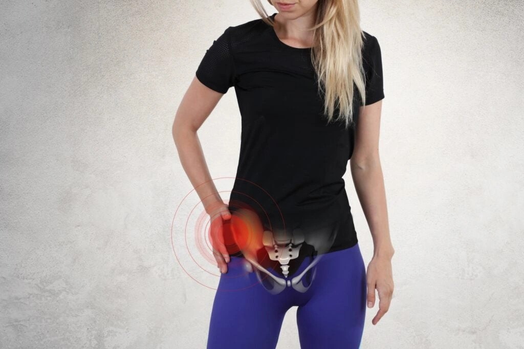
The Theater Sign describes anterior knee pain that worsens after prolonged sitting, especially with the knees bent. Specifically, this pain appears in the front of your knee when you stand after sitting for an extended period with your knees in a flexed position. The name comes from the classic scenario: you settle into a movie theater seat, watch a two-hour film, and then struggle with knee pain when standing up to leave.
This type of discomfort shows up because your patella (kneecap) stays compressed against your thigh bone when your knee remains bent. The longer you sit, the more pressure builds up in this joint space. When you finally stand, that accumulated pressure and stiffness creates the characteristic pain behind the kneecap.
How the theater sign differs from other knee pain
Theater sign pain has distinct characteristics that set it apart from other knee conditions. The pain concentrates in the front of your knee, right behind or around the kneecap, rather than on the sides or back of the joint. More importantly, the timing matters. This pain specifically appears after periods of rest with bent knees, not during activity.
Other knee pain typically worsens with movement or impact. Arthritis pain, for instance, tends to worsen throughout the day with use. Ligament injuries hurt during specific movements or weight-bearing. Theater sign pain does the opposite. It hits you after inactivity and often improves once you start moving around. The first few steps after standing hurt the most, but the discomfort usually decreases as you walk.
Common situations that trigger theater sign
Movie theaters aren’t the only place this pain strikes. You’ll notice it after long flights or car rides when your knees stay bent in cramped spaces. Office workers experience it after hours at a desk. Even activities you enjoy can trigger it: sitting through a concert, attending a long church service, or playing video games for extended sessions.
The pattern remains consistent across all these scenarios. Your knee stays flexed for 30 minutes or longer, and pain greets you when you try to stand. Some people notice it after kneeling for gardening or home repairs. Others feel it after sitting cross-legged on the floor. The common thread is always the same: prolonged sitting with bent knees followed by that uncomfortable moment when you straighten your leg and put weight on it.
Understanding this pattern helps you identify whether your knee pain fits the theater sign profile. If your knee hurts primarily when standing after sitting, rather than during walking or running, you’re dealing with a different mechanism than standard activity-related knee pain. This distinction becomes important when figuring out the right approach to address the problem.
Why does your knee hurt after sitting?
Several biological mechanisms work together to create that uncomfortable sensation when you stand up after sitting. Understanding these processes helps explain why your knees protest after inactivity.
Pressure buildup in the patellofemoral joint
Sitting keeps your knees in a bent position for extended periods, and this puts continuous pressure on your kneecap and surrounding tissues. When your knee stays flexed, your patella presses against the groove in your thigh bone with constant force. This sustained compression increases pressure within the patellofemoral joint space.
The longer you sit, the more this pressure accumulates. According to a 2016 study, about 50 percent of people with patellofemoral pain have problems with prolonged sitting when their knees remain bent. This pressure doesn’t just cause immediate discomfort. It restricts the normal gliding motion of your kneecap, and when you finally stand, your patella needs to readjust its position. That adjustment process triggers the pain you feel during those first few steps.
Reduced blood flow during prolonged sitting
Blood circulation around your knee joint slows down significantly when you sit for extended periods without movement. This reduced circulation leads to stiffness and discomfort when you attempt to stand or walk. Sitting for long hours causes blood flow throughout your body to decrease, particularly affecting your lower extremities.
Research supports the premise that excessive sitting and the consequent repeated exposure to reduced leg vascular shear stress perturbs the endothelium. When blood flow decreases, your muscles and joints receive less oxygen and fewer nutrients. This sluggish circulation contributes to that heavy, stiff feeling in your knees. The reduced blood flow also affects how quickly your body can remove metabolic waste products from the joint area, compounding the stiffness problem.
Cartilage compression and irritation
Prolonged sitting without movement reduces the flow of synovial fluid, which lubricates your joint. This fluid normally cushions and nourishes the cartilage in your knee. When you remain stationary, the cartilage under your kneecap becomes irritated due to continuous compression without adequate lubrication. The cartilage acts as a shock absorber, but extended pressure without movement prevents it from receiving fresh synovial fluid.
Repeated stress combined with inactivity can contribute to early cartilage degeneration, particularly in people above 35, overweight individuals, or those with previous knee injuries. The compression creates a cycle: less movement means less lubrication, which increases friction, which causes more irritation. Gentle movement keeps your joint lubricated and prevents stiffness.
Muscle stiffness and tightness
Your quadriceps at the front of your thigh and hamstrings at the back are key muscle groups involved in knee movement and stability. During sitting, these muscles stay in a relaxed or shortened state. Prolonged sitting leads to tightness in these areas, indirectly affecting your knee’s range of motion and overall stability.
When you sit for extended periods without movement, your muscles and tendons stiffen. During sitting, tendons generally relax, but prolonged inactivity causes tightness or stiffness in adjacent muscles. Sedentary habits weaken your quadriceps and hip muscles, and these muscles play a role in supporting your knee joint. Weak muscle support increases strain on your knee when you change posture, hence the sharp discomfort when standing after long periods of inactivity.
Patellofemoral pain syndrome: The main culprit
What is patellofemoral pain syndrome?
Patellofemoral pain syndrome (PFPS) stands as one of the most common causes of anterior knee pain. Medical professionals also call it runner’s knee or jumper’s knee, though you don’t need to be an athlete to develop this condition. In truth, experts estimate that around one-third of people who visit healthcare providers with knee pain have PFPS.
PFPS describes pain in the front of the knee and around the patella, or kneecap. The condition occurs when nerves sense pain in the soft tissues and bone around the kneecap, including the tendons, the fat pad beneath the patella, and the synovial tissue that lines the knee joint. Your patella normally fits into a groove in your femur and slides smoothly along that space when you move your knee. With PFPS, something affects how your patella moves and makes it painful.
How PFPS causes pain after sitting
The connection between PFPS and theater sign comes down to abnormal tracking of the kneecap. Excessive overload and abnormal tracking of the patella are among the main mechanisms behind PFP symptoms. When your patella has abnormal alignment, it may track laterally within the trochlear groove of the femur, causing increased stress and pressure on specific areas of the patellofemoral articular cartilage.
In some cases, a condition called chondromalacia patella is present, which involves the softening and breakdown of the articular cartilage on the underside of the kneecap. While there are no nerves in articular cartilage itself, damage to the cartilage can lead to inflammation of the synovium and pain in the underlying bone. This explains why sitting with bent knees for extended periods creates that dull ache behind your kneecap.
Other symptoms of runner’s knee
Pain on the front of the knee after sitting for a long period of time with your knees bent represents just one symptom of PFPS. The condition typically produces a dull, aching pain in the front of the knee that usually begins gradually and is frequently activity-related.
You might experience pain during exercise and activities that repeatedly bend the knee, such as climbing stairs, running, jumping, or squatting. Pain related to a change in activity level or intensity, playing surface, or equipment also signals PFPS. Popping or crackling sounds in your knee when climbing stairs or when standing up after prolonged sitting are common. The pain and stiffness can make it difficult to climb stairs, kneel down, and perform other everyday activities.
Risk factors for developing PFPS
The latest research suggests patellofemoral pain pathophysiology is a combination of biomechanical, behavioral, and psychological factors. Overusing your knees through repeated stress, such as jogging, squatting, and climbing stairs, can cause PFPS. Weak or tight muscles around your knee, especially your quad muscles, might not be able to support your knee properly.
Problems with the alignment of the legs between the hips and ankles may result in a kneecap that shifts too far toward the outside or inside of the leg. Muscular imbalances or weaknesses, particularly in the quadriceps muscles at the front of the thigh and the muscles that externally rotate and move the hip away from your body, contribute to poor tracking.
Certain demographics face higher risk. Athletes or physically active people who run, jump or squat frequently develop PFPS more often, as do people who do physical work. Women, teenagers, and adults age 20 to 40 also show increased susceptibility.
Other causes of knee pain after sitting
While PFPS accounts for most theater sign cases, several other conditions can cause knee pain after prolonged sitting. Recognizing these alternatives helps you understand when your symptoms might signal something different.
Early signs of knee arthritis
Osteoarthritis is the most common type of arthritis affecting the knee, and it gets worse with age. Early knee arthritis symptoms usually include pain and swelling, though other symptoms may develop later in the course of the disease. Pain that comes and goes for six months can be a symptom of knee arthritis. Feeling pain in your knees after certain activities such as long car rides, walking for extended periods, or sitting cross-legged is another sign of arthritis.
Knee stiffness after waking up is another common sign of knee arthritis. You may experience limited range of motion in the knees for several minutes after waking as the joints warm up. Many people notice sounds coming from one or both knees while they move or fully extend the joint. The pain tends to be worse when you move your joint or at the end of the day. Your joints may feel slightly stiff after rest, but this usually wears off quickly as you get moving.
Patellar tendinitis and bursitis
Patellar tendinitis causes irritation and inflammation of the patellar tendon, which runs from the kneecap to the shinbone. Pain is the first symptom of patellar tendinitis, most often between the kneecap and where the tendon joins the shinbone. Patellar tendinitis usually feels like a dull ache at the front of your knee, just below your kneecap.
Knee bursitis happens when one or more small fluid-filled sacs near the knee joint become inflamed. The affected portion of your knee might feel warm, tender and swollen. A direct blow to the knee can cause symptoms to come on fast, but knee bursitis often stems from friction and irritation of the bursae, occurring with jobs that require kneeling on hard surfaces.
Meniscus tears and cartilage damage
A meniscus tear usually happens when you twist your knee while playing sport, but it can also happen from more minor injuries such as twisting when standing up. Symptoms include knee pain or tenderness, stiffness or swelling around your knee, difficulty bending, straightening or moving your knee, and a crunching or clicking feeling when you move your knee.
When stiffness signals a bigger problem
Swelling that doesn’t go down after two days could mean internal damage like a torn ligament or fluid accumulation inside the joint. Pain that makes it hard to stand or walk may indicate a fracture or severe soft tissue injury. Pain with fever or warmth could signal an infection inside the joint that needs urgent care.
What to do about knee pain after prolonged sitting
Addressing knee discomfort after inactivity requires a multi-pronged approach combining immediate relief, preventive exercises, and habit modifications.
Immediate relief strategies
Rest, ice, compression, and elevation (RICE) helps with knee pain caused by minor injury or arthritis flare. Apply ice wrapped in a towel for 15 to 20 minutes three or four times a day. Stand up and walk for 2 to 3 minutes every 30 to 45 minutes. Gentle movement keeps your joint lubricated and prevents stiffness.
Stretches and exercises to prevent theater sign
Stretching addresses muscular imbalances by improving strength and flexibility. Sit back in your chair with a straight back, then straighten and raise one leg, holding for a slow count to 10. Repeat 10 times with each leg. Try calf raises and ankle rotations to enhance flexibility around your knees. Performing stretching and strengthening exercises four to five times a week helps ease pain and improve range of motion.
Changes to your sitting habits
Position your knees at or slightly below hip level to promote better circulation. Set your chair height so your feet rest flat on the floor with thighs parallel to the ground. Stand up and stretch every 20 to 30 minutes. Avoid sitting for more than 6 to 8 hours daily.
When to see a doctor for knee pain
Consult an orthopedic specialist if pain lasts more than a few weeks or affects daily life. Seek medical attention for severe or persistent pain, significant swelling or bruising, inability to bear weight, or knee deformity.
Conclusion
Knee pain after sitting doesn’t have to disrupt your daily life. Given these points about theater sign and patellofemoral pain syndrome, you now understand why your knees protest after movie marathons or long flights. The good news? Simple changes make a real difference.
Take frequent breaks during extended sitting, especially if your job keeps you at a desk. Perform the stretches and exercises I’ve outlined four to five times weekly. These habits address the root causes rather than just masking symptoms.
If your pain persists beyond a few weeks or worsens despite these measures, consult an orthopedic specialist. After all, early intervention prevents minor knee issues from becoming chronic problems.
FAQs
Q1. What causes knee pain when standing up after sitting for a long time?
When you sit with bent knees for extended periods, your kneecap presses continuously against your thigh bone, building up pressure in the joint. This compression, combined with reduced blood flow, muscle stiffness, and decreased joint lubrication, creates pain when you finally stand up and straighten your legs.
Q2. What are the early warning signs of knee arthritis?
Early knee arthritis typically presents as pain and swelling that comes and goes over several months. You may notice stiffness after waking up that improves with movement, discomfort after activities like long car rides or extended walking, limited range of motion, and clicking or popping sounds when moving your knee.
Q3. How can I relieve knee pain caused by prolonged sitting?
Stand up and walk for 2-3 minutes every 30-45 minutes to keep your joints lubricated. Apply ice wrapped in a towel for 15-20 minutes several times daily if needed. Adjust your chair height so your knees are at or slightly below hip level with feet flat on the floor, and perform regular stretching exercises to maintain flexibility.
Q4. Why is it difficult to walk immediately after sitting down for a while?
Muscle tightness, reduced circulation, joint stiffness, and weakened supporting muscles all contribute to difficulty walking after sitting. Your quadriceps and hamstrings remain in a shortened state during prolonged sitting, and reduced blood flow means less oxygen reaches your muscles and joints, making those first steps particularly challenging.
Q5. When should I see a doctor for knee pain after sitting?
Consult an orthopedic specialist if your knee pain persists for more than a few weeks, significantly affects your daily activities, or is accompanied by severe swelling, inability to bear weight, knee deformity, fever, or warmth around the joint. These symptoms may indicate a more serious underlying condition requiring medical attention.
Consult with Dr manu for Best orthopedic doctor in Mohali

Dr. Manu Mengi is a best orthopedic doctor in Mohali, specializing in joint pain, arthritis, and sports injuries. With qualifications in orthopedics and advanced training in joint replacement, he provides effective care for bone and joint conditions, helping patients improve mobility and manage pain with the right treatment approach.