Importance of protein after 40
Why deficiency is becoming common
Roughly 1 in 3 people over 50 fall short of their daily protein needs, and the consequences extend far beyond simple nutrition. Protein deficiency after 40 is a hidden culprit behind persistent muscle weakness, nagging knee pain, and frustratingly slow recovery times. Around age 30, we start to lose 3-5% of our muscle mass every decade, and sarcopenia affects 10-20% of older adults. In this guide, I’ll walk you through why protein deficiency becomes more common after 40, the warning signs to watch for, and how adequate protein intake can help you maintain strength, support joint health, and improve recovery as you age.
Why Protein Deficiency Becomes More Common After 40
Your body’s relationship with protein changes after 40, and understanding these shifts explains why deficiency becomes so prevalent. Multiple biological factors converge during middle age, creating a perfect storm for inadequate protein status.
Reduced Protein Absorption With Age
Aging brings alterations to protein digestion that directly impact amino acid availability. The digestive organs themselves change, affecting how efficiently your body breaks down and absorbs protein. Dentition decreases and mastication patterns shift, which can affect particle size reduction during chewing. These changes mean the protein you consume doesn’t get utilized as effectively as it once did.
In reality, your digestive system slows down as you age. Gastric emptying becomes sluggish, making you feel fuller for longer periods. This physiological change creates a cascade effect: you eat less frequently, consume smaller portions, and consequently take in less protein overall.
Reduced Appetite and Dietary Changes Between 15% and 30% of older adults experience appetite loss, a condition known as anorexia of aging. Your appetite involves a complex interplay of the brain, digestive system, hormones, and sensory nerves. As these systems shift with age, hunger signals naturally diminish.
Hormonal Changes and Muscle Loss
Age-related hormonal changes contribute to sensory impairment, reducing your sense of taste and smell. A recent study from the University of Plymouth suggests that the hormone signaling fullness may become overstimulated in elderly individuals. Sweet, salty, and umami tastes tend to decline with age, while sour and bitter flavors become more pronounced. Eating becomes less appealing when food doesn’t taste the way it used to.
Oral and dental problems compound these issues. Tooth loss, ill-fitting dentures, and dry mouth affect your ability to chew and swallow without pain. When eating becomes unpleasant or difficult, you naturally resist mealtimes.
Higher Protein Requirements After 40
Here’s the paradox: approximately 46% of individuals ages 51 and older don’t meet daily protein recommendations. While your appetite decreases, your protein requirements actually increase. Adequate protein is vital for preserving muscles, yet many older adults fall short.
Current dietary guidelines recommend 0.8 g/kg body weight daily, but emerging evidence shows this is insufficient for preventing sarcopenia. Studies indicate that 1.0 to 1.2 g/kg body weight per day is more effective for preserving lean muscle mass, functional performance, and overall strength. Lower protein intake makes building muscle mass more challenging, accelerating the decline that begins naturally around age 30.
Hormonal changes affecting protein metabolism
Hormonal shifts after 40 have a profound impact on protein metabolism. The decline in testosterone begins around the third to fourth decade in men, a phase known as andropause. Total testosterone levels decrease at approximately 1% per year, while free testosterone drops at about 2% annually. These reductions influence insulin sensitivity, with lower testosterone levels associated with higher glucose levels during oral glucose tolerance tests.
By the same token, growth hormone (GH) and insulin-like growth factor 1 (IGF-1) decline with age, termed “somatopause”. Both testosterone and GH are important regulators of muscle mass and strength during aging. The decline in hormone production commonly associated with age may play a critical role in the increased fat mass and decreased lean tissue that occurs over time. These metabolic processes directly affect how your body utilizes dietary protein, making adequate intake even more necessary.
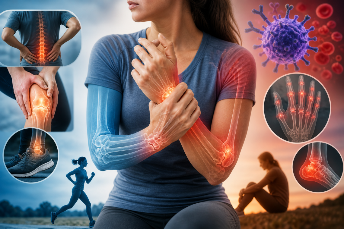
Signs and Symptoms of Protein Deficiency in Adults Over 40
Low protein levels develop gradually, and symptoms often get dismissed as normal aging. That’s why recognizing these warning signs matters for your health after 40.
Persistent fatigue and low energy levels
Feeling sluggish even after a full night’s sleep signals something more than just tiredness. Protein helps manage blood sugar and energy production, so when intake drops, your stamina declines noticeably. Daily tasks like cooking, showering, or cleaning become exhausting rather than routine activities. You might find yourself needing more rest breaks throughout the day or struggling to maintain focus during work hours. This persistent low energy stems from your body’s inability to sustain normal metabolic functions without adequate protein stores.
Muscle weakness and loss of strength
One of the most noticeable signs involves muscle loss. Your muscles break down faster than they can rebuild, leaving you weaker over time. You might notice clothes fitting looser around your arms or legs, or find yourself having more trouble getting up from a chair. Studies indicate adults ages 65 years and older should consume at least 0.5 grams of protein per pound of body weight. Grip strength diminishes, making simple actions like opening jars or carrying groceries more challenging. This weakness differs from temporary tiredness because it persists regardless of rest.
Brittle hair, dry skin, and weak nails
Protein supports the production of elastin, keratin, and collagen, which are key components for healthy hair, skin, and nails. When your hair thins unexpectedly, your nails split easily, or your skin feels perpetually dry, these changes point toward insufficient protein intake. Hair may lose its thickness and break more readily during brushing. Nails become soft, crack easily, or develop ridges. Skin loses elasticity and appears dull or flaky despite moisturizing efforts.
Slow wound healing and frequent infections
Scrapes and bruises taking longer to fade indicate protein deficiency affecting tissue repair. Protein is involved in the production of collagen and cytokines, which are essential for repairing skin and tissue. The healing process slows considerably when protein levels drop. Additionally, protein fuels the production of antibodies that help your body fight bacteria and viruses. Without sufficient intake, you experience more illnesses and infections while taking longer to recover from them.
Unexplained weight loss or muscle wasting
Weight loss that happens when you’re not trying to lose weight, even while eating high-calorie meals, raises serious concerns. Research shows 40% of people with cancer have cachexia when first diagnosed, and 70% with advanced cancer develop this condition. A healthcare provider may suspect protein deficiency if you’ve lost 5% or more of your weight within six to 12 months. This differs from intentional weight loss because muscle mass decreases while fat may also diminish, creating a wasted appearance.
Swelling in hands, feet, or legs (edema)
Protein deficiency causes fluid to build up in your hands, abdomen, legs, or feet. This happens when low protein levels affect how your body regulates fluids. The swelling, called edema, results from reduced human serum albumin levels, the most abundant protein in blood plasma. Albumin maintains oncotic pressure, a force that draws fluid into blood circulation. When albumin drops, fluid accumulates in tissues instead, causing visible puffiness and discomfort in affected areas.
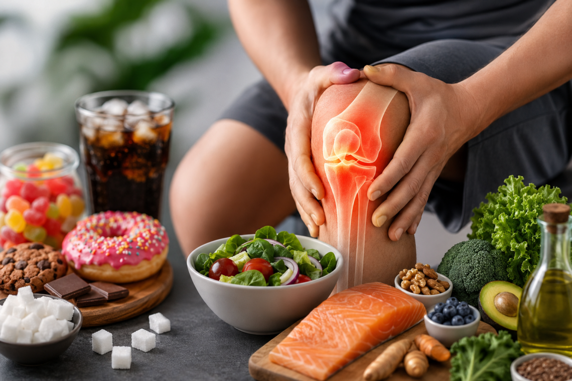
The Link Between Protein Deficiency and Knee Pain
Knee pain after 40 often traces back to inadequate protein intake, though most people never make this connection. Understanding how protein deficiency affects joint health reveals why that persistent ache in your knees might stem from what’s missing on your plate.
How protein supports joint tissue and cartilage
Collagen accounts for 30% of your body’s protein. This structural protein provides the foundation for connective tissues throughout your body, including the cartilage that cushions your knee joints. Type II collagen makes up about 90% of the collagen in cartilage, forming the essential matrix that allows joints to move smoothly.
Cartilage comprises about 60% collagen, creating a firm tissue that surrounds bones and absorbs shock from high-impact movements. Protein-rich foods contain amino acids like glycine, proline, and hydroxyproline that support collagen production and help your body repair cartilage damage. Without adequate protein intake, your body struggles to maintain this protective cushioning layer.
Muscle weakness increasing stress on knee joints
Weakness of the periarticular knee muscles initiates dynamic joint instability, resulting in asymmetric stress on the joint. When muscles surrounding your knee lack strength, they fail to properly stabilize the joint during movement. This instability forces the knee to compensate in ways that increase wear on cartilage and other tissues.
Both activation deficit and atrophy contribute to quadriceps weakness. Muscle impairments in people with osteoarthritis aren’t limited to quadriceps but involve multiple muscle groups around the knee. As a result, joints lose muscular support, worsening joint pain over time. Eating adequate protein helps slow this process down.
Reduced collagen production affecting joint health
Your body produces less collagen as you age, and existing collagen breaks down at a faster rate. The collagen quality also diminishes compared to when you were younger. Women experience a significant reduction in collagen production after menopause, while everyone sees a decline after age 60.
This breakdown means joints lose their structural integrity. Stiffness develops in tendons and ligaments, which become less flexible. Joint pain or osteoarthritis emerges due to worn cartilage. Amino acids from protein-rich foods strengthen the ligaments and tendons that anchor muscles to bones, supporting joint mobility and stability while lowering injury risk.
Inflammation and slower recovery from joint strain
Inflammation drives much of the joint pain experienced with osteoarthritis. Some protein sources help lower inflammation, particularly oily fish like salmon, which also contain omega-3 fats that carry additional anti-inflammatory properties.
Protein plays a fundamental role in tissue repair and the regeneration of blood vessels. Whether recovering from major surgery or a small scratch, protein is essential for healing. Consuming the right amount of protein aids in quicker recovery from joint strain. Without sufficient protein, the body takes longer to repair damaged joint tissues, extending pain and limiting mobility.
How Low Protein Causes Slow Recovery After Exercise
Exercise creates micro-tears in muscle fibers, and protein determines how quickly those tears heal. When protein intake falls short after 40, recovery stalls and performance plateaus.
Protein’s role in muscle repair and rebuilding
Protein helps repair micro-tears in muscle fibers caused by resistance training. Your body uses 20 different amino acids to build protein, but it can only synthesize 11 of them on its own. The other nine must come from your diet.
Branch-chain amino acids (BCAAs), particularly leucine, activate specific pathways that directly stimulate muscle protein synthesis. Leucine triggers the cellular machinery responsible for healing muscles. Research suggests consuming between 20 and 40 grams of protein post-workout optimizes muscle recovery. While doctors usually recommend taking protein one to two hours after exercise, total daily protein intake matters more than precise timing.
Why recovery time increases with age
Older adults experience anabolic resistance, meaning they require higher protein intake to maintain muscle function. The muscle protein synthetic response to food intake is blunted in older versus younger adults. This reduced responsiveness to meal ingestion represents a key factor in the loss of muscle mass.
Physical activity makes skeletal muscle tissue more sensitive to the anabolic properties of amino acid or protein administration. A single bout of resistance-type exercise increases the sensitivity of skeletal muscle tissue to protein feeding for at least 24 hours following cessation of exercise. Correspondingly, older individuals may experience a greater rise in post-prandial muscle protein synthesis following meals consumed one to two days following exercise.
Impact on sarcopenia and age-related muscle loss
Sarcopenia is a progressive condition characterized by the decline of skeletal muscle mass, strength, and physical function. Inadequate protein intake, along with hormonal imbalances, reduced physical activity, and chronic inflammation, accelerates muscle loss. Studies indicate that an intake of 1.0 to 1.2 grams per kilogram body weight per day is more effective for preserving lean muscle mass, functional performance, and overall strength.
Connection between protein intake and workout performance
Total daily caloric and protein intake over the long term play the most crucial dietary roles in facilitating adaptations to exercise. Ingestion of milk-based protein following a damaging eccentric resistance protocol helps attenuate the expected decrements in strength and repeated sprint ability from 24 to 72 hours following the bout. Good nutrition, especially adequate protein and energy intake, can help limit and treat age-related declines in muscle mass, strength, and functional abilities.
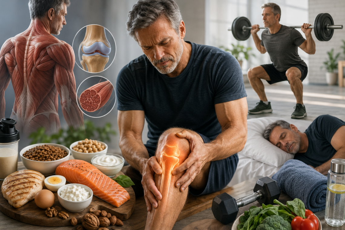
How to Prevent and Treat Muscle Atrophy After 40
Preventing muscle atrophy after 40 requires a strategic approach that combines adequate nutrition with targeted physical activity. Note that the standard recommendations often fall short for middle-aged adults.
Daily protein requirements for adults over 40
Between ages 40 and 50, your protein needs increase to about 1.0-1.2 grams per kilogram or 75-90 grams per day for a 165-pound person. Experts in the field of protein and aging recommend a protein intake between 1.2 and 2.0 g/kg/day or higher for elderly adults. People who exercise regularly have higher needs, about 1.1-1.5 grams per kilogram. Those who regularly lift weights or train for running or cycling events need 1.2-1.7 grams per kilogram.
General recommendations are to consume 15-30 grams of protein at each meal. Studies show that higher intakes in one sitting, more than 40 grams, are no more beneficial than consuming the recommended 15-30 grams at one time.
Best protein sources for middle-aged adults
Animal-based options include beef (3 ounces, 27 grams), chicken breast (3 ounces, 26 grams), eggs (3 medium, 21 grams), tuna (5-ounce can, 20 grams), and salmon (3 ounces, about 17 grams). For instance, cottage cheese provides 14 grams per half cup, while Greek yogurt offers 10-16 grams.
Plant-based protein sources deliver lentils (1 cup, 18 grams), beans (1 cup, 16 grams), peanuts (2 ounces, 14 grams), and nut butter (2 tablespoons, 7 grams). If you feel you need more protein, consider adding more beans, lentils, soy or seafood rather than processed supplements.
Combining protein with strength training exercises
Combining exercise and protein supplementation is the most effective method for improving muscle mass, strength, and physical function in older adults. Supplementing the diet with protein plus a regimen of heavy resistance exercise leads to the most improvement in muscle mass and strength in healthy older adults. Together, the two approaches can significantly aid muscle growth in older adults.
High-protein meal and snack ideas
Options like hard-boiled eggs, string cheese, jerky sticks and protein bars are protein-packed and packable. Add a protein source to your pasta, salad or stir fry. Top your salad with nuts or seeds. Add nuts or seeds to cereal or oatmeal. Add beans or lentils to soup. Add cheese to a sandwich, wrap or burger.
When to consider protein supplements
Protein supplements are probably best taken twice daily, if possible soon after exercise, in doses that achieve protein intakes of 30 grams or more. Research suggests that whey protein is particularly effective for building muscle in older adults, more so than either plant-based proteins or casein. For individuals who are avoiding dairy, plant-based protein powder options like soy isolate protein can also be beneficial.
Tracking your protein intake effectively
Tracking protein intake becomes a practical necessity rather than an optional measure. It enables you to identify shortfalls early, adjust meal composition, and ensure that the weight lost is predominantly fat rather than muscle. Distributing protein evenly across meals (25-30g per meal) optimizes muscle protein synthesis and helps meet daily targets.
Whey vs Plant Protein
Whey protein is commonly used for faster muscle recovery and repair because it contains all essential amino acids. Plant protein is a good alternative for people who follow vegetarian or dairy-free diets.
Lifestyle Tips to Prevent Muscle Loss
Strength Training
Regular strength training exercises help maintain muscle mass, improve joint support, and reduce the risk of age-related muscle loss.
Sleep and Recovery
Good-quality sleep is essential for muscle repair and recovery. Poor sleep can slow healing and increase fatigue levels.
Hydration and Balanced Nutrition
Proper hydration and balanced nutrition support muscle function, energy levels, and joint health. Combining protein with vitamins, minerals, and healthy fats helps maintain overall wellness after 40.
Muscle Weakness and Fatigue
Low protein can cause muscle weakness, tiredness, and low energy. It may also increase the need for joint pain treatment.
Knee Pain and Joint Weakness
Weak muscles increase pressure on joints, leading to knee pain and stiffness. Many people seek knee pain treatment and orthopedic consultation for relief.
Hair Fall and Weak Nails
Protein deficiency may cause hair fall, weak nails, and dry skin.
Slow Recovery After Exercise
Low protein slows muscle recovery after workouts and sports activities. This is common on many sports injury page topics.
Frequent Body Aches and Low Energy
Poor protein intake can cause body aches, weakness, and low stamina. It also affects overall health discussed in bone health blogs.
Conclusion
Protein deficiency after 40 creates a ripple effect that extends from your muscles to your joints and recovery time. As I’ve shown throughout this guide, your body requires more protein as you age, not less, with needs increasing to 1.0-1.2 grams per kilogram daily.
The good news? You can reverse muscle weakness, reduce knee pain, and speed up recovery by prioritizing protein-rich foods at every meal. Indeed, combining adequate protein intake with regular strength training delivers the most powerful results for maintaining muscle mass and joint health.
Start tracking your daily protein today and aim for 25-30 grams per meal. Your muscles, joints, and overall vitality will thank you for it.
Key Takeaways
After 40, your protein needs increase while absorption decreases, creating a perfect storm for deficiency that affects muscle strength, joint health, and recovery speed.
• Adults over 40 need 1.0-1.2g protein per kg body weight daily (75-90g for 165lb person) – significantly more than standard recommendations • Protein deficiency causes muscle weakness that increases knee joint stress, while reduced collagen production directly impacts cartilage health • Combining 25-30g protein per meal with regular strength training is the most effective strategy for preventing muscle loss after 40 • Warning signs include persistent fatigue, slow wound healing, brittle hair/nails, and unexplained muscle weakness beyond normal aging • Track daily protein intake and distribute evenly across meals to optimize muscle protein synthesis and maintain strength as you age
The solution is straightforward: prioritize protein-rich foods at every meal and pair adequate intake with resistance exercise to maintain muscle mass, support joint health, and improve recovery times throughout middle age and beyond.
FAQs
Q1. What are the consequences of having low protein intake?
Low protein intake leads to several noticeable effects including persistent fatigue, muscle weakness and loss of strength, brittle hair and weak nails, slow wound healing, frequent infections, and unexplained weight loss. You may also experience swelling in your hands, feet, or legs due to fluid buildup. These symptoms develop gradually and are often mistaken for normal aging, but they indicate your body isn’t getting enough protein to maintain essential functions.
Q2. Can protein deficiency cause knee pain?
Yes, protein deficiency can contribute to knee pain in multiple ways. When you don’t consume enough protein, your muscles weaken and fail to properly stabilize your knee joints, increasing stress on the joint itself. Additionally, low protein reduces collagen production, which is essential for maintaining healthy cartilage that cushions your knees. Strong muscles from adequate protein intake support and stabilize joints better, lowering the risk of knee pain from muscle loss.
Q3. How much protein should adults over 40 consume daily?
Adults over 40 need approximately 1.0-1.2 grams of protein per kilogram of body weight daily, which equals about 75-90 grams for a 165-pound person. This is significantly higher than standard recommendations. Experts suggest distributing this intake across meals, consuming 25-30 grams of protein per meal for optimal muscle protein synthesis. Those who exercise regularly may need even more, ranging from 1.2-1.7 grams per kilogram depending on activity level.
Q4. How long does it take to recover from protein deficiency?
Recovery from protein deficiency varies depending on severity and individual factors. Once you increase your protein intake to recommended levels (1.0-1.2g per kg body weight), you may notice improvements in energy levels within a few weeks. However, rebuilding muscle mass and strength takes longer, typically several months of consistent adequate protein intake combined with strength training exercises. The key is maintaining proper protein consumption over the long term rather than expecting quick fixes.
Q5. Why does recovery time after exercise increase with age?
Recovery time increases with age due to a condition called anabolic resistance, where older adults require higher protein intake to maintain muscle function. The muscle protein synthesis response to food intake becomes blunted compared to younger adults, meaning your body doesn’t respond as efficiently to dietary protein. Additionally, natural declines in hormones like testosterone and growth hormone affect how your body utilizes protein for muscle repair, making adequate protein intake even more critical for proper recovery after 40.
Q6. What are the common signs of protein deficiency after 40?
Common signs include muscle weakness, fatigue, slow recovery after exercise, increased body aches, hair fall, reduced stamina, frequent hunger, and gradual loss of muscle mass. Many people also notice worsening knee pain or difficulty climbing stairs.
Q7. How much protein do adults over 40 need
After 40, the body naturally starts losing muscle mass, a process called Sarcopenia. Protein helps maintain muscles, supports joint stability, improves recovery, and reduces weakness associated with aging.
Q8. Can protein deficiency cause knee pain?
Yes. Weak muscles around the knee provide less support to the joint. This can increase stress on the knee structures and worsen pain, especially while climbing stairs, squatting, or standing for long periods.
Q9. How much protein do adults over 40 need?
Most adults over 40 generally require around 1.0 to 1.2 grams of protein per kilogram of body weight daily. People who exercise regularly, recover from illness, or have muscle loss may need even more under medical guidance.
Q10. Why do muscles become weaker with age?
Aging reduces muscle-building hormones, physical activity, and protein absorption efficiency. Without adequate protein intake and resistance exercise, muscles gradually shrink and weaken over time.
Q11. Can protein deficiency slow recovery?
Yes. Protein is essential for tissue repair, muscle healing, and recovery after injury or surgery. Low protein intake may delay healing and prolong soreness or weakness.
Q12. Are protein supplements necessary after 40?
Good protein sources include eggs, fish, chicken, paneer, curd, milk, lentils, chickpeas, soy products, nuts, seeds, and protein supplements if needed.
Q13. Can vegetarians become protein deficient?
Vegetarians can meet their protein needs, but they must combine multiple protein sources carefully. Relying mainly on carbohydrates without adequate pulses, dairy, soy, or nuts may increase the risk of deficiency.
Q14. Does protein deficiency cause fatigue and low energy?
Yes. Protein helps maintain muscle function, energy balance, and metabolism. Low protein intake may lead to tiredness, weakness, and reduced physical endurance.
Q15 . Is muscle loss after 40 reversible?
In many cases, yes. Adequate protein intake, strength training, sleep, and proper nutrition can significantly improve muscle strength and slow age-related muscle loss.
Q16. Can low protein affect balance and increase fall risk?
Yes. Weak muscles reduce body stability and coordination, increasing the risk of falls, especially in older adults.
Q17. Are protein supplements necessary after 40?
Not always. Many people can meet their protein requirements through diet alone. However, supplements may help individuals who struggle to consume enough protein through food.
Q18. Can someone be overweight but still protein deficient?
Yes. A person may consume excess calories but still have inadequate protein intake. This can lead to “hidden malnutrition,” where body weight is high but muscle quality is poor.
Q19. Does protein help with exercise recovery and soreness?
Yes. Protein supports muscle repair after workouts and may reduce post-exercise soreness when combined with proper hydration and recovery.
Q20. When should someone consult a doctor about possible protein deficiency?
Medical advice should be considered if there is persistent weakness, unexplained muscle loss, severe fatigue, poor recovery, repeated falls, or ongoing joint pain despite lifestyle improvements.

Dr. Manu Mengi is a best orthopedic doctor in Mohali, specializing in joint pain, arthritis, and sports injuries. With qualifications in orthopedics and advanced training in joint replacement, he provides effective care for bone and joint conditions, helping patients improve mobility and manage pain with the right treatment approach.