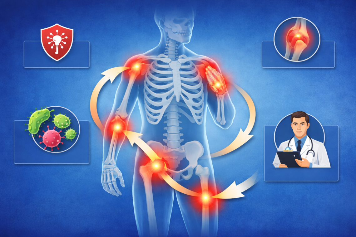
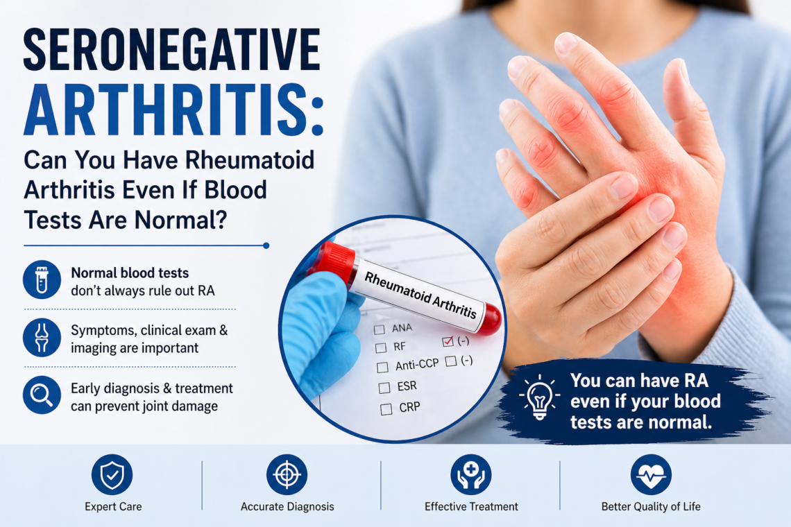
Why Do My Hands Go Numb at Night and How to Stop It From Happening – Dr. Manu Mengi

Why do my hands go numb at night? This question crosses your mind when you wake up with tingling fingers or a pins-and-needles sensation. Numbness in the hands is a frequently reported symptom, and nighttime hand numbness is more than just an occasional annoyance. It’s a signal from your body that something may be off.

In this article, I’ll walk you through common causes of hand numbness during sleep, including carpal tunnel syndrome, nerve compression, and poor sleep positions. I’ll also show you practical solutions to stop hand numbness at night and when you should see a doctor.

Common Causes of Nighttime Hand Numbness

Several medical conditions can cause your hands to go numb while you sleep. Understanding these causes helps you identify what’s happening and take appropriate action.

Carpal Tunnel Syndrome

Carpal tunnel syndrome occurs when the median nerve gets compressed in the narrow passageway on the front of your wrist. This compression causes tingling and numbness as the most common symptoms, along with weakness in grip strength. Repetitive hand motions like typing on a keyboard or using machinery may trigger it. Women develop carpal tunnel syndrome three times more often than men. Symptoms usually start slowly and often appear at night, sometimes waking you from sleep. Pain or tingling may feel like it’s coming from inside your hand or wrist, creating a sensation that makes you want to shake your hands.

Cervical Radiculopathy (Pinched Nerve in Neck)

A pinched nerve in your neck causes radiating pain, weakness, and numbness down your arm. Cervical radiculopathy affects approximately 85 out of 100,000 people. In over half of cases, the C7 nerve root is affected, while about a quarter of cases involve the C6 nerve root. Age-related wear and tear to spinal disks creates bone spurs and bulging disks that narrow the space in your cervical spine and place pressure on nerve roots. In younger people, a sudden injury resulting in a herniated disk is the most common cause. The pain typically starts at the neck and travels down the arm, described as burning or sharp.

Ulnar Nerve Compression

Ulnar nerve entrapment affects your ulnar nerve in your arm, causing numbness and tingling in your pinky and ring fingers. Two types exist: cubital tunnel syndrome at the elbow and Guyon’s canal syndrome at the wrist. Cubital tunnel syndrome is the second-most common peripheral neuropathy affecting your upper limbs. Sleeping with your elbows bent for long periods can cause or worsen symptoms. Ganglion cysts cause up to 40% of Guyon’s canal syndrome cases, while another 45% occur for no apparent reason. Signs come on gradually and may worsen at night or during activities that stretch or pressure your elbow or wrist.

Poor Sleep Position

Pressure on your hands from your sleeping posture is a likely cause of waking up with numb hands. When you sleep on your arm or hand, or in a position that puts pressure on a nerve, temporary lack of blood flow creates numbness or pins and needles. Changing your position typically provides enough relief for your symptoms. Sleeping with arms and elbows bent puts more pressure on nerves and increases numbness risk.

Thoracic Outlet Syndrome

Thoracic outlet syndrome develops when nerves or blood vessels in the lower neck and upper chest area are irritated, injured, or compressed. Neurogenic thoracic outlet syndrome affects 1 in 40,000 people annually. Numbness in the forearm, hand, and fingers are common symptoms of nerve compression, which can also cause pain in parts of your neck, shoulder, arm, or hand. Most people receive a diagnosis in their 30s, with repetitive stress injuries from sports and sudden trauma to the neck being common causes.

Diabetes and Peripheral Neuropathy

Diabetes causes high blood sugar that, over time, injures nerves throughout your body. Approximately half of all people with diabetes have some form of nerve damage, including peripheral neuropathy and carpal tunnel syndrome. Studies show that up to 50% of people with diabetes have peripheral neuropathy. High blood sugar damages small blood vessels that supply nerves with oxygen and nutrients. Symptoms include tingling, numbness, sharp stabbing pains, and a buzzing sensation. Peripheral neuropathy symptoms often worsen at night.

Which Fingers Go Numb? Understanding Nerve Patterns

The specific fingers that go numb provide clues about which nerve is compressed. Different nerves control sensation in different parts of your hand, creating distinct numbness patterns that help identify the underlying problem.

Thumb, Index, and Middle Finger Numbness

Numbness in your thumb, index, and middle fingers points to median nerve compression. Carpal tunnel syndrome creates this specific pattern because the median nerve provides sensation to these fingers and part of your ring finger. When tested, your small finger should have normal feeling if carpal tunnel is the cause. The median nerve sends touch, pain, and temperature sensations from the bottom side of your thumb, index, middle fingers, and part of your ring finger to your brain. Specifically, it affects the palm side of these fingers and the nail bed side of your index and middle fingers.

Symptoms typically worsen at night and often wake you from sleep. You might experience shock-like sensations radiating to these fingers or feel pain traveling up your forearm toward your shoulder. While other fingers and even your whole hand can sometimes feel numb, the classic pattern affects only the thumb, index, middle, and half of the ring finger following median nerve distribution. Numbness in all five fingers, particularly including the pinky, suggests additional nerve involvement or alternative diagnoses.

Pinky and Ring Finger Numbness

Tingling and numbness in your pinky and ring fingers indicates ulnar nerve entrapment. The ulnar nerve is responsible for sensation in your fourth and fifth fingers, part of your palm, and the underside of your forearm. This nerve travels from your neck to the tips of your pinky and ring fingers, helping you feel things with these fingers and move your hand.

The location of compression determines the exact pattern. Cubital tunnel syndrome at the elbow causes numbness on both the palm and back of your hand, but only on the little finger side. It affects the front and back of your little finger, plus the side of your ring finger next to your little finger. There is no numbness or tingling in your forearm or elbow since different nerves control those areas. Guyon’s canal syndrome at the wrist creates numbness and tingling on the pinky finger side, but the back of your hand usually stays normal.

Early signs include tingling in your ring and pinky fingers, particularly at night. The tingling can become so intense that it turns painful. Some people wake up at night because their fingers are numb. These symptoms happen more often when your elbow is bent, such as during phone calls or driving.

All Fingers and Both Hands

Symmetrical numbness in both hands starting from fingertips signals systemic conditions like diabetic neuropathy. This pattern progresses upward in a stocking-glove distribution and remains constant rather than fluctuating with position or activity. If numbness involves your forearm, thumb, index, or long fingers simultaneously with ulnar symptoms, a different condition is likely. Cervical radiculopathy creates variable patterns that change with neck position, distinguishing it from nerve compression at the wrist or elbow.

How to Stop Hand Numbness at Night: Immediate Solutions

Fortunately, you can take several immediate steps to reduce or eliminate hand numbness while you sleep. These practical solutions address the most common triggers.

Adjust Your Sleep Position

Sleep position is the primary reason hands go numb at night. Laying on your side, stomach, or with arms above your head bends your wrists in ways that cut off circulation and compress nerves.

Back sleeping offers the best option to minimize hand numbness. Keep your arms at your sides or rest them on your stomach. This position maintains straight wrists and reduces median nerve pressure.

If you prefer side sleeping, modifications help. Place your upper hand on your thigh and avoid tucking the other hand underneath your pillow, as your head’s weight pressures the nerves. Keep your elbows as straight as possible. Side sleeping shows a strong association with carpal tunnel syndrome development[57]. Hugging a pillow or body pillow prevents you from curling into the fetal position, which increases tunnel pressure. A pillow between your legs supports proper hip and spine alignment.

Use a Wrist Splint at Night

Wearing a splint at night holds your wrist in a neutral position, the position where carpal tunnel pressure is lowest. The splint prevents your hand from bending while you sleep, which worsens symptoms.

Studies show that wearing a splint can improve symptoms within a few weeks. Research demonstrates that splints combined with tendon and nerve gliding exercises improve carpal tunnel syndrome more than splints alone. Most people adjust to wearing a splint without sleep disruption. According to research, no particular splint type proves more effective than others, so you can try different options if one feels uncomfortable.

Improve Your Workspace Ergonomics

Daytime habits affect nighttime symptoms. Position your keyboard directly in front of you so wrists and forearms align and shoulders stay relaxed. While typing, keep wrists straight with upper arms close to your body and hands at or slightly below elbow level.

Set your monitor at arm’s length, 20 to 40 inches from your face, with the top at or slightly below eye level. Adjust your chair height so feet rest flat on the floor with thighs parallel to the ground.

Try Hand and Wrist Exercises

Specific exercises relieve pressure on the median nerve. Performing these consistently for six to eight weeks helps alleviate symptoms. Apply heat for 15 minutes before exercising, then ice for 20 minutes afterward to prevent inflammation.

Wrist extension stretches inner forearm muscles. Hold one arm straight out, bend your wrist back like making a stop sign, and gently pull your palm toward your body with the other hand. Hold for 15 seconds and repeat five times per arm.

When to See a Doctor About Hand Numbness

Recognizing when hand numbness requires professional medical attention protects you from potential complications. While mild, temporary numbness often resolves on its own, certain symptoms demand immediate action.

Frequency and Duration of Symptoms

Hand numbness that lasts more than a few hours warrants prompt medical attention. If you notice numbness coming and going repeatedly, seek care rather than waiting. Numbness that doesn’t go away, gets worse, or keeps coming back can signal a more serious condition.

Contact your provider specifically if numbness has no obvious cause, like your hand falling asleep from pressure. Pain in your neck, forearm, or fingers accompanying numbness also requires evaluation.

Muscle Weakness and Loss of Grip

Progressive weakness signals advancing nerve damage. Dropping things due to hand weakness indicates late-stage carpal tunnel syndrome, where tingling and pain have been present for an extended period before weakness develops. Pain that gets worse instead of better needs medical assessment.

Watch for difficulty coordinating finger movements. An obvious physical deformity of your hand or arm requires immediate attention.

Numbness During Daytime Activities

Numbness that interferes with everyday activities means you should talk to your doctor. If symptoms started after an injury or illness, speak with a provider.

Numbness spreading to other parts of your body needs evaluation. Dizziness, muscle spasms, or other unusual symptoms accompanying hand numbness warrant a medical visit.

Bilateral Hand Numbness

Seek immediate medical attention if sudden hand numbness comes with weakness, inability to move, difficulty speaking, dizziness, confusion, or a sudden headache. Call 911 if you cannot control arm or leg movement or have lost bladder or bowel control.

Numbness in other parts of your body alongside hand numbness, paralysis, confusion, slurred speech, vision loss, or rash require emergency care. These symptoms may indicate a heart attack or stroke[75]. In fact, sudden numbness with slurred speech or a change in vision signals a medical emergency.

Long-Term Prevention and Self-Care Strategies

Building sustainable habits prevents hand numbness from returning once you’ve addressed immediate symptoms. These long-term strategies create lasting relief.

Sleep Position Best Practices

Consistent sleep positioning requires deliberate practice. According to sleep specialists, if you start in one position but move throughout the night to one that causes numbness, investing in a brace keeps your elbow and wrist straight. Once you adapt to back sleeping, you can stop using the brace.

Pillow selection matters for nerve health. Your pillow should support your head and neck without causing abnormal spine bending. Stretching and massaging your wrists and hands before bed improves circulation and reduces tension.

Daily Hand and Neck Stretches

Regular stretching prevents nerve irritation and maintains mobility. Neck stretches done consistently can help relieve everyday pain and stiffness while preventing future problems. Many neck stretches work well in the workplace since they don’t require much space or equipment.

For carpal tunnel prevention, perform wrist exercises for three to four weeks under physical therapist guidance. Ice and heat therapy complement your stretching routine. Use heat therapy for tight neck and arm muscles, and ice for tender, inflamed ones.

Managing Underlying Health Conditions

Diabetes control directly impacts nerve health. Keep your A1C under 7% and maintain blood pressure below 140/90 mmHg or within your provider’s target range. Exercise helps lower blood sugar, improves blood flow, and keeps your heart healthy. Work up to 150 minutes of moderate or 75 minutes of vigorous aerobic exercise weekly.

Workplace and Activity Modifications

Breaking up repetitive tasks protects your nerves. Set alarms every 30 minutes while sitting to remind yourself to walk around. Apply the 20-20-20 rule: every 20 minutes, look 20 feet away for 20 seconds. Take a one to two minute break each hour to stretch your hands, wrists, and shoulders.

Conclusion

Hand numbness at night disrupts your sleep and signals underlying nerve issues that deserve attention. Most important, you now understand what causes those tingling fingers and how to address them effectively.

Start by adjusting your sleep position and wearing a wrist splint at night. These simple changes often provide relief within a few weeks. Likewise, improving your workspace ergonomics prevents symptoms from worsening during the day.

Pay attention to warning signs that require medical care, such as persistent numbness, muscle weakness, or symptoms that interfere with daily activities. Early intervention prevents nerve damage from progressing and helps you get back to restful, uninterrupted sleep.

Key Takeaways

Understanding the causes and solutions for nighttime hand numbness can help you achieve better sleep and prevent nerve damage progression.

• Sleep position matters most – Back sleeping with arms at your sides prevents nerve compression better than side or stomach sleeping positions.

• Wrist splints provide immediate relief – Wearing a neutral-position splint at night reduces carpal tunnel pressure and improves symptoms within weeks.

• Finger patterns reveal the cause – Thumb/index/middle numbness indicates median nerve issues, while pinky/ring numbness suggests ulnar nerve compression.

• Seek medical help for persistent symptoms – Numbness lasting hours, muscle weakness, or daytime interference requires professional evaluation to prevent permanent damage.

• Daily ergonomics prevent recurrence – Proper workspace setup, regular breaks every 30 minutes, and consistent hand stretches address root causes during waking hours.

The key to stopping nighttime hand numbness lies in combining immediate position adjustments with long-term prevention strategies, while recognizing when professional medical intervention becomes necessary.

FAQs

Q1. How can I prevent my hands from going numb while I sleep?

The most effective approach is to sleep on your back with your arms at your sides or resting on your stomach. This position keeps your wrists straight and reduces pressure on nerves. If you prefer side sleeping, avoid tucking your hand under your pillow and consider hugging a body pillow to prevent curling into positions that compress nerves.

Q2. What’s the fastest way to relieve hand numbness?

Wearing a wrist splint at night that holds your wrist in a neutral position can provide relief within a few weeks. Additionally, changing your sleep position, shaking out your hands, and performing gentle wrist stretches can offer immediate temporary relief when numbness occurs.

Q3. Can hand numbness become a permanent condition?

While some cases of hand numbness can be fully treated with proper intervention, others may result in lasting effects. Early treatment is crucial—if medical care at least prevents the condition from worsening, that’s considered a positive outcome. Permanent nerve damage is possible if symptoms are ignored for extended periods.

Q4. Should I be concerned if my hands go numb frequently at night?

Yes, persistent nighttime hand numbness warrants attention. While occasional numbness from sleeping on your arm is normal, frequent episodes could indicate conditions like carpal tunnel syndrome, ulnar nerve compression, or cervical radiculopathy. If numbness lasts more than a few hours, occurs repeatedly, or is accompanied by weakness, you should consult a healthcare provider.

Q5. Why do specific fingers go numb instead of my entire hand?

Different nerves control sensation in different fingers, creating distinct numbness patterns. Numbness in your thumb, index, and middle fingers typically indicates median nerve compression (carpal tunnel syndrome), while numbness in your pinky and ring fingers suggests ulnar nerve entrapment. These patterns help identify which nerve is affected and guide appropriate treatment.

Q.6 Why do my hands go numb at night while sleeping?

This usually happens due to pressure on nerves during sleep. Common causes include poor wrist position, carpal tunnel syndrome, or compression of nerves in the neck or elbow.

Q.7 Is it normal for hands to go numb during sleep?

Occasional numbness can be normal if you sleep in an awkward position. However, frequent or persistent numbness may indicate an underlying nerve compression issue.

Q.8 Why do I wake up with numb fingers?

When you sleep, prolonged pressure on nerves or reduced blood flow can cause your fingers to feel numb or tingly upon waking.

Q.9 Can carpal tunnel syndrome cause hand numbness at night?

Yes. Carpal tunnel syndrome commonly causes numbness, tingling, and pain in the thumb, index, and middle fingers, often worsening at night.

Q.10 Which sleeping position can cause hand numbness?

Sleeping with your wrists bent, hands under your head, or lying on your arm can compress nerves and lead to numbness.

Q.11 Why does shaking my hand relieve the numbness?

Shaking your hand improves blood flow and reduces pressure on the nerve, temporarily relieving symptoms.

Q.12 Can a neck problem cause hand numbness at night?

Yes. Conditions like cervical radiculopathy can compress nerves in the neck, causing numbness or tingling in the hands, especially at night.

Q.13 Can diabetes or vitamin deficiency cause numb hands at night?

Yes. Diabetes can damage nerves (neuropathy), and deficiencies like Vitamin B12 can also cause numbness and tingling in the hands.

Q.14 How can I prevent hand numbness while sleeping?

• Keep wrists in a neutral position
• Avoid sleeping on your arms
• Use a supportive pillow
• Consider a wrist splint if needed
• Maintain good neck posture

Q.15 When should I see a doctor for hand numbness at night?

Consult a doctor if:
• Numbness is frequent or worsening
• It is associated with weakness or loss of grip
• Symptoms persist during the day
• It affects daily activities

Hand numbness during sleep can signal nerve issues. Consult Dr Manu Mengi, Best orthopedic doctor in Mohali, and discover effective treatments and prevention tips.

Why Does My Back Pain Increase When I Stand Too Long? Causes and Relief Tips – Dr. Manu Mengi

Standing for just 2 hours can trigger lower back pain in 50% of healthy individuals due to muscle fatigue. You’re not alone if you find yourself asking, “Why does my back pain increase when I stand too long?” Indeed, 84% of adults worldwide face this issue at some point in their lives. Lower back pain when standing is often mechanical in nature, caused by factors including muscle strain, poor posture, and spinal compression. In this guide, we’ll explore the causes of prolonged standing back pain and provide practical relief tips to help you prevent back pain while standing.

What Happens to Your Spine When You Stand Too Long

Your spine faces a relentless opponent every moment you’re upright. Gravity never stops pulling downward, and when you stand for extended periods, this invisible force creates a cascade of mechanical changes in your back.

The mechanics of standing and spinal compression

Your spine isn’t a rigid pole holding you up. In reality, it’s a living suspension system built for balance, flexibility, and endurance. The S-shaped curves in your neck and lower back act like springs, distributing gravitational load evenly across the entire structure. When these natural curves maintain their proper shape, pressure spreads uniformly through each segment.

Each disk between your vertebrae functions as a shock absorber, filled with fluid and designed to handle compressive forces from above. When your alignment stays healthy, pressure distributes evenly through the disk. Lose that alignment, and the pressure becomes uneven, concentrating in small areas instead of spreading across the entire surface. This creates tiny tears and fluid loss over time.

Your muscles, tendons, and fascia form what’s known as tensegrity, a system where tension and compression balance each other. This allows you to stand upright, move efficiently, and resist fatigue. When the right joints, particularly in the mid-back and ribs, move properly, your nervous system can shift from constant bracing to true balance.

How gravity affects your lower back

Gravity compresses your spine, but it also organizes it when your body stacks correctly. An aligned spine lets gravity “seat” the joints naturally. A forward head or rigid rib cage, by contrast, creates shear forces your tissues must resist throughout the day. Over time, your nervous system learns that tension equals safety, and the cycle perpetuates itself.

A one-inch forward shift of the head adds roughly 10 pounds of extra force to the neck. Multiply that by thousands of hours annually, and those joints begin breaking down. Standing activities can irritate the lumbar spine when forces don’t travel cleanly through your body.

Every step pushes force up through your feet into your legs, pelvis, and spine. If your arches collapse or your hip stiffens, the upward force doesn’t travel cleanly. It detours into the low back or mid back. When alignment falters, gravity becomes a 24-hour workout. Every muscle holding you upright burns energy constantly. With proper spinal alignment, gravity passes cleanly through the structure, keeping energy cost low. When posture collapses, the muscles of your neck, back, and pelvis must continuously fight to prevent you from falling forward.

Why muscles fatigue during prolonged standing

Prolonged standing effectively reduces the blood supply to the muscles, resulting in the acceleration of the onset of fatigue. A work environment requiring extended standing was associated with fatigue of the lower extremity muscles, such as those of the anterior leg. Prolonged standing contributes to both psychological fatigue and muscle fatigue among workers.

Muscles that “hold on for dear life” create bracing patterns: clenched glutes, shrugged shoulders, tight jaw. Over time, your body confuses bracing with stability, and posture becomes tightness. Good alignment lowers unnecessary muscle tone so the right muscles can do the right job at the right time. The fix isn’t more tension but better stacking plus more movement options.

Common Causes of Back Pain When Standing Too Long

Standing demands that your spine support your entire body weight without the relief that sitting or lying down provides. This continuous load exposes underlying spinal conditions that might otherwise remain dormant. Several medical conditions explain why your back pain increases when you stand too long.

Muscle strain and ligament sprain

Muscle strains occur when you stretch or tear muscle fibers beyond their limit. These injuries happen suddenly during forceful movements or develop gradually through repetitive motions without adequate rest. Strains range from mild fiber breaks to complete tears requiring surgical intervention.

Chronic strains develop over time from repeating the same movement or maintaining awkward positions during prolonged standing. On account of weakened muscle fibers, your back becomes vulnerable to pain with ordinary use. Previous injuries increase your susceptibility to future strains in the same area.

Facet joint irritation

Facet joints connect your vertebrae and allow your spine to bend and twist. These small joints bear significant load when you stand upright. Facet joint irritation causes 15 to 41% of chronic low back pain cases.

The pain feels like a dull, aching sensation that wraps around your lower back. It worsens when you arch backward or stand straight, activities that compress these joints. Bending forward typically provides relief. The pain can radiate to your buttocks, thighs, or groin, staying on the same side as the affected joint.

Degenerative disk disease

Degenerative disk disease isn’t actually a disease but a condition where spinal disks wear down. Everyone’s disks degenerate over time as a natural part of aging. Almost everyone has some disk degeneration after age 40, even without symptoms.

The cushioning between vertebrae loses water content and becomes thinner. When these cushions wear away, bones can start rubbing together. This contact leads to back pain in about 5% of adults. The pain can be sharp, aching, or stiff, and it gets worse after bending, twisting, or lifting. Standing for extended periods intensifies the discomfort as gravity compresses already compromised disks.

Spinal stenosis pain while standing

Spinal stenosis narrows the space around your spinal cord, compressing nerves that branch from it. Pain begins in your buttocks and extends down your leg, sometimes reaching your foot. A heavy feeling develops in your legs, potentially leading to cramping.

In particular, standing for long periods or walking downhill worsens the symptoms. Sitting, leaning forward, or walking uphill lessens the pain. This pattern occurs because forward-leaning positions open the spinal canal, relieving pressure on compressed nerves.

Herniated or bulging disk

More than 3 million people in the U.S. develop a herniated disk annually. The soft, gel-like center of a disk pushes through cracks in the firmer outer layer, pressing on spinal nerves. This condition most often affects the lower back.

Standing loads the spine vertically, increasing pressure on the damaged disk. The leaked material presses harder against nearby nerves, causing lower back pain, tingling, numbness in legs and feet, and muscle weakness.

Compression fractures

Compression fractures are small breaks in vertebrae that cause the bone to collapse. An estimated 1 to 1.5 million compression fractures happen every year in the United States. These fractures occur most commonly in the thoracic spine.

Pain gets worse when you stand or walk but lessens when you lie down. Standing forces the fractured vertebra to bear weight it can no longer support. The pain typically starts gradually and worsens over time.

Factors That Make Standing Back Pain Worse

Beyond the structural issues in your spine, several controllable factors amplify back pain when you stand for extended periods. Recognizing these aggravators allows you to address them directly.

Poor standing posture

Poor posture increases mechanical stress in the lower back, which directly leads to pain. When you stand with misaligned posture, your spine loses its natural support system. Standing with your hips tilted forward, slouching with rounded shoulders, or developing a flat back where your spine loses its natural curvature all strain your lower back muscles. Your balance center shifts, forcing your muscles to work harder just to keep you upright. These unhealthy postures create an uncomfortable position that, when maintained too long, triggers muscle strain.

Wearing unsupportive footwear

The shoes you wear determine how forces travel up through your legs, hips, and spine. High heels push your body weight forward, forcing your lower spine to arch more than normal. Completely flat shoes or flip-flops lack cushioning, allowing repeated impact on the spine with each step. Over time, these misalignments contribute to muscle fatigue, disk compression, and chronic lower back pain.

Shoes with insufficient arch support cause overpronation (inward rolling of the foot) or supination (when the foot rolls outward). Either condition creates a domino effect that results in a misaligned spine. Old shoes with worn-out soles lose their ability to cushion and stabilize your movements. Uneven soles may cause one side of your body to bear more weight than the other, disturbing spinal alignment.

Weak core and glute muscles

Increased sedentary jobs and sitting time lead to muscle imbalances and weak core musculature, putting your low back at increased risk of injury. The deep core muscles are meant to endure prolonged activation to support and stabilize the spine. When you slouch in a chair all day, the core remains relatively inactive.

The gluteal muscles become lengthened and weak, a condition known as “glute amnesia”. These muscles are then unable to do their job in regular activities of daily living, forcing other muscles, including those in the low back, to compensate. Weak gluteal muscles contribute to lower back pain by passing their work to the low back.

Flat feet and arch problems

People with flat feet are 4.5 times more likely to experience chronic lower back pain. When you stand and your feet are well supported by healthy arches, it encourages proper alignment in your legs. When you have flat feet, your arches collapse and your ankles can roll inward. This upsets the alignment up through your legs and into your hips and lower back, leading to joint pain and muscle strain as your body compensates for the misalignments.

Weight and body mechanics

Your body weight affects how forces distribute through your spine when standing. Excess weight increases the load your spine must support, intensifying compression on already stressed structures. The way you move and position yourself further influences this distribution pattern.

Stress and emotional factors

Stress leads to inflammation, muscle spasms, and tension in your back. Muscle tension occurs when stress causes the muscles in your back to tense up, leading to stiffness and pain. Chronic stress triggers inflammation throughout the body, including in the back. Stress makes the body more sensitive to pain. During stressful times, your blood vessels may constrict, reducing blood flow to your back muscles and causing pain. Prolonged stress makes people more susceptible to back injury and back pain.

How to Get Relief from Back Pain After Standing

When back pain hits after prolonged standing, you need quick, effective strategies to find comfort.

Shift your weight and change positions

Moving your body weight from one leg to the other relieves pressure on fatigued muscles. This simple action prevents one side from bearing continuous load. If possible, prop one foot on a low stool or step, alternating feet every few minutes to redistribute spinal compression.

Use heat or cold therapy

For chronic back pain lasting more than four weeks, apply heat to relax tissues and decrease pain signals. Use a heating pad or adhesive wrap that provides several hours of low-level warmth. Apply heat for 10 to 15 minutes before activity to prepare muscles for movement.

Ice works best immediately after new soreness develops. Wrap ice packs in a towel to protect your skin from frostbite. Apply cold therapy for up to 20 minutes at a time. After one day of icing, switch to heat to encourage tissue healing.

Take movement breaks

Standing stretches can provide lower back pain relief in as little as 30 seconds. Brief movement interrupts the static load on your spine, allowing fresh blood flow to reach tired muscles.

Gentle stretches for immediate relief

Hold each stretch for 30 seconds or as long as comfortable. Try these positions: lie on your back, pull one knee toward your chest, hold five seconds, then repeat with the other leg. For rotational relief, keep shoulders flat on the floor and slowly roll bent knees to one side, holding 5 to 10 seconds. Child’s Pose stretches your lower back by sitting on your heels and leaning forward with arms extended.

Over-the-counter pain relief options

Acetaminophen reduces pain with fewer side effects than other options. Take no more than 3,000 mg in 24 hours to avoid liver damage. NSAIDs like ibuprofen reduce swelling around irritated disks or joints. Tell your provider if you take over-the-counter pain relievers for more than two weeks.

How to Prevent Back Pain While Standing

Prevention targets the root causes of standing-induced back pain before symptoms develop. Implementing these strategies protects your spine from prolonged standing stress.

Strengthen your core muscles

Core muscles act as your body’s natural support system, reducing stress on the spine and protecting spinal disks from compression and injury. Strong core muscles distribute forces evenly across the spine, whereas weak cores force the lower back muscles to work harder. Engage your core muscles even when you’re not exercising. A quick 10-minute routine done a couple of times a week strengthens these stabilizing muscles.

Improve your standing posture

Think of a straight line passing through your body from ceiling to floor, with ears, shoulders, hips, knees, and ankles aligned vertically. Keep your shoulders back, pull your stomach in, and distribute weight mostly on the balls of your feet. Hold your pelvis level and resist standing on tiptoe.

Choose supportive footwear

Shoes with a stiff sole and curved rocker bottom work best for prolonged standing. A stiff sole absorbs walking impact, transferring ground reaction forces away from your foot. Flexible shoes don’t provide the support your foot needs for extended periods. Choose shoes with a wide toe box and arch support insoles to improve balance and foot function.

Use anti-fatigue mats on hard surfaces

Anti-fatigue mats reduced low back pain in pain developers from 6.8 mm to 3.6 mm on a visual analog scale. These mats facilitate subtle movements at the foot-floor interface, providing selective benefit to individuals prone to developing standing-induced back pain.

Practice exercises to build endurance

Standing stretching exercises may provide lower back pain relief in as little as 30 seconds. Regular physical activity strengthens the core and increases stability.

Manage your weight

Excess weight weakens abdominal muscles, causes pelvic and spine problems, and contributes to low back pain. Maintaining a healthy weight protects spine health and prevents weight-related spine disorders.

Conclusion

Back pain from prolonged standing stems from mechanical stress on your spine, particularly when gravity compresses disks, irritates facet joints, and fatigues supporting muscles. Now that we’ve identified the underlying causes, you can address them directly rather than simply enduring the discomfort.

Relief doesn’t require complicated interventions. In fact, simple strategies like shifting your weight, taking movement breaks, and applying heat or cold therapy provide immediate comfort. For lasting results, focus on strengthening your core, improving your posture, and choosing supportive footwear.

Your spine responds quickly to positive changes. Start with one or two prevention strategies today, and you’ll likely notice improvements within days.

Key Takeaways

Understanding why your back hurts after standing and implementing targeted relief strategies can significantly improve your comfort and spinal health.

• Prolonged standing compresses spinal disks and fatigues muscles – gravity continuously loads your spine, causing mechanical stress that triggers pain in 50% of healthy individuals after just 2 hours.

• Poor posture and weak core muscles amplify standing back pain – misaligned spine forces muscles to work harder, while weak glutes and core create compensation patterns that strain the lower back.

• Immediate relief comes from weight shifting, movement breaks, and heat/cold therapy – simple position changes and 30-second stretches can provide quick pain relief by redistributing spinal pressure.

• Prevention requires core strengthening, proper footwear, and posture awareness – building muscle endurance and maintaining spinal alignment protects against future standing-induced back pain.

• Anti-fatigue mats and supportive shoes reduce standing stress – proper footwear with arch support and cushioned surfaces can cut back pain severity nearly in half for pain-prone individuals.

The key to managing standing back pain lies in addressing both immediate symptoms and underlying causes through targeted movement, strengthening, and ergonomic improvements.

FAQs

Q1. Why does standing for extended periods cause back pain?

Standing for long periods compresses your spinal disks and fatigues the muscles supporting your spine. Gravity continuously pulls downward, creating mechanical stress on your lower back. When you maintain an upright position without movement breaks, muscles must work constantly to keep you balanced, leading to fatigue and pain. Poor posture, weak core muscles, and improper weight distribution can intensify this discomfort.

Q2. How quickly can standing trigger lower back pain?

Back pain can develop surprisingly fast when standing. Research shows that just 2 hours of standing can trigger lower back pain in 50% of healthy individuals due to muscle fatigue and spinal compression. The onset varies depending on factors like your posture, core strength, footwear, and any underlying spinal conditions you may have.

Q3. What immediate steps can I take to relieve back pain after standing?

For quick relief, shift your weight from one leg to the other and change positions frequently. Apply heat therapy to relax tight muscles or use ice for new soreness. Take brief movement breaks and perform gentle stretches like pulling your knee to your chest or doing Child’s Pose for 30 seconds. Over-the-counter pain relievers like acetaminophen or ibuprofen can also help reduce discomfort.

Q4. Can my shoes really affect my back pain when standing?

Yes, footwear plays a significant role in standing-related back pain. Shoes with poor arch support, high heels, or completely flat soles alter how forces travel through your legs and into your spine. Unsupportive footwear can cause misalignment that forces your lower back muscles to compensate. Choose shoes with a stiff sole, curved rocker bottom, wide toe box, and proper arch support for prolonged standing.

Q5. How can I prevent back pain from developing when I need to stand for long periods?

Prevention focuses on strengthening your core muscles, which act as your spine’s natural support system. Maintain proper standing posture by keeping your ears, shoulders, hips, and knees aligned vertically. Wear supportive footwear and use anti-fatigue mats on hard surfaces. Practice regular core-strengthening exercises, take frequent movement breaks, and manage your weight to reduce stress on your spine.

Q6. Why does my back pain increase when I stand too long?
When you stand for prolonged periods, your spinal muscles fatigue and your lumbar joints (especially facet joints) take more load. Over time, this leads to stiffness, muscle strain, and pain.

Q7. Why does my back feel better when I sit or bend forward?
Sitting or bending forward reduces pressure on the lumbar spine and facet joints. It also relaxes overworked back muscles, which is why pain often decreases.

Q8. Is it normal to have back pain after standing for long hours?
Occasional discomfort can be normal, especially in people with weak core muscles or poor posture. However, persistent or severe pain may indicate an underlying issue like disc degeneration or spinal stenosis.

Q9. Can poor posture while standing cause back pain?
Yes. Slouching, leaning to one side, or standing with an exaggerated arch in the lower back increases strain on muscles and joints, leading to pain.

Q10. Which conditions can cause back pain while standing?
Common causes include:
• Muscle fatigue or weakness
• Facet joint arthritis
• Lumbar spinal stenosis
• Degenerative disc disease
• Postural imbalance

Q11. Does standing too long damage the spine?
Standing alone doesn’t usually damage the spine, but prolonged standing with poor posture can accelerate wear and tear, especially in people with pre-existing spine issues.

Q12. How can I prevent back pain when I have to stand for long hours?
• Maintain good posture
• Shift weight frequently
• Use supportive footwear
• Take short sitting breaks
• Strengthen core muscles

Q13. Can flat feet cause back pain when standing?
Yes. Flat feet alter body alignment and can increase stress on the lower back, especially during prolonged standing.

Q14. What exercises help reduce back pain from prolonged standing?
Core strengthening, stretching (especially hamstrings and hip flexors), and lower back stabilization exercises are very effective in reducing pain.

Q15. When should I see a doctor for back pain from standing?

You should consult a doctor if:

  • Pain persists for more than a few weeks
  • Pain is severe or worsening
  • There is numbness, tingling, or weakness in the legs
  • Pain interferes with daily activities

Consul with Dr. Manu Mengi for all the Back problems he is the best orthopedic doctor in Mohali

Joint Cracking Sounds: What’s Normal and When You Should Actually Worry – Dr. Manu Mengi

If you’ve ever heard your knees pop during a squat or felt your knuckles crack, you’ve probably wondered: why do my joints crack or make sounds, and is it dangerous? You’re not alone. Up to 54% of people crack their knuckles, and many don’t understand why joints pop and crack in the first place. The good news is that studies show joint noise alone does not lead to injury or damage. However, understanding what causes these sounds and when they signal a problem can help you distinguish between harmless popping and something that needs medical attention.

What causes joint cracking sounds

Your joints contain a lubricating fluid called synovial fluid that keeps bones moving smoothly against each other. When you stretch or move a joint, you create a change in pressure within the joint capsule. This pressure change causes dissolved gasses in the synovial fluid (primarily carbon dioxide at about 80%, along with oxygen and nitrogen) to rapidly come out of solution and form a bubble. The sound you hear is either from the bubble forming or collapsing back into the fluid.

Gas bubbles in synovial fluid

The bubble formation process, known as cavitation, happens when joint surfaces separate and the volume inside the joint capsule increases. The existing synovial fluid can’t fill this expanding space, creating negative pressure[18]. Gasses dissolved in the fluid quickly liberate and fill the empty space, producing that familiar cracking or popping sound.

Research using real-time MRI in 2015 showed that bubbles actually remain in the fluid after a joint cracks, suggesting the sound occurs when the bubble forms rather than when it collapses. This explains why you can’t immediately crack the same joint again. The refractory period lasts about 20 minutes while gasses slowly reabsorb back into the synovial fluid.

Tendons and ligaments moving over bones

Joints are held together by ligaments, while tendons connect muscles to bones. These tissues sit close to bony structures and sometimes shift position during movement. When a tendon or ligament slides over a bone prominence and then snaps back into place, it creates a clicking or snapping sound.

You might notice this sensation in your ankle when rotating it or in your knee while climbing stairs. The snapping occurs more frequently if joints are tight or after periods of inactivity. While this movement is typically harmless, it can sometimes indicate tightness or misalignment.

Cartilage changes with movement

Cartilage cushions the ends of bones where they meet in a joint. When cartilage wears down or becomes damaged, bones can rub together with increased friction. This contact produces a grinding or crunching sensation called crepitus, which feels different from a clean pop.

Crepitus often appears as a rough, repeated noise every time you move the joint. The sound can signal cartilage breakdown, particularly in conditions like osteoarthritis where bone spurs develop and rub against each other during movement. Reduced synovial fluid also contributes to these grinding sounds by allowing more friction between cartilage surfaces.

Age-related joint changes

Joints naturally make more noise as you age. Cartilage wears away as part of normal aging, making joint surfaces rougher. These rougher surfaces create more noise when they rub against each other, even in otherwise healthy joints[13].

The changes don’t necessarily indicate a problem. Your body simply experiences natural wear over time, particularly if you’re over 30. As long as the sounds aren’t accompanied by pain, they’re usually part of normal joint aging.

Is joint cracking normal or harmful

Most joint cracking is completely normal and harmless. Millions of people experience popping, clicking, and cracking in their joints every day without any negative health consequences. The real question isn’t whether joints should make noise, but rather when that noise signals an actual problem.

Joint popping without pain is harmless

In general, crepitus alone is not a cause for concern. Joints naturally make some noise during movement, and this becomes more common as you age. A joint crack can reach volumes as loud as 83 decibels, about the same as a running garbage disposal. Despite the alarming sound, the noise itself is safe and can even help restore range of motion and decrease pain.

Research shows that knee crepitus alone has no negative effect on function or physical activity level. The important distinction is pain. As long as joint noises occur without pain, they’re OK. Your body is simply going through normal mechanical processes. Studies confirm that in the absence of pain, swelling, or other joint symptoms, these sounds are probably nothing to be concerned about.

One interesting finding shows that many people develop negative thoughts about their joints making noise and self-limit their activity as a result. Patients with crepitus self-report more pain and lower function, however no differences were found in walking or knee strength for those with or without crepitus. This suggests the psychological impact of joint sounds often exceeds the physical reality.

Does cracking knuckles cause arthritis

The belief that cracking your joints is bad for them is wrong. Contrary to popular belief, studies have shown no direct link between joint cracking and arthritis. Arthritis is more commonly caused by genetics, aging, or injury. Several studies that compared rates of hand arthritis among habitual knuckle-crackers and people who didn’t crack their knuckles reached the same conclusion: cracking your knuckles doesn’t raise your risk for arthritis.

The most convincing evidence comes from a California physician who conducted an experiment on himself over his lifetime. He regularly cracked the knuckles of only one hand, and after decades of this behavior, x-rays showed no difference in arthritis between his hands. This experiment lasted over 50 years. The researcher found no difference between his left hand and the uncracked knuckles of his right hand.

A larger study published in 1990 examined 74 people who regularly cracked their knuckles compared to 226 people who didn’t. The incidence of arthritis was the same in both groups. However, habitual knuckle crackers were more likely to have hand swelling and lower grip strength. Chronic knuckle-cracking may lead to reduced grip strength, and there are at least two published reports of injuries suffered while people were trying to crack their knuckles.

Joint noises in athletes and active people

You can work out even if you have crepitus. Especially if you have no pain with the movements, you can continue to do your workouts. The noise you hear does not always mean anything is getting worse or injured. Studies have shown that crepitus is more common in those with other diagnoses associated with knee pain, but knee crepitus alone doesn’t prevent physical activity.

Many people get fearful when they hear noise in their joints and worry about hurting themselves or causing damage. Therefore, they stop doing activities. This fear-based response is unnecessary when the joint noise is painless. Recent findings indicate that knee crepitus is not linked to the need for knee replacement down the line.

Different types of joint sounds and what they mean

Not all joint sounds are created equal. The type of noise your body makes can tell you whether you’re experiencing normal joint function or something that warrants attention.

Popping sounds

A sharp pop typically indicates air moving within the joint space. When you hear a distinct popping noise, air is seeping into the soft tissues around the joint, like your kneecap. The bubbles burst when you bend the joint, creating that characteristic sound. You might notice these pops when you bend your knee or elbow.

Popping can affect any joint in your body, and the sound may be muffled or easily heard. Knees are the most common location where people experience this type of noise, but it happens in hands, feet, lower back, hips, and shoulders as well. The key indicator of a harmless pop is its painless nature.

Clicking or snapping sounds

Clicking differs from popping in both quality and cause. When you hear a click or snap, tendons or ligaments are moving over the bony surfaces of your joint. This snapping occurs due to irregularities and intermittent contact between these structures.

On the other hand, clicking can sometimes involve discomfort. Tension or misalignment in tendons or ligaments produces audible sounds during stretching, twisting, or other motions that strain these tissues. In your ankle, for example, tendons on the side will pop as they move back and forth, though these usually don’t hurt much. Your kneecap and meniscus can shift slightly during movement, creating clicks as fluid shifts and pressure differentials change.

Grinding or crunching sounds (crepitus)

Grinding sounds signal a different situation altogether. When you hear a grinding, rubbing, crunching, or crackling sound from your knee, damaged or deteriorated cartilage is likely present. This type of crepitus feels rough and repeated every time you move the joint.

The grinding occurs because your bones are rubbing against each other, usually caused by arthritis. Crunching sounds in your knee when you climb stairs or kneel, or crackling and grinding sensations when you move your shoulder, typically indicate cartilage breakdown. Unlike harmless pops, crepitus may be a symptom of osteoarthritis, tendonitis, meniscal tears, or other forms of joint damage.

Cracking sounds during exercise

During workouts or repetitive movements, you might hear joints cracking more frequently. These sounds happen because the muscles you’re using are tight. Muscle tightness creates friction on the bone, leading to noise generation.

When stretching produces a loud pop followed by relief in the area, the stretching has done its job. This particular scenario signals that tension has released, though it implies you should incorporate more stretching into your routine to prevent muscle tightness and potential injuries.

When joint cracking becomes a concern

While most joint sounds are benign, certain symptoms transform harmless popping into a medical concern. Pain changes everything. If cracking is coupled with consistent pain or swelling, that signals something is wrong. According to medical guidelines, there are only two scenarios where popping requires a healthcare visit: experiencing chronic pain in the joint or pain specifically after it cracks.

Joint cracking with pain or swelling

Pain can indicate a torn ligament or tendon, damaged cartilage, or even a pinched nerve. When crepitus comes with pain, you should consult a doctor to evaluate the cause. Swelling around the joint, whether occasional or continual, compounds the concern. These symptoms together suggest underlying damage that won’t resolve on its own.

Several conditions produce painful crepitus. Osteoarthritis causes cartilage to rub away, leaving bones unprotected and creating inflammation. When bones rub and grind, it causes pain and stiffness that usually gets worse with activity. Patellofemoral pain syndrome creates crepitus along with pain behind the kneecap, often from suddenly increasing activity levels through running, squatting, or jumping. Torn cartilage from sports injuries, falls, or accidents represents another possible cause of painful crepitus.

Sudden changes in joint sounds after injury

Joint cracking can occur due to sports injuries or accidents. Cartilage damage causes bones to rub together and make noise, producing sound along with pain, soreness, and swelling. In the event that you notice new or different sounds following an injury, seek medical evaluation without delay.

Joints that lock or feel unstable

Joint instability happens when tissues such as muscles, ligaments, and bones weaken and can no longer keep bones properly aligned. Symptoms include repeated dislocation, tenderness, and a feeling that the joint gives out. Chronic instabilities present with mechanical symptoms such as locking, catching, clicking, or giving way, particularly with twisting movements.

Locked knee occurs when you cannot bend or straighten your knee. True locked knee happens when something physically prevents the knee from straightening, while pseudo locked knee results from severe pain triggering muscle spasms.

Persistent grinding with stiffness

Persistent grinding combined with stiffness indicates wear and tear. Your doctor will move the joint to test the strength and looseness of surrounding tissues. If conservative treatments don’t help, surgery may repair ligaments so the joint regains stability.

How to reduce or prevent joint cracking

You can reduce how often your joints crack by keeping them active and healthy. The principle is simple: “motion is lotion”. The more you move, the more your body lubricates itself. When you sit or lie around for extended periods, fluid in your joints doesn’t circulate. Regular activity stimulates synovial fluid production, which reduces friction and minimizes the popping sounds.

Regular stretching and movement

Moving every 20 to 30 minutes helps reduce stiffness and preserve joint motion. Low-impact exercises work particularly well for managing joint sounds. Swimming, cycling, and yoga improve flexibility without stressing your joints. These activities increase bone density, promote circulation that delivers nutrients to cartilage, and release joint lubricant that reduces wear and tear.

Warming up before exercise prepares your body and prevents injury. Similarly, cooling down afterward reduces joint stiffness and soreness. Aim for at least 30 minutes of exercise daily, three days a week.

Strengthening muscles around joints

Strength training builds muscle mass around joints, which improves range of motion and reduces inflammation. Progressive resistance training two to three times weekly supports joint health and slows age-related decline. Exercises like resistance band work, squats, lunges, and shoulder presses strengthen the muscles that stabilize your joints. Stronger muscles reduce the load on joints themselves, helping prevent degeneration.

Staying hydrated for joint lubrication

Your cartilage consists of 65% to 80% water. Water stimulates production of synovial fluid that cushions your joints and acts like a shock absorber. When you’re dehydrated, your body can’t create sufficient synovial fluid, leading to more friction and pain. Dehydration also increases chronic inflammation in joints. Drink at least eight glasses of water daily.

When you should see a doctor

See a healthcare provider if you experience chronic pain in a joint or pain specifically after it cracks. You should also get help if you feel so much pressure in a joint that you have to pop it to feel comfortable.

Conclusion

Joint cracking might sound alarming, but overall, it’s just your body doing what bodies do. As long as your joints pop and crack without pain or swelling, you have nothing to worry about. The myth about knuckle cracking causing arthritis has been thoroughly debunked, so you can put that concern to rest.

Pay attention to what your body tells you. Pain changes everything. Equally important are signs like swelling, locking, or instability that accompany the sounds. These symptoms warrant a doctor’s visit.

Keep your joints healthy through regular movement, strength training, and proper hydration. Motion truly is lotion for your joints, and staying active will help you maintain mobility for years to come.

Key Takeaways

Understanding joint sounds can help you distinguish between normal body mechanics and potential health concerns that need attention.

• Joint cracking without pain is completely harmless – it’s caused by gas bubbles forming in synovial fluid during movement • Knuckle cracking does not cause arthritis – decades of research have debunked this common myth entirely • Pain changes everything – seek medical attention if joint sounds come with pain, swelling, or instability • Grinding sounds (crepitus) may indicate cartilage wear, especially when accompanied by stiffness or discomfort • Regular movement and hydration reduce joint cracking by keeping synovial fluid circulating and joints lubricated

The key distinction is simple: painless popping is your body’s normal function, while painful joint sounds signal the need for professional evaluation. Stay active, stay hydrated, and listen to your body’s signals.

FAQs

Q1. When should joint cracking be a cause for concern?

Joint cracking becomes concerning when it’s accompanied by pain, swelling, or joint instability. If you experience chronic pain in a joint or pain specifically after it cracks, you should consult a healthcare provider. Additionally, seek medical attention if your joint locks, feels unstable, or if you notice sudden changes in joint sounds following an injury.

Q2. Can vitamin deficiencies cause joints to crack?

While vitamin C deficiency can affect joint health by impairing collagen synthesis and tissue repair, most joint cracking sounds are caused by gas bubbles in synovial fluid, tendons moving over bones, or cartilage changes. Maintaining proper nutrition supports overall joint health, but painless joint cracking is typically a normal mechanical process rather than a sign of vitamin deficiency.

Q3. How can I reduce or prevent my joints from cracking?

Regular movement and exercise are the most effective ways to reduce joint cracking. Staying active stimulates synovial fluid production, which lubricates joints and reduces friction. Low-impact exercises like swimming, cycling, and yoga improve flexibility, while strength training builds supportive muscle around joints. Drinking at least eight glasses of water daily also helps maintain proper joint lubrication.

Q4. Is it true that cracking your knuckles causes arthritis?

No, this is a myth that has been thoroughly debunked by research. Multiple studies, including one where a physician cracked only one hand’s knuckles for over 50 years, found no difference in arthritis rates between knuckle-crackers and non-crackers. While habitual knuckle cracking may lead to reduced grip strength, it does not increase your risk of developing arthritis.

Q5. What causes the different sounds joints make during movement?

Different joint sounds indicate different mechanisms: popping occurs when gas bubbles form in synovial fluid; clicking or snapping happens when tendons or ligaments move over bones; and grinding or crunching (crepitus) suggests cartilage wear or bones rubbing together. During exercise, cracking sounds often result from tight muscles creating friction on bones. The type of sound, combined with whether pain is present, helps determine if it’s normal or requires medical attention.

Q6. Why do my joints crack or make sounds?

Joint sounds usually occur due to gas bubbles bursting within the joint fluid, movement of tendons or ligaments over bones, or minor joint surface irregularities. Most of the time, this is completely normal.

Q7. Is joint cracking dangerous?

In the absence of pain, swelling, or stiffness, joint cracking is generally harmless and does not indicate damage.

Q8. Does cracking knuckles cause arthritis?

No. Studies have not shown a direct link between knuckle cracking and arthritis. It may irritate surrounding tissues if done excessively, but it does not cause joint damage.

Q9. What is crepitus in joints?

Crepitus refers to grinding, clicking, or crackling sounds in a joint. It can be normal or may be associated with cartilage wear in some cases.

Q10. Why do my knees crack when I squat or climb stairs?

This can be due to patellofemoral joint movement, tight muscles, or minor cartilage changes. It is usually not concerning unless associated with pain.

Q11 Why does my shoulder make popping sounds?

Shoulder popping often occurs when tendons or ligaments move over bony structures. It is common in people with tight or imbalanced shoulder muscles.

Q12. Why do joints crack more as I get older?

With age, joint cartilage can wear down and joint fluid changes, leading to more noticeable sounds during movement.

Q13. Is it okay if my joints crack without pain?

Yes. Painless joint cracking is usually normal and does not require treatment.

Q14. When should I worry about joint cracking?

You should seek medical advice if joint sounds are accompanied by pain, swelling, stiffness, locking, or reduced movement.

Q15. Can joint cracking be prevented?

You may not completely stop it, but maintaining good joint health with regular exercise, stretching, and strengthening can reduce excessive joint noises.

consult with Dr. Manu Mengi for all types of jointpain and bones surgery Best orthopedic doctor in Mohali

Why Does Heel Pain Disappear After Walking? Understanding This Common Symptom – Dr. Manu Mengi

Understanding why heel pain disappears after walking helps you manage this common condition that affects 1 in 10 people at some point in their lives.

• Morning heel pain occurs because your plantar fascia tightens and cools during rest, then stretches suddenly when you stand

• Walking increases blood flow and warms the tissue, making it more flexible and reducing pain within minutes

• First-step pain that improves with movement is the hallmark symptom of plantar fasciitis, the most common cause of heel pain

• Simple morning stretches in bed, supportive footwear, and night splints can significantly reduce morning heel pain

• Pain that persists throughout the day or includes burning/tingling may indicate other conditions requiring different treatment

The key to managing heel pain lies in breaking the cycle of overnight tightening through proper stretching, supportive footwear, and maintaining tissue flexibility. Most cases improve with conservative treatments, making invasive procedures unnecessary for the majority of sufferers.

Have you ever wondered why your heel pain disappears after walking for a few minutes, even though those first steps in the morning feel unbearable? You’re not alone. Around 1 in 10 people will develop plantar fasciitis at some point throughout their life, and more than 2 million people in the U.S. are treated for it each year. This condition is the most common cause of heel pain, characterized by sharp discomfort when you first stand up. In fact, this distinctive pattern is known as first-step pain, and understanding why it happens can help you manage plantar fasciitis symptoms more effectively. We’ll explore what causes heel pain after rest and why movement brings relief.

The Morning Heel Pain Phenomenon: What You’re Experiencing

Heel pain first step in morning explained

That first step out of bed can feel like stepping on glass. The sharp, jolting sensation under your heel isn’t just typical stiffness. You might notice the pain while you’re still lying in bed, or it might strike the moment your foot touches the floor. The discomfort concentrates under the inner part of your heel, near where your arch attaches.

This sharp morning heel pain stands out from general foot aches in several distinct ways. The tenderness stays localized under the heel rather than spreading across your entire foot. Furthermore, the intensity peaks specifically during those first few steps after waking or after you’ve been sitting for extended periods. Many people describe the sensation as stabbing pain that catches them off guard each morning.

Why the pain feels worst after rest

Poor blood supply to your heel and foot area during rest explains why symptoms intensify in the morning. While you sleep, circulation to these areas becomes limited, which sets the stage for that brutal wake-up pain. The plantar fascia, that thick band of tissue running along your foot’s sole, shortens and tightens throughout the night. When you suddenly stand and stretch this tightened tissue, it responds with sharp discomfort.

Achilles tendinitis can produce similar morning symptoms. The band of tissue connecting your calf muscle to your heel bone can become inflamed, resulting in stiffness and pain in the heel area. In the light of restricted circulation during rest, these symptoms worsen overnight.

Other conditions mimic this pattern too. Stress fractures cause pain that develops gradually over days or weeks, often accompanied by swelling. Whereas plantar fasciitis pain eases after a few minutes of walking, stress fracture pain persists throughout the day. Hypothyroidism can also trigger morning heel pain through disrupted chemicals and hormones that lead to inflammation and swelling in your feet, ankles, and heels.

The pattern of pain throughout the day

The pain typically follows a predictable cycle. Those first few steps hurt the most. After walking around for several minutes, the discomfort subsides. Most people find relief within just a few minutes of activity.

The pain may return, though. After prolonged standing or extended activity, that familiar ache can creep back. Climbing stairs or working out might trigger increased pain afterward, even if you didn’t feel discomfort during the actual activity. This creates a frustrating pattern where rest brings stiffness, movement provides relief, but too much activity reignites the problem.

Stiffness in your foot or heel can make walking a challenge, particularly after waking up or sitting for long stretches. The tissue needs those initial minutes of movement to warm up and regain flexibility. Once warmed up, your heel might feel relatively normal until your next period of inactivity.

Why Heel Pain Disappears After Walking

The relief you feel after those first painful steps isn’t just in your head. Several biological processes kick in the moment you start moving, and they work together to ease your discomfort.

The plantar fascia tightens during rest

Your plantar fascia doesn’t maintain the same length throughout the day. While you sleep or sit for extended periods, this thick band of tissue becomes still and cool. The ligament shortens and contracts during these inactive hours, similar to how a rubber band left in cold temperatures loses its stretch.

The discomfort you feel stems from tiny tears in the plantar fascia that develop when tension or stress on the soft tissue becomes too great. When you suddenly stand after hours of rest, you’re forcing this tightened, cool tissue to stretch instantly. In effect, you’re pulling on tissue that hasn’t prepared for the load, which triggers that sharp, stabbing sensation.

Blood flow increases with movement

Movement changes everything about how blood reaches your feet. Walking gets blood flowing to your tendons, ligaments, and muscles. This increased circulation delivers oxygen and nutrients to the affected tissue, which helps reduce inflammation and supports the healing process.

The contrast between rest and activity makes a noticeable difference. During sleep, circulation to your heel area becomes limited. Once you start walking, your heart pumps blood more vigorously to your lower extremities. This surge in blood flow brings warmth and essential nutrients to the plantar fascia.

The tissue warms up and becomes more flexible

As you begin to walk around, you’ll notice that your heel pain decreases because the ligament has had a chance to warm up. Think of your plantar fascia like cold taffy. When it’s cool, it’s stiff and prone to tearing. Warmth makes it pliable and able to stretch without damage.

The warming process happens gradually with each step. Your muscles generate heat through contraction, and increased blood flow delivers additional warmth to the tissue. This combination makes the plantar fascia more flexible and better able to handle the stress of supporting your body weight. Hence, the pain subsides as the tissue reaches a more optimal temperature for function.

How long does the relief typically last

The pain usually goes away after walking for a few minutes. For most people, this relief comes quickly. As you get up and move, the pain normally decreases within just a few minutes of activity.

The relief isn’t permanent, though. You’ll find that the pain improves once you’ve walked around for a short time. However, the discomfort can return after you’ve walked for an extended amount of time. This creates a challenging balance where too little movement causes stiffness, but too much activity can reignite inflammation and pain.

Similarly, sitting down for another long stretch will restart the cycle. Your plantar fascia cools and tightens again during the next period of inactivity, which means you’ll likely experience that same sharp pain when you stand up. The pattern repeats throughout the day, with each period of rest followed by temporary relief through movement.

Is This First-Step Pain a Sign of Plantar Fasciitis?

Understanding plantar fasciitis symptoms

Plantar fasciitis stands as the most common cause of heel pain, affecting 10% of the general population at some point in their lives. The hallmark symptom is throbbing pain on the medial plantar heel that worsens with your first steps after rest. This pain typically decreases after further walking, though it can return with continued weight bearing.

Your doctor can identify plantar fasciitis through physical examination. Palpation of the medial calcaneal tuberosity, the bony prominence on the inner part of your heel, typically causes sharp, stabbing pain. Passive dorsiflexion, where your toes are gently pulled upward toward your shin, often elicits pain as well.

Heel spurs appear in approximately 50% of patients with plantar fasciitis, but they don’t correlate well with symptoms. These bony growths can also be found in people without plantar fasciitis. The spurs form as your bone’s response to traction forces from the plantar fascia and other foot muscles. Most people don’t feel pain from heel spurs, so their presence doesn’t necessarily explain your discomfort.

Other conditions that cause similar pain

Achilles tendinitis produces heel pain that shares similarities with plantar fasciitis but follows a different pattern. While both conditions worsen after rest due to limited circulation, Achilles tendinitis causes pain or discomfort throughout the day. In contrast, plantar fasciitis pain typically subsides after a few minutes of walking.

Stress fractures of the calcaneus develop from repetitive overload to your heel. Pain usually begins after increasing weight-bearing activities or switching to a harder walking surface. The discomfort initially occurs only during activity but can later appear at rest. You may notice swelling, and your doctor can perform a calcaneal squeeze test, which produces pain when the sides of your heel bone are compressed.

Nerve compression presents with burning, tingling, or numbness alongside heel pain. Tarsal tunnel syndrome results from compression of the posterior tibial nerve as it courses toward your heel. Patients with this condition describe pain with tingling sensations around the plantar and medial aspect of the heel. Lumbar radiculopathy at the L4-S2 levels can also cause heel pain, even without associated low back pain.

Heel pad syndrome produces a deep, bruise-like pain in the middle of your heel that can be reproduced with firm palpation. Pain intensifies when walking barefoot, on harder surfaces, or for prolonged periods.

Key differences to watch for

The anatomic location of your pain provides diagnostic clues. Plantar fasciitis pain concentrates at the medial calcaneal tubercle on the bottom inner part of your heel. Whereas tarsal tunnel syndrome typically feels most intense when standing and walking after long periods of rest, plantar fasciitis pain improves with activity.

Stress fracture pain persists throughout the day rather than improving with movement. You’ll experience point tenderness at the fracture site and pain when your heel is squeezed from side to side. Nerve-related heel pain brings additional symptoms like burning or tingling that plantar fasciitis doesn’t cause.

What Triggers Heel Pain After Sitting or Sleeping

Several specific factors trigger that sharp heel pain after sitting or sleeping. Understanding these triggers helps you identify patterns in your own discomfort and make adjustments to reduce morning stiffness.

Foot position during inactivity

The way you rest your feet in bed causes the plantar fascia ligament to tighten during sleep. Your foot naturally settles into a pointed position while you lie down, similar to how your foot would look when pressing a gas pedal. This shortened position allows the plantar fascia to contract and cool over several hours.

The muscles in your feet might feel tight while lying in bed. This isn’t just stiffness from being still. The tissue has literally shortened during your hours of rest. When you suddenly stand and force your foot into a flexed position, you’re stretching tissue that has spent the night in a relaxed, contracted state.

The role of the Achilles tendon

Tightness in the calf muscle increases pull on the tendon and the heel, which can irritate both the tendon and the fascia. Your Achilles tendon connects your calf muscle to your heel bone, handling about 93% of the plantar flexion force. Tight or weak calf muscles put you at higher risk for both Achilles tendinitis and heel pain.

Achilles tendinitis causes stiffness or tenderness in your tendon, along with leg weakness and swelling around the affected area. You may notice more discomfort after you’re active or the day after exercising, as you climb stairs or go uphill, and in the morning with improvement throughout the day.

Impact of footwear choices

Your shoe choices directly affect heel stress. Flat shoes with no support, ballet flats, flip-flops, and sandals provide little to no arch support or heel cushioning. Footwear without support causes the plantar fascia to stretch excessively with each step, increasing strain on the heel.

Worn-out shoes lose their supportive structure over time as cushioning thins and the heel counter weakens. High heels shift body weight forward, placing excessive pressure on the forefoot while tightening the Achilles tendon. When you switch back to flat shoes again, tension increases at the heel, triggering pain.

Weight and activity level factors

Patients with heel pain had a higher BMI (30.4) than those without heel pain (28.2). Excess body weight puts more pressure on your heels. Being on your feet all day for work, playing sports, or exercising on hard surfaces like warehouse floors or sidewalks can irritate your plantar fascia. Standing for long periods, especially on concrete floors, increases your likelihood of developing heel pain.

Managing Morning Heel Pain and Start-Up Pain

Most heel pain gets better over time with nonsurgical treatments that focus on easing discomfort, improving flexibility, and reducing stress on your heel.

Stretching exercises before getting out of bed

Before your feet hit the floor, spend a few minutes stretching while still in bed. Start with a point and flex warmup by sitting upright with legs extended, pointing your toes down, then flexing them back up 10 times. This warms up the soft tissues in your feet and prepares them for deeper stretches.

Next, wrap a belt or towel around the ball of one foot. Sit straight and pull the foot into a flexed position using your arm strength, holding for 20-30 seconds. Repeat 5-10 times per foot. Finish by massaging the bottom of your foot, working your thumbs from the inner arch to the outer edge for about five minutes.

Using night splints

Night splints keep your foot at a 90-degree angle overnight, preventing the plantar fascia and Achilles tendon from tightening. By maintaining this stretched position during sleep, night splints significantly reduce morning pain severity. Research shows consistent use over several weeks leads to noticeable improvements in mobility and pain relief.

Choosing proper footwear

Buy shoes with good arch support, thick soles, and extra cushioning. Avoid flats and replace worn athletic shoes before they stop supporting your feet. Over-the-counter or custom orthotics can take pressure off your heel. Proper footwear prevents excessive plantar fascia stretching with each step.

Ice and rest strategies

Apply ice twice daily for 15-20 minutes to reduce inflammation. Skip icing first thing in the morning; your feet need to warm up. Try rolling a frozen water bottle under your foot. Rest from high-impact activities like running and switch to low-impact options like swimming or cycling.

When over-the-counter treatments help

Nonsteroidal anti-inflammatory drugs like ibuprofen combined with ice packs ease pain and swelling. Don’t take NSAIDs for more than 10 days without consulting your doctor.

Conclusion

That sharp pain when you first step out of bed follows a predictable pattern. Your plantar fascia tightens during rest, circulation decreases, and the tissue cools. Once you start walking, blood flow increases, the ligament warms up, and flexibility returns. This explains why those first steps hurt so much while movement brings relief.

Above all, understanding this cycle helps you manage symptoms more effectively. Start with simple morning stretches before getting out of bed, invest in supportive footwear, and consider night splints to prevent overnight tightening. Most cases improve with these conservative treatments, so you won’t need invasive procedures. Pay attention to your body’s signals and adjust your routine accordingly for lasting relief.

FAQs

Q1. Why does heel pain feel better after walking for a few minutes?

When you start walking, blood flow increases to your heel area, delivering oxygen and nutrients that reduce inflammation. The plantar fascia tissue also warms up and becomes more flexible with movement, which is why the sharp pain you feel during those first steps gradually subsides after a few minutes of activity.

Q2. What causes the sharp heel pain when taking the first steps in the morning?

During sleep, the plantar fascia—the thick band of tissue along your foot’s sole—tightens and shortens while circulation to your heel decreases. When you suddenly stand and put weight on your foot, this cool, contracted tissue is forced to stretch quickly, causing that stabbing sensation under your heel.

Q3. Is heel pain that improves with walking always plantar fasciitis?

Not necessarily. While plantar fasciitis is the most common cause of heel pain that improves with movement, other conditions like Achilles tendinitis can produce similar symptoms. However, stress fractures cause pain that persists throughout the day rather than improving with activity, and nerve compression typically includes burning or tingling sensations alongside heel pain.

Q4. Can walking long distances make heel pain worse even if it initially feels better?

Yes. While a few minutes of walking provides temporary relief by warming up the tissue and increasing blood flow, prolonged walking or standing can reignite inflammation and pain. This creates a cycle where too little movement causes stiffness, but excessive activity can worsen symptoms and bring the discomfort back.

Q5. What can I do to reduce morning heel pain before getting out of bed?

Try stretching while still in bed by pointing and flexing your toes 10 times to warm up the tissue. Then use a belt or towel around the ball of your foot to gently pull it into a flexed position, holding for 20-30 seconds and repeating 5-10 times per foot. This prepares your plantar fascia for weight-bearing and can significantly reduce that first-step pain.

Consult with Dr. Manu Mengi for all type of Orthopedic problems he is the best Orthopedic doctor in Mohali visit for all kind of joint problems.

Why Does My Neck Hurt After Using the Phone? The Real Cause and How to Fix It – Dr. Manu Mengi

Why does my neck hurt after using the phone? Tanswer might surprise you. People spend an average of three hours and 15 minutes on their phones daily, checking them 58 times each day. What many don’t realize is that looking down at your phone puts up to 60 pounds of force on your neck. This condition, known as text neck or tech neck, occurs when poor posture while using smartphones strains your cervical spine. Neck pain is now the fourth leading cause of disability, with an annual prevalence rate exceeding 30%. In this guide, I’ll explain what causes phone-related neck pain and how you can prevent and treat it.

What Is Text Neck Syndrome?

Text neck syndrome refers to a repetitive stress injury caused by holding your head in a forward position for extended periods. Also called tech neck, this condition manifests as chronic neck or shoulder pain, soreness, or stiffness resulting from poor posture while using technology.

The weight and pressure problem

Your head weighs approximately 5 kilograms. When you hold your head in a neutral position, your neck supports this natural weight without issue. The problem starts when you tilt your head forward to look at your phone.

The load on your neck increases exponentially based on the angle of inclination. At 15 degrees of forward tilt, the force on your neck surges to 12 kilograms. Bend further to 30 degrees, and the pressure rises to 18 kilograms. At 45 degrees, your neck muscles must support 22 kilograms. When you tilt your head 60 degrees forward, which many people do while texting, the load reaches an alarming 27 kilograms[6].

Think about it this way: holding a gallon of milk next to your body feels manageable. A gallon weighs roughly eight pounds, while your head weighs about 10 to 12 pounds. But if you extend your arm to hold that gallon out to your side, you create 72 pounds of pressure at your elbow and 96 pounds at your shoulder. Your neck and upper back muscles feel the same pressure from your head’s weight when you spend extended time looking down at electronic devices.

How your neck supports your head

The cervical spine forms the bony structure of your neck. Its primary function centers on supporting the skull while still allowing movement. The neck remains resilient enough to sustain a five-kilogram weight constantly, yet mobile enough to move your head in several directions.

Your cervical spine supports and cushions loads to the head and neck while allowing rotation and protecting the spinal cord extending from the brain. Large vertebral bodies aren’t necessary at this level considering the relatively small weight-bearing load. Increased range of motion takes priority over vertebral size and rigidity.

Why smartphones create this condition

Smartphones create text neck syndrome through several mechanisms. Looking down at electronic devices causes your neck muscles to strain and your shoulders to slump forward. Your body prioritizes visual comfort over postural comfort. For this reason, you tend to change your posture to accommodate vision with your screen, which leads to musculoskeletal fatigue.

People looking at their phones often stay in a static position for extremely long periods. This actually requires more work and effort from your body than dynamic movement. Staying in this position increases the force and effort on your shoulders, neck, and upper back muscles while putting uneven pressure on your spine.

The sustained head-forward tilt posture may deteriorate the mechanism of muscle reaction efficiency or reduce the stiffness of connective tissues of your cervical spine. Prolonged phone use increases pressure on the disks and ligaments between the cervical vertebrae. Neck flexion, the most common posture during phone use, serves as a risk factor for pain.

Musculoskeletal fatigue and pressure on the nerve supply in your neck can cause pain that also refers to your arms and hands. Weak neck muscles impair your ability to maintain postural stability, leading to abnormal forces on the cervical vertebrae.

Why Looking Down at Your Phone Causes Neck Pain

When you work on a computer or look down at your phone, the muscles in the back of your neck contract to hold your head up. The angle of your head dictates how hard these muscles must work.

The angle effect on your neck muscles

The more you look down, the harder your muscles work to keep your head up. These muscles get overly tired and sore from looking down at smartphones, computers, or tablets all day. When you look down just 45 degrees, your neck muscles do the work of lifting an almost 50-pound bag of potatoes. This puts considerable strain on your neck joints and disks, contributing to them wearing out.

Research shows that neck muscle activity differs at various neck flexion angles. The Cervical Erector Spinae muscle activity increases while the Upper Trapezius muscle activity decreases at increased neck flexion angles. At neck flexion angles of 0 to 15 degrees, the activity of both muscles remains acceptably low. Smartphone users should consider adopting neck flexion angles between 0 and 15 degrees during smartphone use, as there is an association between this neck flexion angle range and reduced muscle activity.

The muscles initially strain to hold your head up. But the muscles tighten and, as they do, they put more pressure on the disks. This makes the disks wear out faster, and as the disks wear out, they can bulge or even rupture.

Forward head posture explained

Bending your head forward at a 45-degree angle to look at a cellphone or tablet can dramatically increase your chances of developing forward head posture. This condition occurs when the neck and upper back muscles are strained due to the altered forward position of the head relative to the shoulder. During smartphone use, the maintenance of head-forward posture decreases cervical lordosis of the lower cervical vertebrae and creates a posterior curve in the upper thoracic vertebrae to maintain balance. This is known as forward head posture or turtle neck posture, which may cause musculoskeletal disorders such as upper crossed syndrome.

Forward head posture contributes to the onset and perpetuation of neck and back pain syndromes, with further loss of cervical spine extension. Near maximal head angles exceeding approximately 40 degrees may trigger the cervical flexion-relaxation phenomenon, potentially aggravating neck issues by shifting load from active musculature to passive structures.

Static positions vs. dynamic movement

People looking at their phones often stay in a static or same position for extremely long periods. This actually requires more work and effort from your body than dynamic movement. Prolonged postural maintenance can cause shoulder and neck disorders. Muscle fatigue occurs mainly from functional decline due to metabolite accumulation or weakened neural signaling under repetitive or prolonged loading.

Visual comfort over postural comfort

While using a cellphone, you bring it closer to your body to maintain visual comfort and see the screen more clearly. Your body wants visual comfort over postural comfort. For this reason, you tend to change your posture to accommodate vision with your screen, which can cause musculoskeletal fatigue. Over time, staying in this position increases the force and effort on your shoulders, neck, and upper back muscles and puts uneven pressure on your spine.

Tech Neck Symptoms You Shouldn’t Ignore

Recognizing tech neck symptoms early can prevent long-term damage. Research shows that 58.3% of university students experienced neck pain within a one-year period, with neck pain affecting between 10% to 20% of adults.

Pain in the neck and upper back

You might experience pain in one specific spot, with intensity increasing when touched. This resembles a jab that makes you wince and instinctively move your neck. Some people describe the sensation as a persistent ache, while others report stabbing or burning pain. The discomfort often spreads from one side of your neck to your shoulders and back, creating a dull throbbing sensation.

The pain typically worsens when your neck moves forward while speaking on the phone. You may also notice increased discomfort when looking down continuously to text or play games. This pain can be either axial, felt mostly in your neck, or radicular, shooting into your shoulders or arms.

Stiffness and reduced mobility

The muscles in your neck, back, and shoulders may tighten and become rigid, causing pain with every movement. This restricts your mobility and makes simple head movements uncomfortable. Some people report having a hard time looking up after looking down for extended periods.

Headaches and muscle spasms

Spending excessive time looking at your smartphone can cause your neck muscles to spasm suddenly. This painful condition sends neck pain upward, triggering headaches at the base of your skull. Studies confirm that smartphone use increases headache duration and frequency. Mobile phone use causes headaches through factors like eye strain and poor posture. The number of hours you spend on your phone directly links to your headache frequency.

Numbness or tingling in arms

In more severe cases, you might develop numbness, tingling, or weakness extending down into your arms. These symptoms suggest nerve involvement and may indicate cervical radiculopathy, also known as a pinched nerve. This condition typically affects only one side of your body. People with cervical radiculopathy describe the pain as sharp or burning, with symptoms including a pins and needles feeling in fingers or hands.

When to see a doctor

Contact a healthcare provider if neck pain interferes with work or daily activities. Seek urgent medical attention if pain develops after an accident, involves loss of bowel or bladder control, or occurs with fever, headache, and neck stiffness. Additionally, pain traveling down one arm, especially with weakness, numbness, or tingling, requires prompt evaluation. If non-operative measures don’t work or you experience constant numbness or weakness in the arms, consider consulting a specialist.

How to Prevent Tech Neck

Prevention starts with changing how you interact with your devices. Simple adjustments to your phone habits can reduce neck strain significantly and protect your cervical spine from long-term damage.

Hold your phone at eye level

Keeping your phone at eye level helps maintain your cervical spine in a neutral position. When standing and texting, roll your shoulders back and down, then place your left hand on your right rib cage. Your right elbow rests on top of your left hand to support your right arm while you view your phone and swipe with your thumb. Switch sides often to prevent overuse and fatigue.

At your desk, sit tall in your chair and place your elbows on the desk surface. Hold your phone at eye level, push down with your elbows into the desk, and engage your back muscles while bringing your shoulders down and away from your ears. When sitting without a desk, move to the front of your chair and lean forward with a flat back, placing your elbows on top of your knees.

Keep your wrists in a neutral position without excessive bending. Holding your phone at waist or chest level with elbows slightly bent helps maintain this position.

Take regular breaks from your device

Aim for a 5-minute break every 20 to 30 minutes to stretch and move around. Studies show that frequent breaks along with correct posture while using smartphones serve as key entities to prevent text neck syndrome. Taking breaks every 30 to 60 minutes allows you to stand up, stretch your neck and shoulders, or take a short walk to reset your posture.

Maintain proper posture while sitting

Sitting with your back completely straight puts force on the disks in your lower back and requires neck muscles to contract constantly to hold your head up. In other words, a better approach involves reclining your chair 25 to 30 degrees with good lumbar support to prevent slouching.

Ensure your back maintains a neutral spine with your shoulders relaxed but not slouched. Keep your chin slightly tucked and your ears aligned with your shoulders. Your feet should rest flat on the floor with your knees at 90-degree angles.

Use ergonomic computer setup

Place your monitor at or slightly below eye level, roughly an arm’s length away. Lean the screen back 10 to 20 degrees to minimize neck flexion. Position your keyboard and mouse at elbow level so your wrists stay straight. Rest your elbows on a desk or armrests to take the weight off your shoulders and neck.

Change positions frequently

Alternating between sitting and standing throughout the day reduces strain on your neck and back. A standing desk allows better posture control by keeping your screen at eye level. If a standing desk isn’t available, take standing breaks every 30 to 60 minutes to reset your posture and reduce stiffness.

Exercises and Treatments for Phone-Related Neck Pain

Addressing existing neck pain requires a combination of stretching, strengthening, and therapeutic interventions. Neck stretches can improve range of motion and mobility while easing tension.

Neck stretches and range of motion exercises

Chin tucks strengthen your neck muscles and improve spinal awareness. Sit tall with your chin parallel to the floor, then gently draw your head back like you’re making a double chin. Hold for three deep breaths. The trap stretch releases tension in upper trapezius muscles spanning your neck and shoulders. Place one hand on your waist, tilt your head to the opposite side, and gently pull until you feel a comfortable stretch for 20-30 seconds.

Strengthening exercises for neck muscles

Prone retraction builds neck strength. Lie face down with arms at your sides, keep your neck straight, and lift your chin, arms, and knees slightly off the floor. Hold for 2-3 seconds and repeat 10 times for three sets.

Hot and cold therapy

Ice works best for acute injuries, sudden pain, or swelling. Heat relieves chronic stiffness and tension once swelling subsides. Apply either for 20 minutes several times daily.

Physical therapy options

Physical therapy provides significant benefits including pain reduction and posture correction. Therapists guide proper exercise form and develop personalized treatment plans.

Pain relief medications

NSAIDs like ibuprofen reduce inflammation and pain. Muscle relaxants eliminate spasms, while corticosteroids reduce severe inflammation for 7-10 days.

Conclusion

Text neck might seem like a minor inconvenience, but the damage adds up quickly when you ignore it. Your neck wasn’t designed to support 60 pounds of pressure from hours of phone use daily. Now that you understand how phone angles affect your cervical spine, you can take action to protect yourself.

Start by holding your phone at eye level and taking regular breaks every 20 to 30 minutes. Combine these habits with the stretches and strengthening exercises I’ve outlined above. If you experience persistent pain, numbness, or tingling, don’t wait to consult a healthcare provider. Prevention is always easier than treatment.

Key Takeaways

Understanding the mechanics of phone-related neck pain empowers you to take preventive action and protect your cervical spine from long-term damage.

• Looking down at your phone creates up to 60 pounds of pressure on your neck – tilting your head just 60 degrees forward forces your neck muscles to support 27 kilograms instead of the normal 5 kilograms.

• Hold your phone at eye level and take breaks every 20-30 minutes – maintaining neutral spine position and frequent movement breaks are the most effective prevention strategies.

• Text neck symptoms progress from stiffness to nerve damage – early signs include neck pain and headaches, but severe cases can cause numbness and tingling in your arms.

• Simple exercises and posture corrections provide immediate relief – chin tucks, trap stretches, and ergonomic adjustments can reverse existing damage and prevent future problems.

• Seek medical attention for persistent or severe symptoms – pain that interferes with daily activities, numbness in arms, or weakness requires professional evaluation to prevent permanent damage.

The key is consistency in applying these preventive measures rather than waiting for pain to develop. Small daily adjustments to your phone habits can save you from chronic neck problems and expensive treatments down the road.

FAQs

Q1. How can I get rid of neck pain caused by phone use?

To relieve neck pain from phone use, keep your screen at eye level to maintain a neutral spine position. Take breaks every 20 to 30 minutes to stretch and move around. Practice chin tucks and neck stretches regularly, and strengthen your neck and upper back muscles through targeted exercises. Using proper posture while sitting and ensuring your workspace is ergonomically set up can also provide significant relief.

Q2. Can tech neck be reversed or does it cause permanent damage?

Tech neck can be reversed with consistent effort and proper care. Performing regular range of motion exercises—turning your head up, down, left, and right—helps stretch the neck muscles. Combining these movements with posture corrections, strengthening exercises, and ergonomic adjustments can eliminate symptoms. However, if left untreated, prolonged poor posture may lead to more serious conditions requiring medical intervention.

Q3. What are the common symptoms of tech neck?

Tech neck typically presents as chronic neck or shoulder pain, soreness, and stiffness. You may experience a dull ache or sharp, stabbing pain that spreads from your neck to your shoulders and upper back. Other symptoms include reduced mobility, muscle tightness, headaches at the base of the skull, and in severe cases, numbness or tingling that extends down into your arms.

Q4. Why does tilting my head down to look at my phone cause so much strain?

When you tilt your head forward, the weight your neck must support increases dramatically. While your head weighs about 5 kilograms in a neutral position, tilting it 60 degrees forward creates up to 27 kilograms of pressure on your neck muscles and spine. This excessive force strains muscles, puts pressure on disks and ligaments, and can lead to chronic pain and structural damage over time.

Q5. How often should I take breaks from my phone to prevent neck pain?

You should take a 5-minute break every 20 to 30 minutes when using your phone or other devices. During these breaks, stand up, stretch your neck and shoulders, and move around to reset your posture. Alternating between sitting and standing positions throughout the day also helps reduce strain on your neck and prevents the muscle fatigue that comes from maintaining static positions for extended periods.

Consult Dr. Manu Mengi for orthopedic care and treatment of joint, bone, and muscle problems.